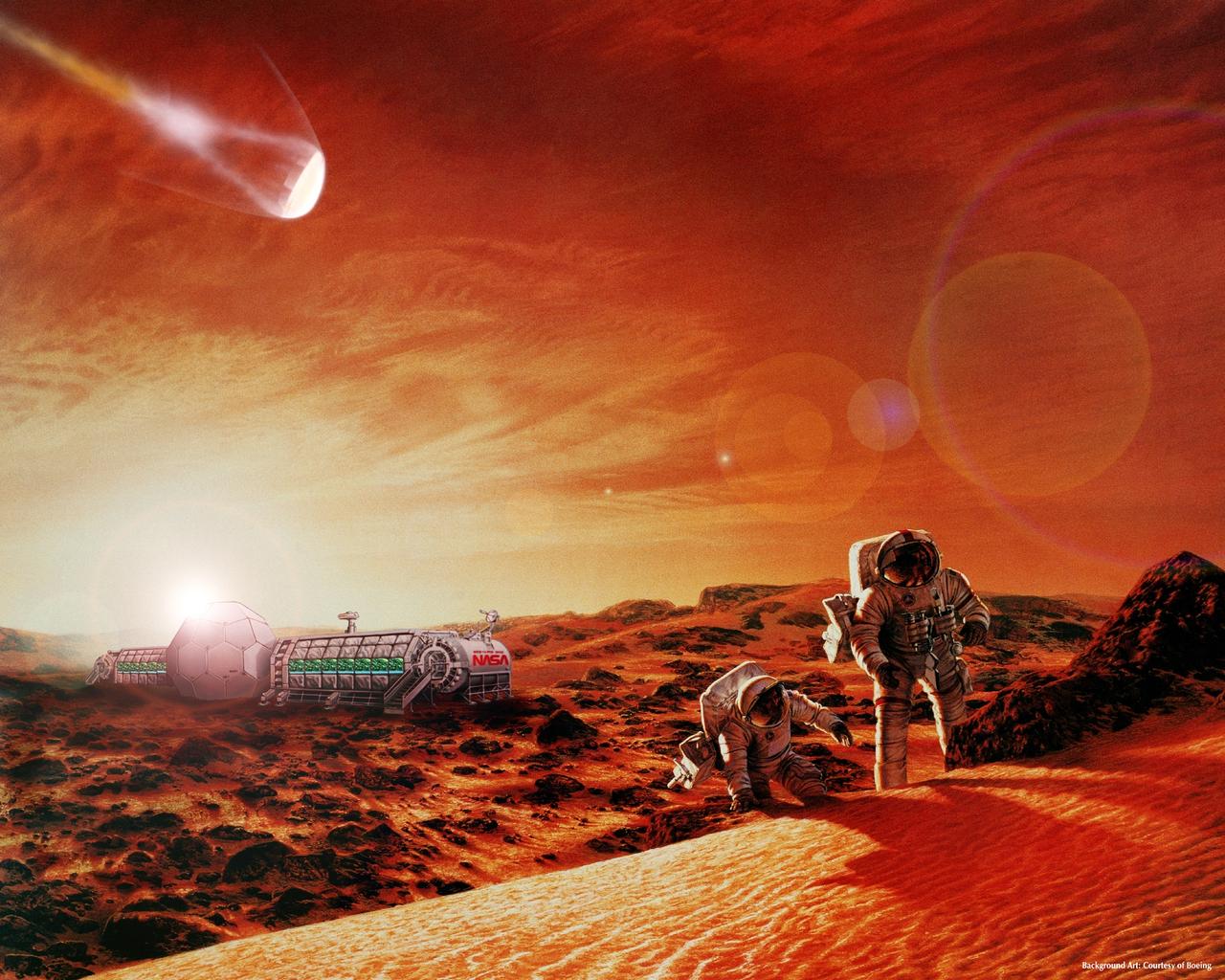
Digital Art by Jae Park Mars Exploraton: composite art showing AEF and CELSS (background art courtesy of Boeing)
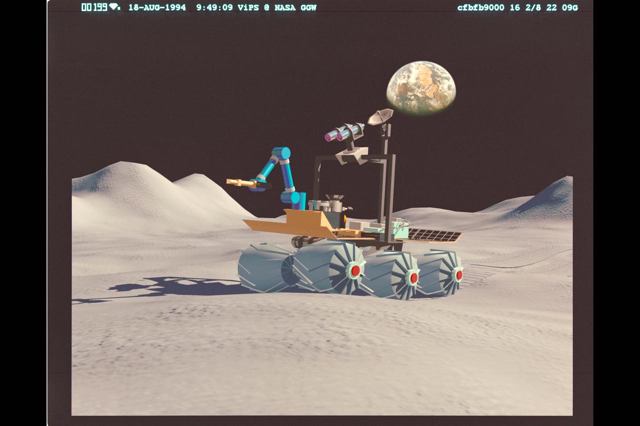
Digital Art by Boris Rabin Telepresence: Russian Rover Marsakhod on Lunar surface
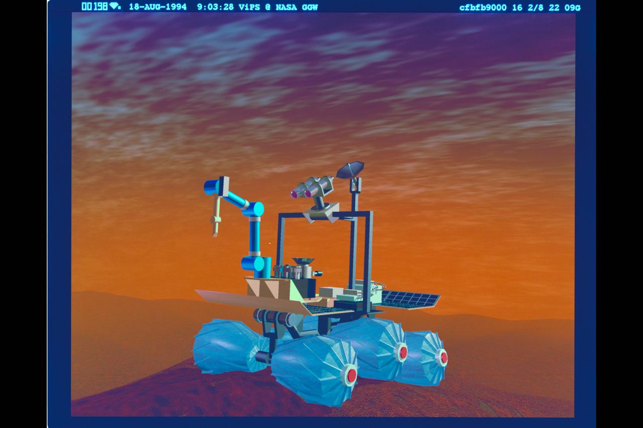
Digital Art by Boris Rabin Telepresence: Russian Rover Marsakhod on Martian surface
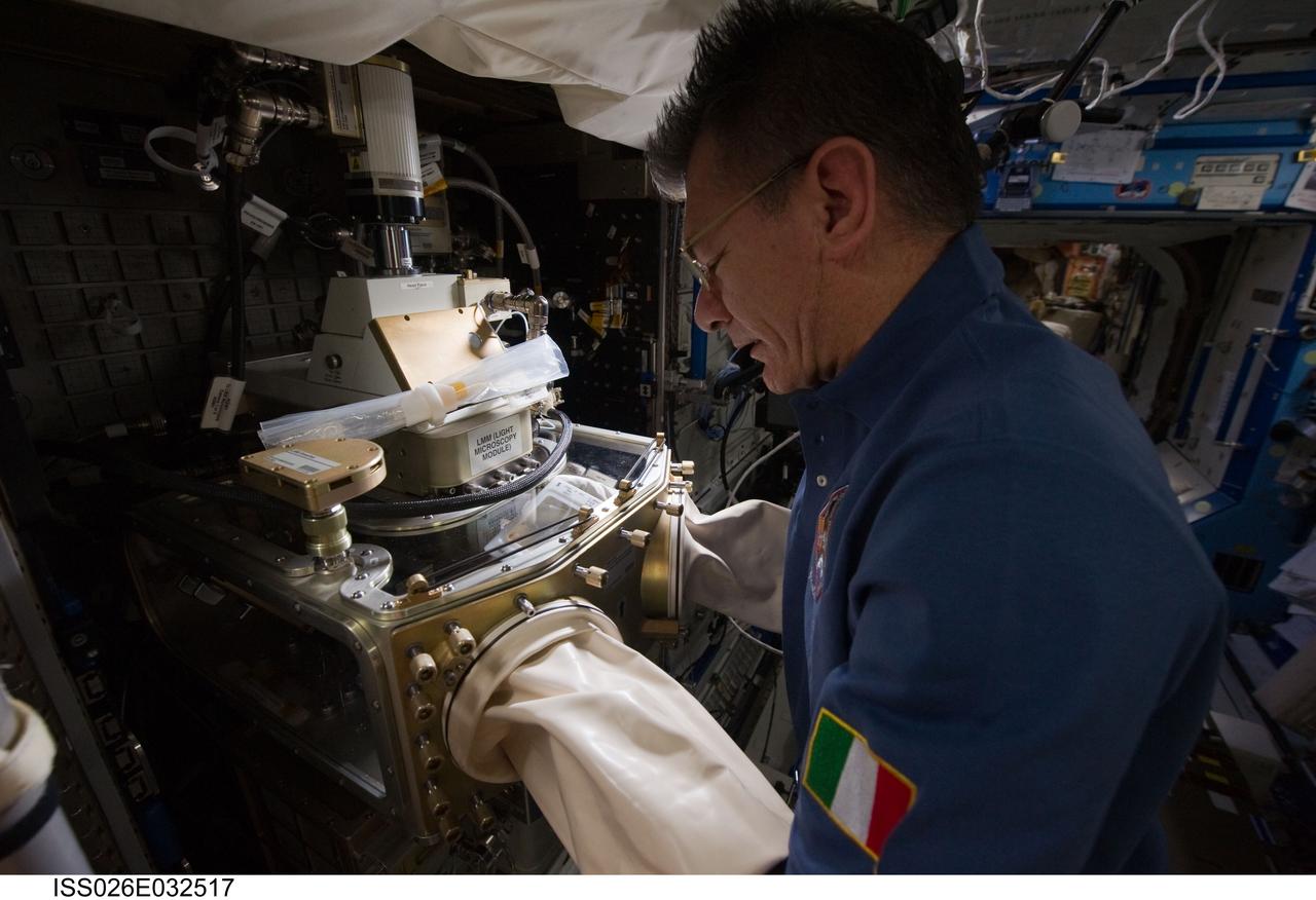
iss026e032517 (3/8/2011) --- European Space Agency (ESA) Paolo Nespoli works with the Light Microscopy Module (LMM) in the U.S. Laboratory. The Light Microscopy Module (LMM) is a modified commercial, highly flexible, state-of-the-art light imaging microscope facility that provides researchers with powerful diagnostic hardware and software onboard the International Space Station (ISS). The LMM enables novel research of microscopic phenomena in microgravity, with the capability of remotely acquiring and downloading digital images and videos across many levels of magnification.
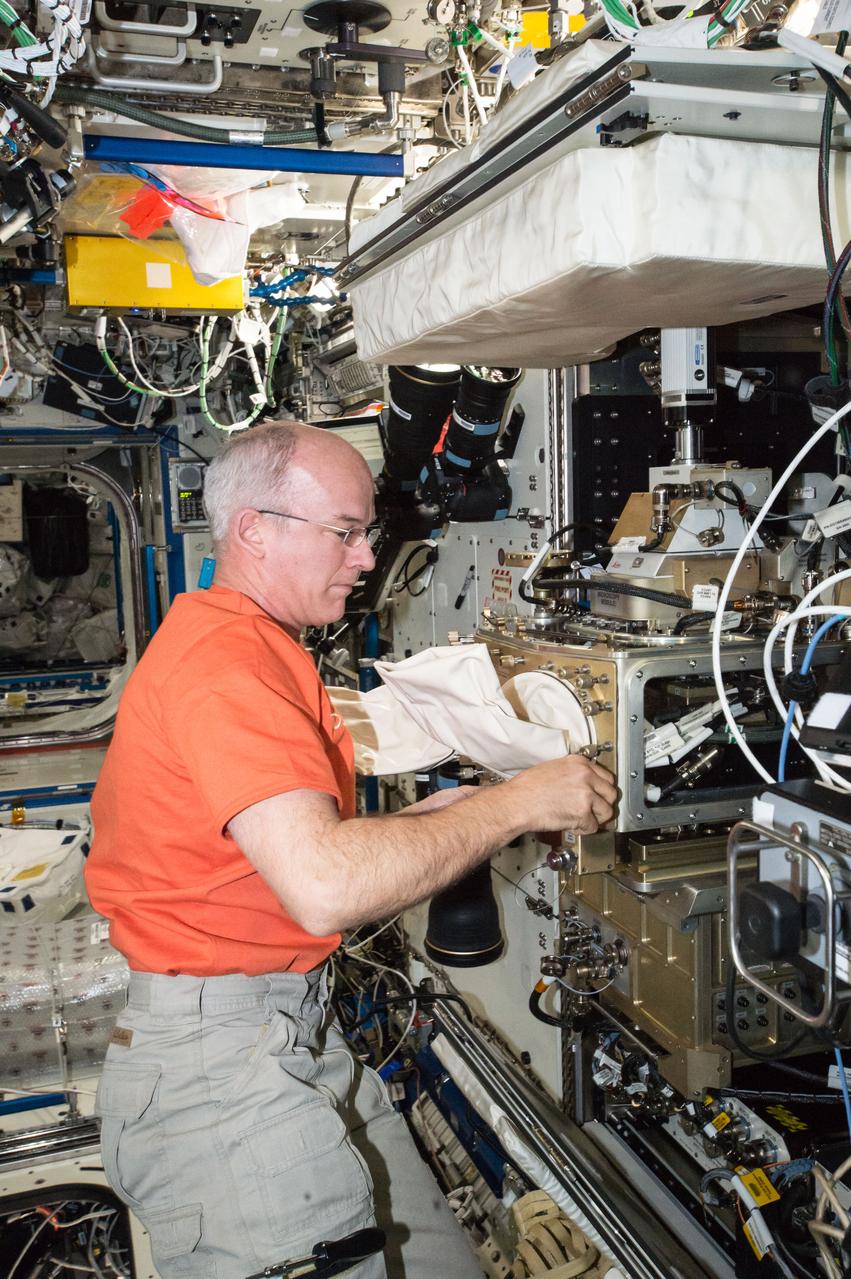
ISS047e066551 (04/18/2016) --- NASA astronaut Jeff Williams configures the station’s Light Microscopy Module (LMM), a modified commercial, highly flexible, state-of-the-art light imaging microscope facility that provides researchers with powerful diagnostic hardware and software. The LMM enables novel research of microscopic phenomena in microgravity, with the capability of remotely acquiring and downloading digital images and videos across many levels of magnification.

See Jupiter's Great Red Spot as you've never seen it before in this new Jovian work of art. Artist Mik Petter created this unique, digital artwork using data from the JunoCam imager on NASA's Juno spacecraft. The art form, known as fractals, uses mathematical formulas to create art with an infinite variety of form, detail, color and light. The tumultuous atmospheric zones in and around the Great Red Spot are highlighted by the author's use of colorful fractals. Vibrant colors of various tints and hues, combined with the almost organic-seeming shapes, make this image seem to be a colorized and crowded petri dish of microorganisms, or a close-up view of microscopic and wildly-painted seashells. The original JunoCam image was taken on July 10, 2017 at 7:10 p.m. PDT (10:10 p.m. EDT), as the Juno spacecraft performed its seventh close flyby of Jupiter. The spacecraft captured the image from about 8,648 miles (13,917 kilometers) above the tops of the clouds of the planet at a latitude of -32.6 degrees. https://photojournal.jpl.nasa.gov/catalog/PIA21777 . - Enhanced image by Mik Petter (CC-NC-SA) based on images provided courtesy of NASA/JPL-Caltech/SwRI/MSSS
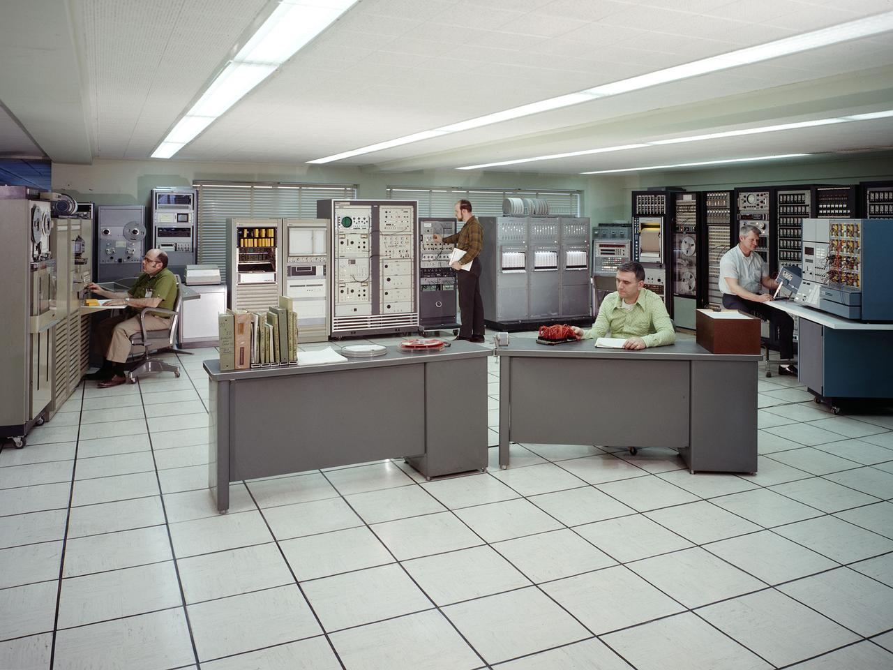
The test data recording equipment located in the office building of the 10-by 10-Foot Supersonic Wind Tunnel at the NASA Lewis Research Center. The data system was the state of the art when the facility began operating in 1955 and was upgraded over time. NASA engineers used solenoid valves to measure pressures from different locations within the test section. Up 48 measurements could be fed into a single transducer. The 10-by 10 data recorders could handle up to 200 data channels at once. The Central Automatic Digital Data Encoder (CADDE) converted this direct current raw data from the test section into digital format on magnetic tape. The digital information was sent to the Lewis Central Computer Facility for additional processing. It could also be displayed in the control room via strip charts or oscillographs. The 16-by 56-foot long ERA 1103 UNIVAC mainframe computer processed most of the digital data. The paper tape with the raw data was fed into the ERA 1103 which performed the needed calculations. The information was then sent back to the control room. There was a lag of several minutes before the computed information was available, but it was exponentially faster than the hand calculations performed by the female computers. The 10- by 10-foot tunnel, which had its official opening in May 1956, was built under the Congressional Unitary Plan Act which coordinated wind tunnel construction at the NACA, Air Force, industry, and universities. The 10- by 10 was the largest of the three NACA tunnels built under the act.

The high-tech art of digital signal processing (DSP) was pioneered at NASA's Jet Propulsion Laboratory (JPL) in the mid-1960s for use in the Apollo Lunar Landing Program. Designed to computer enhance pictures of the Moon, this technology became the basis for the Landsat Earth resources satellites and subsequently has been incorporated into a broad range of Earthbound medical and diagnostic tools. DSP is employed in advanced body imaging techniques including Computer-Aided Tomography, also known as CT and CATScan, and Magnetic Resonance Imaging (MRI). CT images are collected by irradiating a thin slice of the body with a fan-shaped x-ray beam from a number of directions around the body's perimeter. A tomographic (slice-like) picture is reconstructed from these multiple views by a computer. MRI employs a magnetic field and radio waves, rather than x-rays, to create images.

The high-tech art of digital signal processing (DSP) was pioneered at NASA's Jet Propulsion Laboratory (JPL) in the mid-1960s for use in the Apollo Lunar Landing Program. Designed to computer enhance pictures of the Moon, this technology became the basis for the Landsat Earth resources satellites and subsequently has been incorporated into a broad range of Earthbound medical and diagnostic tools. DSP is employed in advanced body imaging techniques including Computer-Aided Tomography, also known as CT and CATScan, and Magnetic Resonance Imaging (MRI). CT images are collected by irradiating a thin slice of the body with a fan-shaped x-ray beam from a number of directions around the body's perimeter. A tomographic (slice-like) picture is reconstructed from these multiple views by a computer. MRI employs a magnetic field and radio waves, rather than x-rays, to create images. In this photograph, a patient undergoes an open MRI.

The high-tech art of digital signal processing (DSP) was pioneered at NASA's Jet Propulsion Laboratory (JPL) in the mid-1960s for use in the Apollo Lunar Landing Program. Designed to computer enhance pictures of the Moon, this technology became the basis for the Landsat Earth resources satellites and subsequently has been incorporated into a broad range of Earthbound medical and diagnostic tools. DSP is employed in advanced body imaging techniques including Computer-Aided Tomography, also known as CT and CATScan, and Magnetic Resonance Imaging (MRI). CT images are collected by irradiating a thin slice of the body with a fan-shaped x-ray beam from a number of directions around the body's perimeter. A tomographic (slice-like) picture is reconstructed from these multiple views by a computer. MRI employs a magnetic field and radio waves, rather than x-rays, to create images.

The high-tech art of digital signal processing (DSP) was pioneered at NASA's Jet Propulsion Laboratory (JPL) in the mid-1960s for use in the Apollo Lunar Landing Program. Designed to computer enhance pictures of the Moon, this technology became the basis for the Landsat Earth resources satellites and subsequently has been incorporated into a broad range of Earthbound medical and diagnostic tools. DSP is employed in advanced body imaging techniques including Computer-Aided Tomography, also known as CT and CATScan, and Magnetic Resonance Imaging (MRI). CT images are collected by irradiating a thin slice of the body with a fan-shaped x-ray beam from a number of directions around the body's perimeter. A tomographic (slice-like) picture is reconstructed from these multiple views by a computer. MRI employs a magnetic field and radio waves, rather than x-rays, to create images.