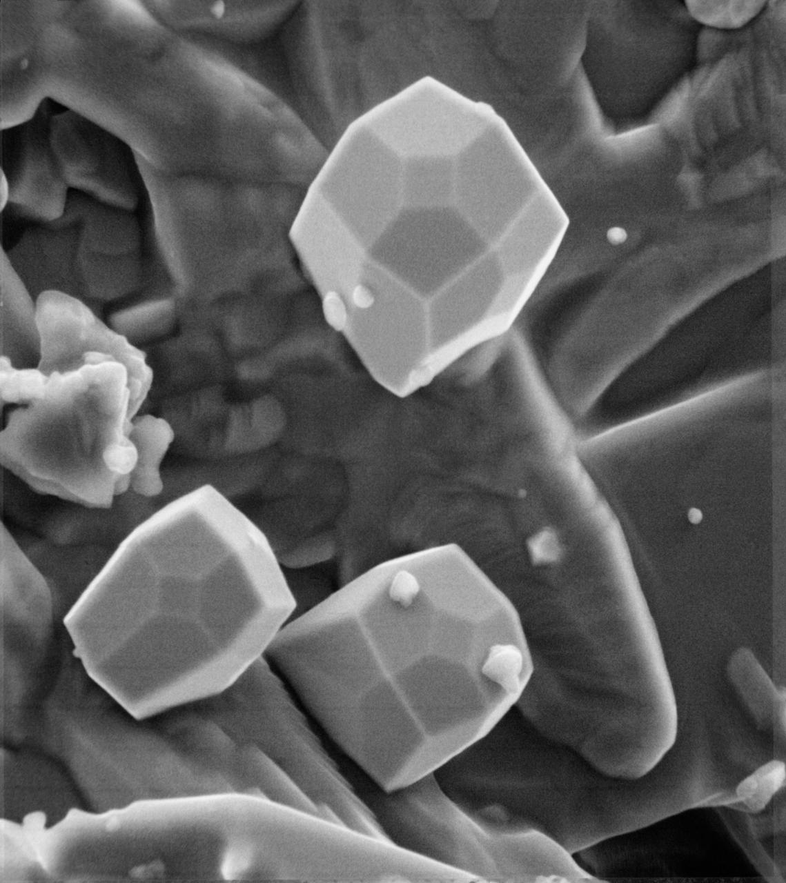
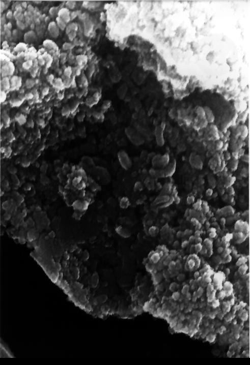
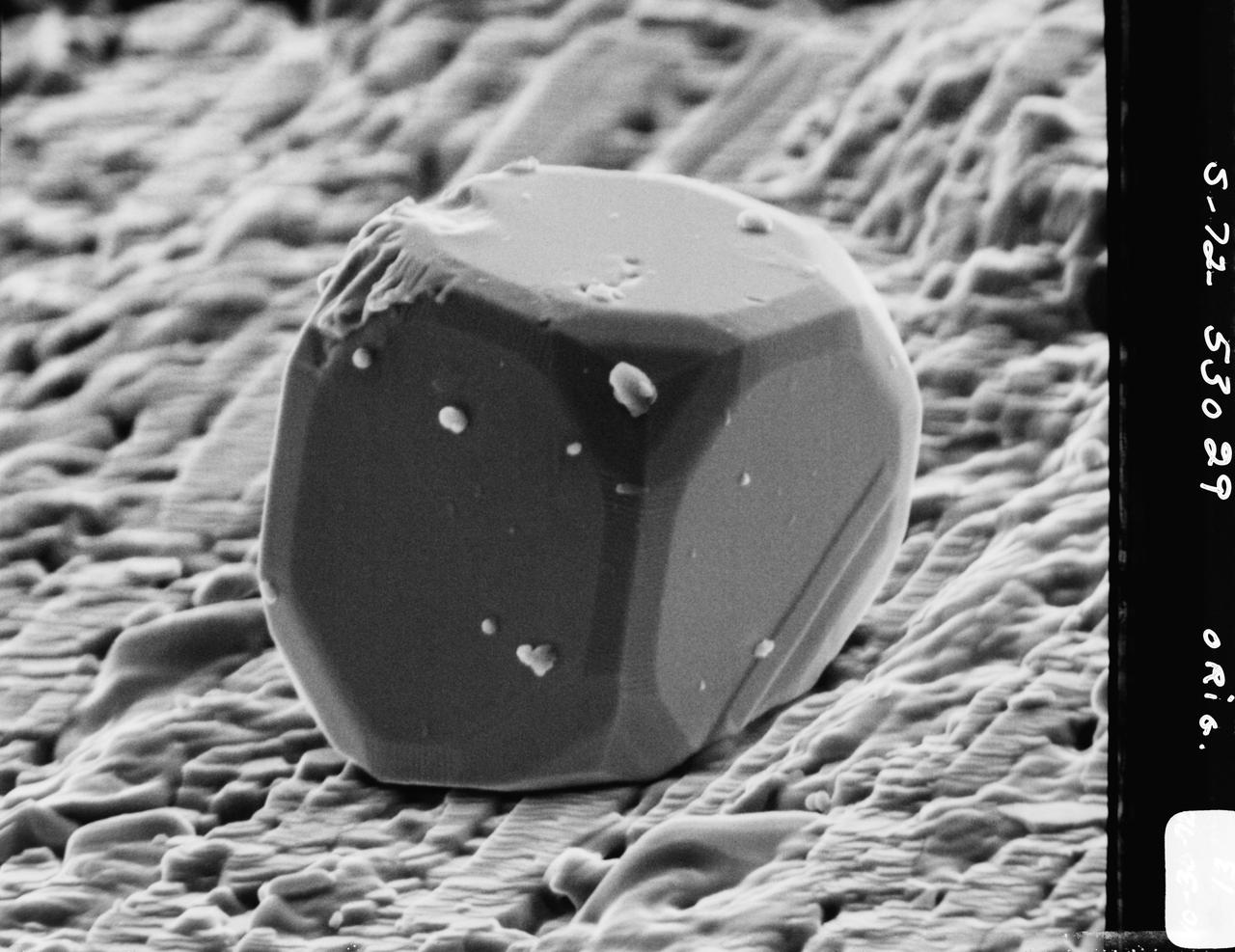
A scanning electron microscope photograph of iron crystals which grow in a small vug or cavity in a recrystallized breccia (fragmented rock) from the Apollo 15 Hadley-Apennino lunar landing site. The largest crystal is three microns across. Perfectly developed crystals such as these indicate slow formation from a hot vapor as the rock was cooling. The crystals are resting on an interlocking lattice of pyroxene (calsium-magnesium-iron silicate).
This electron microscope image is a close-up of the center part of photo number S96-12301. http://photojournal.jpl.nasa.gov/catalog/PIA00284
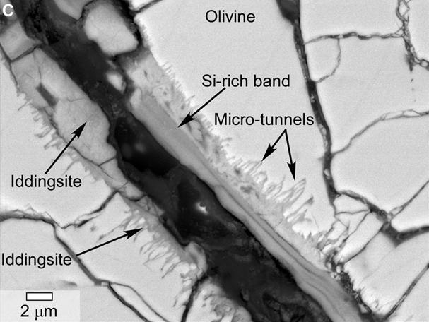
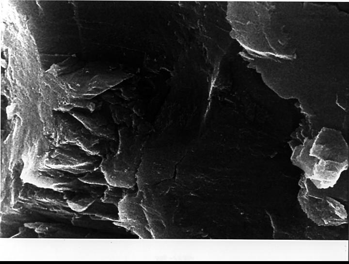
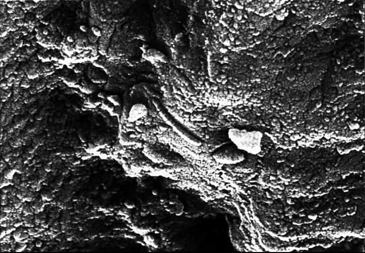
This scanning electron microscope image of a polished thin section of a meteorite from Mars shows tunnels and curved microtunnels.
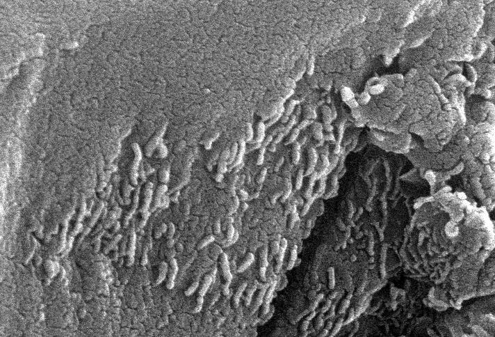
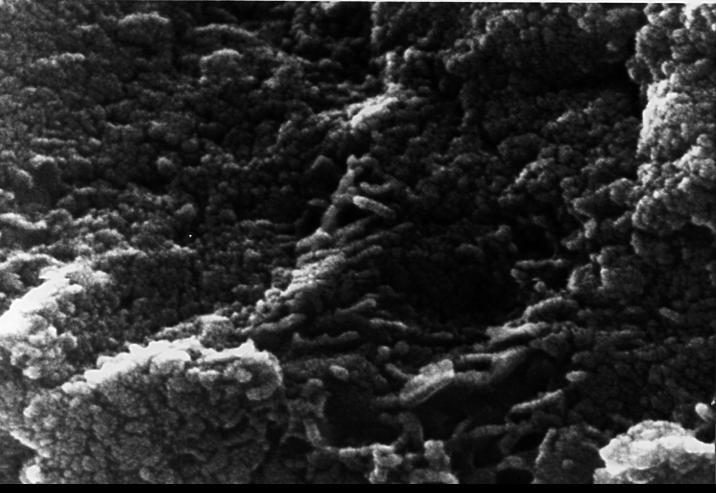
This electron microscope image shows extremely tiny tubular structures that are possible microscopic fossils of bacteria-like organisms that may have lived on Mars more than 3.6 billion years ago. http://photojournal.jpl.nasa.gov/catalog/PIA00285
This electron microscope image shows egg-shaped structures, some of which may be possible microscopic fossils of Martian origin as discussed by NASA research published in the Aug. 16, 1996. http://photojournal.jpl.nasa.gov/catalog/PIA00286
In the center of this electron microscope image of a small chip from a meteorite are several tiny structures that are possible microscopic fossils of primitive, bacteria-like organisms that may have lived on Mars more than 3.6 billion years ago. http://photojournal.jpl.nasa.gov/catalog/PIA00283
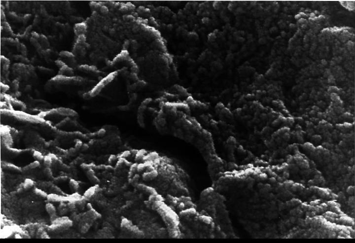
This scanning electron microscope image shows speroidal features embedded in a layer of iddingsite, a mineral formed by action of water, in a meteorite that came from Mars.
This electron microscope image shows tubular structures of likely Martian origin. These structures are very similar in size and shape to extremely tiny microfossils found in some Earth rocks. http://photojournal.jpl.nasa.gov/catalog/PIA00287
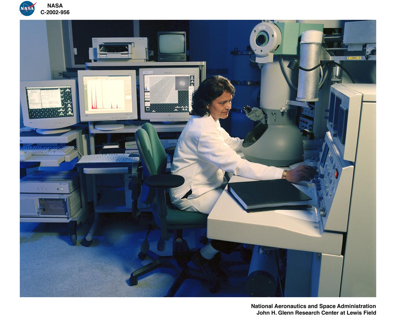
TRANSMISSION ELECTRON MICROSCOPE / TEM
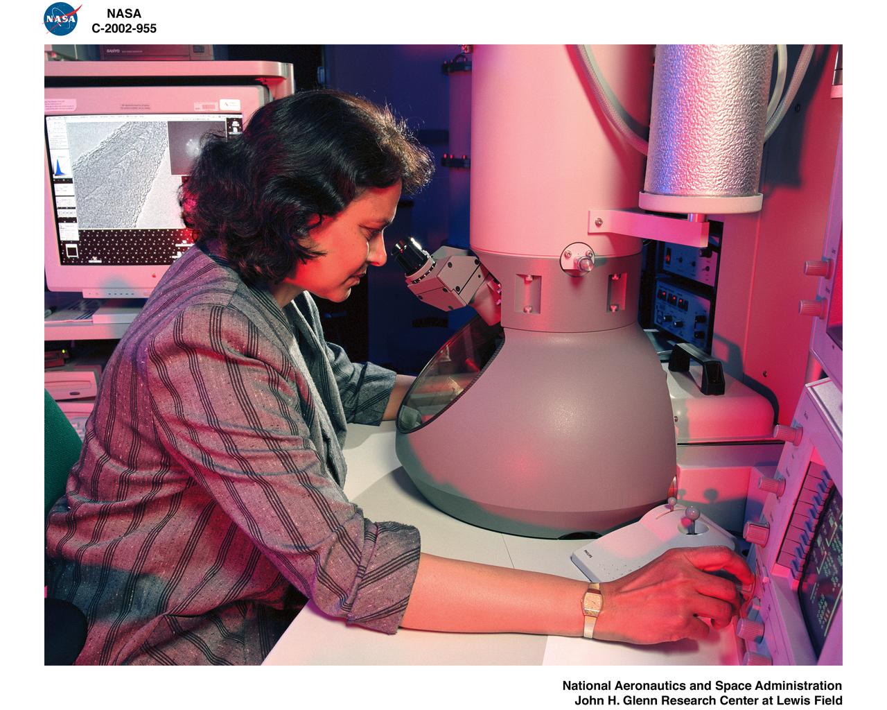
TRANSMISSION ELECTRON MICROSCOPE / TEM
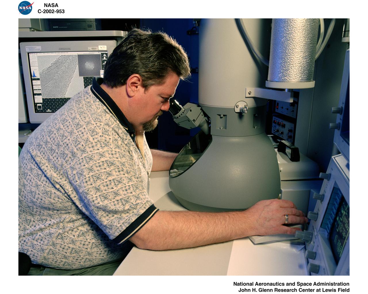
TRANSMISSION ELECTRON MICROSCOPE / TEM
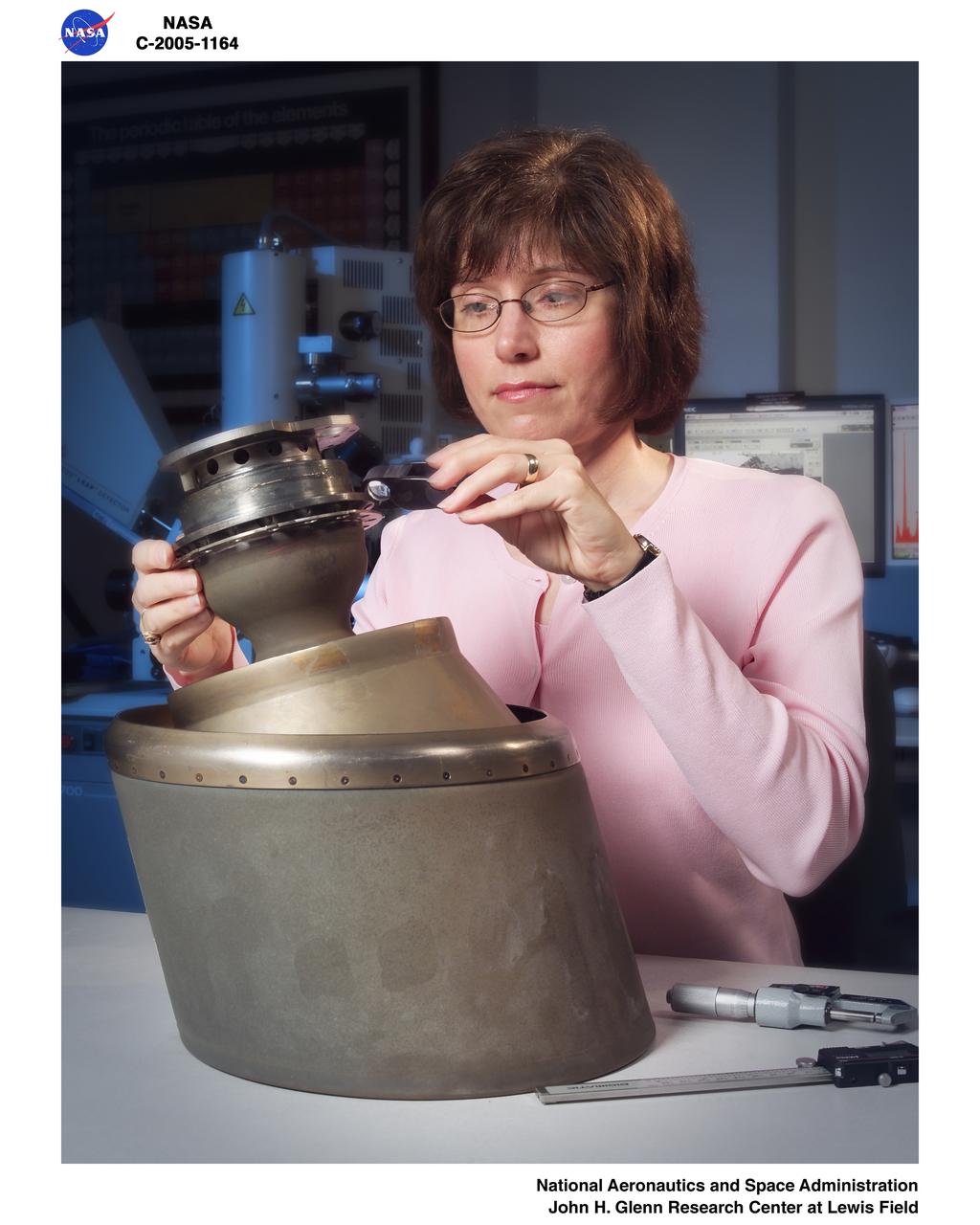
Reaction Control System Thruster examined in the electron optics lab Near Field Emission Scanning Electron Microscope
This high-resolution scanning electron microscope image shows an unusual tube-like structural form that is less than 1/100th the width of a human hair in size found in meteorite ALH84001, a meteorite believed to be of Martian origin. http://photojournal.jpl.nasa.gov/catalog/PIA00288
PHOTOMICROPHOTOGRAPHY -GEOLOGY (SEM) High magnification and resolution views of lunar, meteorite and terrestrial materials with the Scanning Electron Microscope (SEM).

OVERVIEW OF THE MATERIALS DIAGNOSTIC LABORATORY. THE NEAR END SHOWS THE SURFACE ANALYSIS INSTRUMENTS SUCH AS THE SECONDARY ION MASS SPECTROSCOPE (CLOSEST) AND THE TWO ELECTRON SPECTROSCOPY INSTRUMENTS, WHILE THE FAR END SHOWS THE NEW SCANNING ELECTRON MICROSCOPES
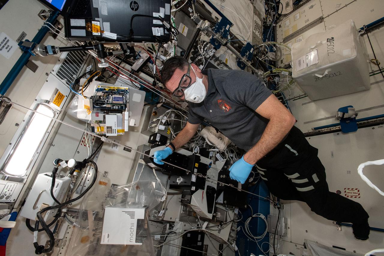
iss065e241981 (Aug. 12, 2021) --- NASA astronaut and Expedition 65 Flight Engineer Shane Kimbrough conducts maintenance on a miniature electron microscope, called Mochii, to support spectroscopic investigations and analyses of microscopic particles aboard the International Space Station.
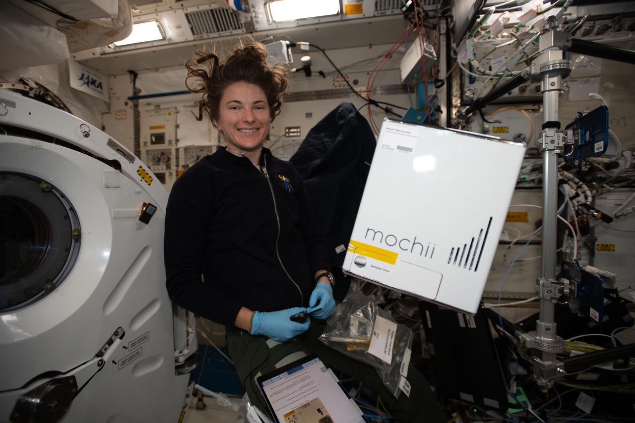
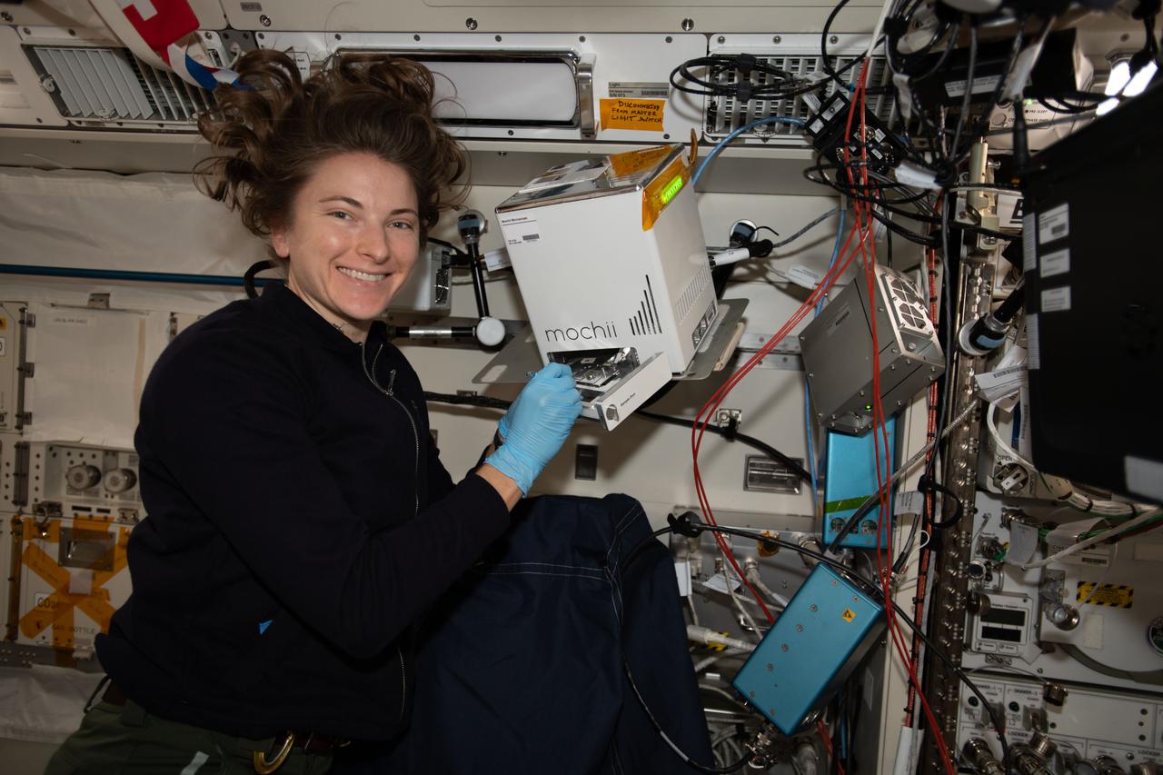
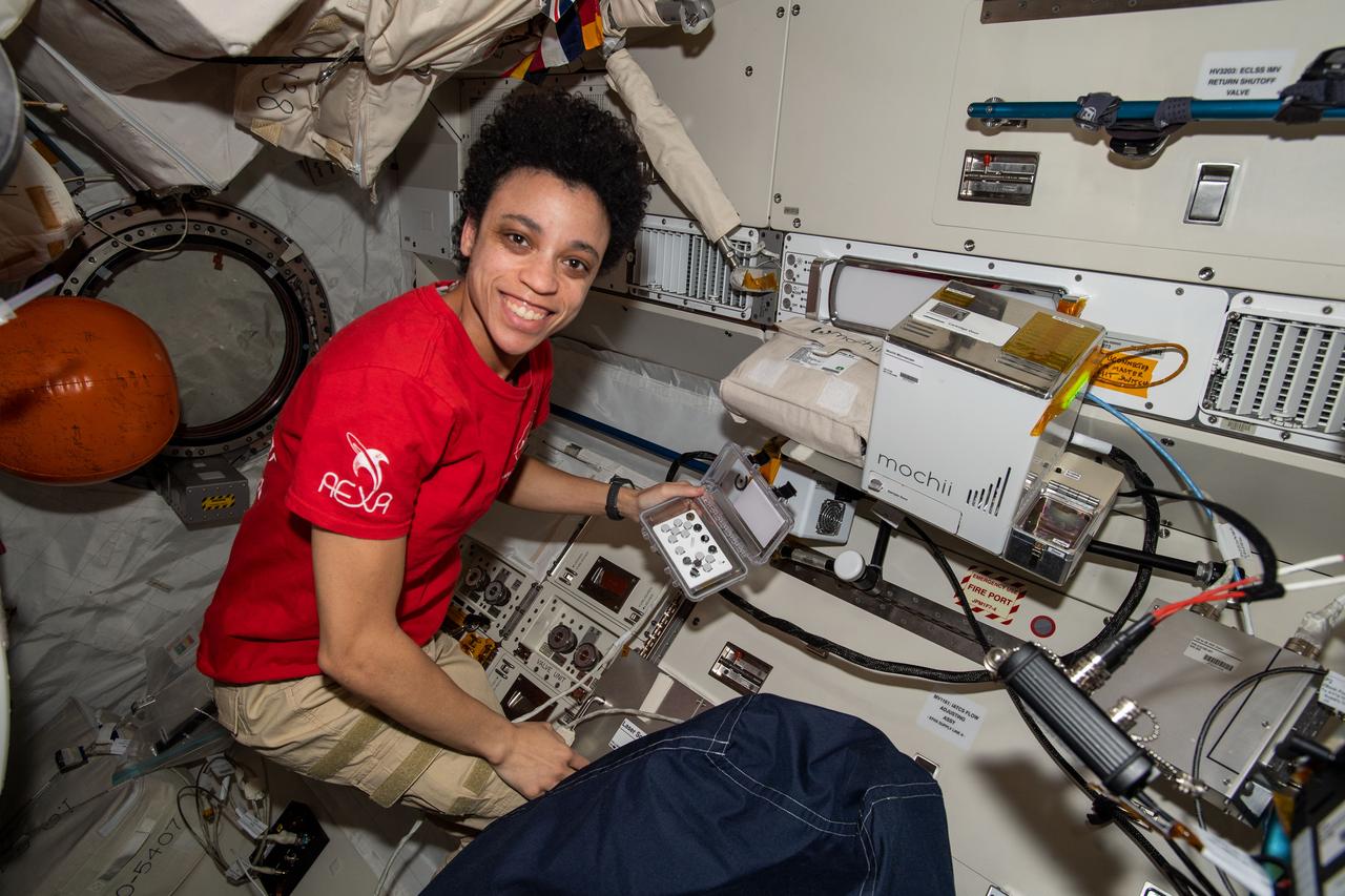
iss066e110531_alt (1/10/2022) --- NASA astronaut Kayla Barron sets up the Mochii microscope. Mochii is a miniature scanning electron microscope (SEM) with spectroscopy to conduct real-time, on-site imaging and compositional measurements of particles on the International Space Station (ISS).
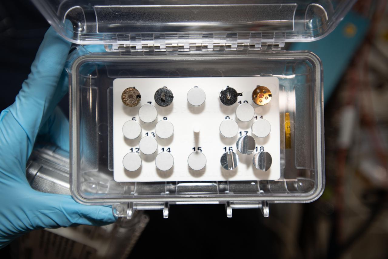
iss066e110547 (1/10/2022) --- A view of the Mochii microscope sample load aboard the International Space Station (ISS). Mochii is a miniature scanning electron microscope (SEM) with spectroscopy to conduct real-time, on-site imaging and compositional measurements of particles on the International Space Station (ISS)
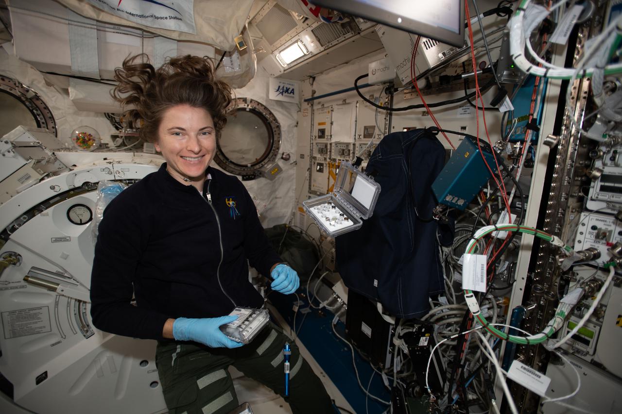
iss066e110556 (1/10/2022) --- NASA astronaut Kayla Barron sets up the Mochii microscope. Mochii is a miniature scanning electron microscope (SEM) with spectroscopy to conduct real-time, on-site imaging and compositional measurements of particles on the International Space Station (ISS).
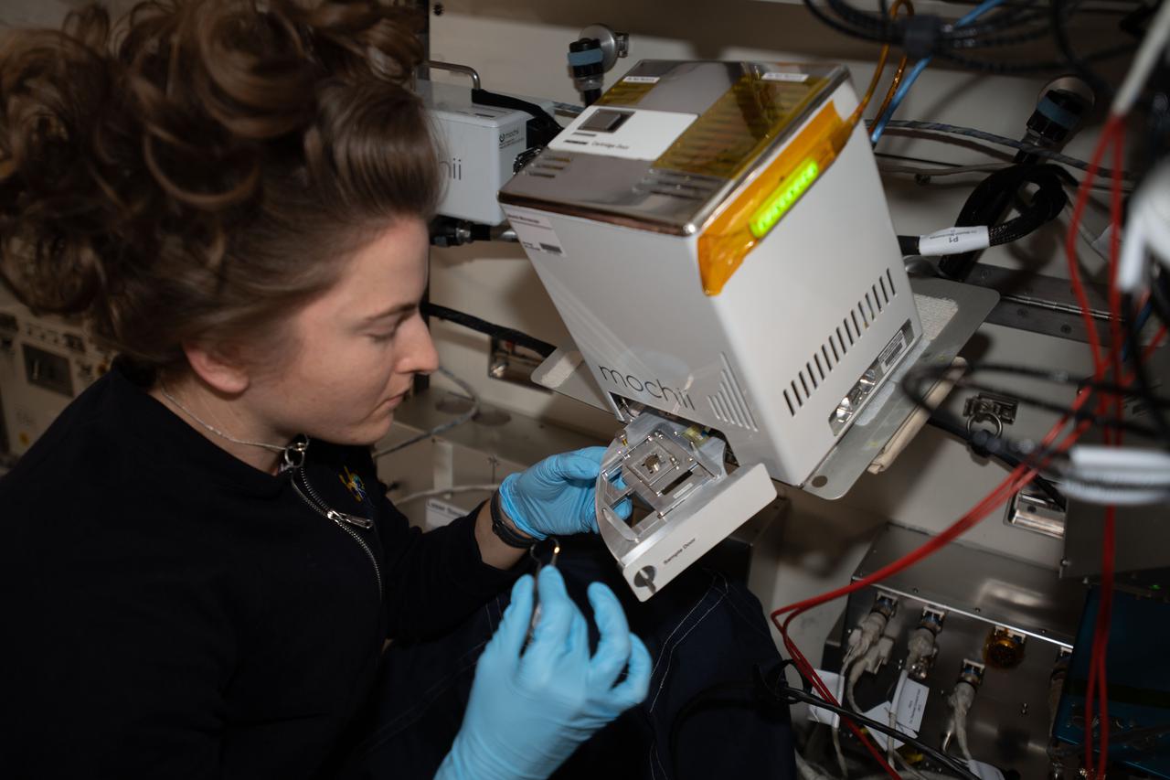
iss066e110566 (1/10/2022) --- NASA astronaut Kayla Barron sets up the Mochii microscope. Mochii is a miniature scanning electron microscope (SEM) with spectroscopy to conduct real-time, on-site imaging and compositional measurements of particles on the International Space Station (ISS).
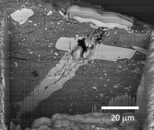
A scanning electron microscope image of a micrometeorite impact crater in a particle of asteroid Bennu material. Scientists found microscopic craters and tiny splashes of once-molten rock – known as impact melts – on the surfaces of samples, signs that the asteroid was bombarded by micrometeorites.
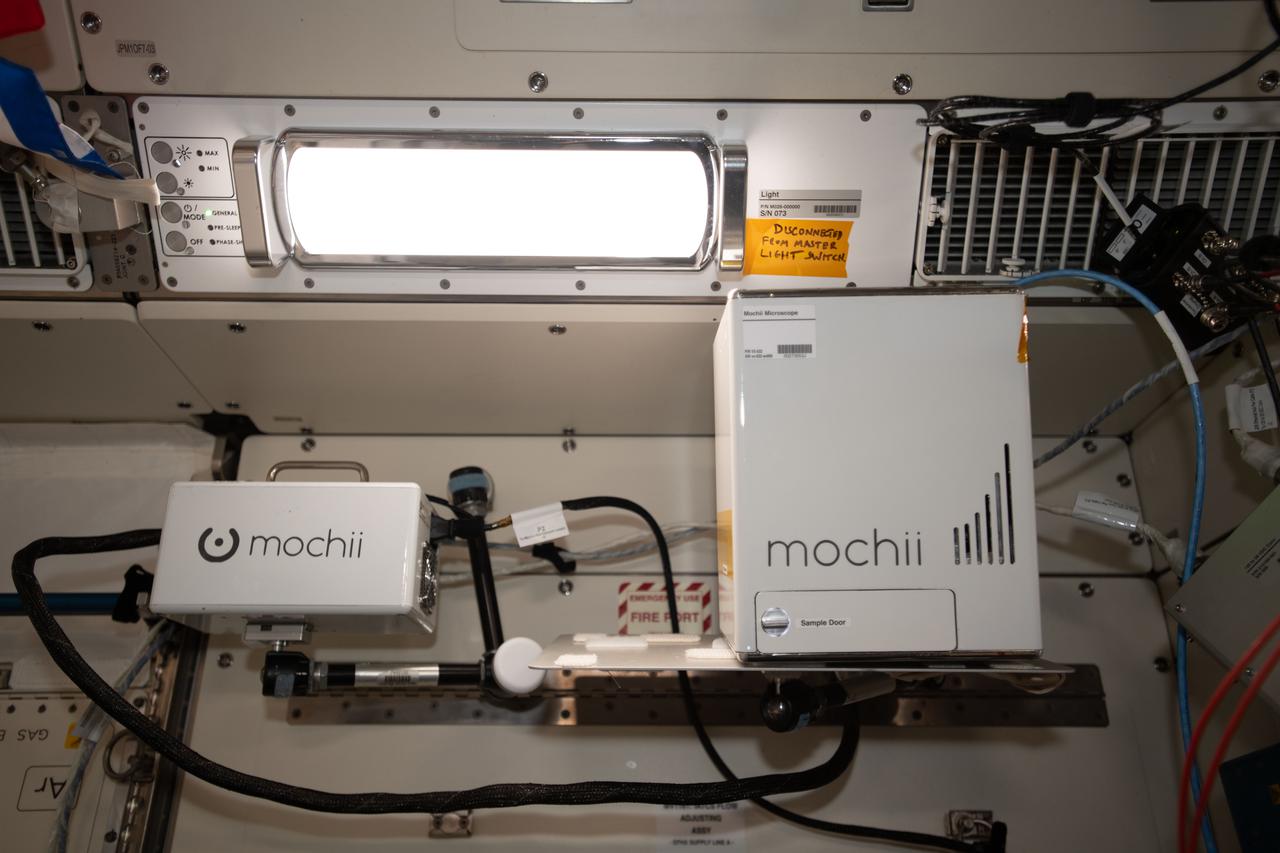
iss066e110545 (1/10/2022) --- A view of the Mochii microscope aboard the International Space Station (ISS. Mochii is a miniature scanning electron microscope (SEM) with spectroscopy to conduct real-time, on-site imaging and compositional measurements of particles on the International Space Station (ISS).
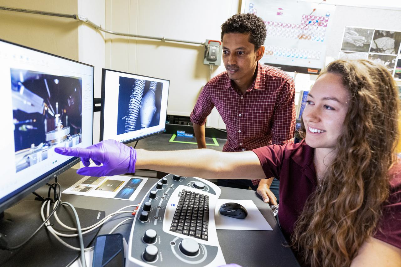
Chemists Misle Tessema (left) and Macy Mullen (right) discuss scanning electron microscope operations inside NASA Engineering’s Analytical Laboratories at Kennedy Space Center in Florida on July 7, 2021. One of seven branches in the NASA Laboratories, Development, and Testing Division, the Analytical Laboratories branch provides microscopic imagery and analysis through the use of a wide variety of microscopic techniques to identify contaminants and other urgent problems associated with aerospace flight hardware, ground support equipment, and related facilities.
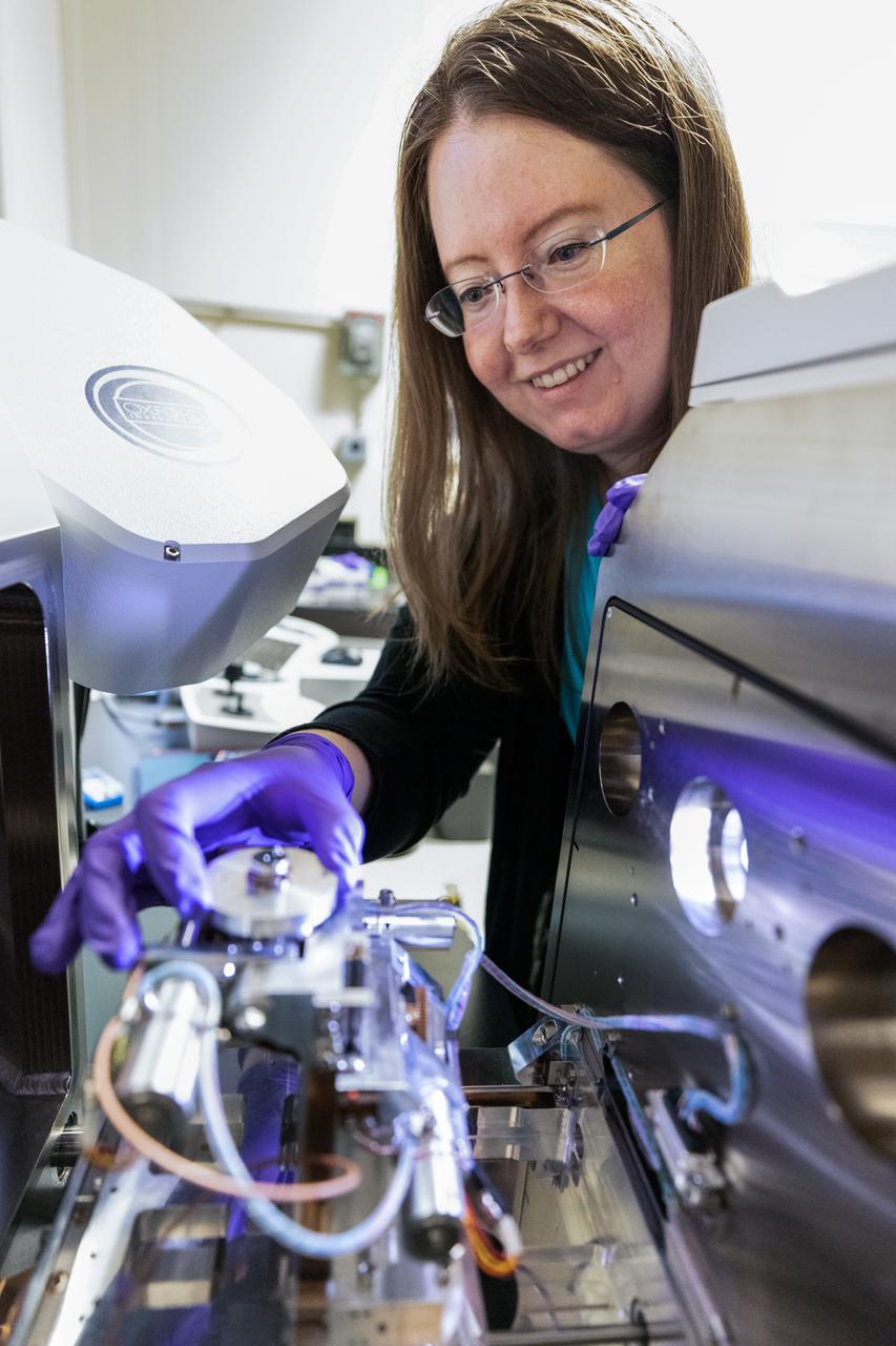
Chemist Athela Frandsen from NASA Engineering’s Analytical Laboratories at Kennedy Space Center in Florida loads a sample into a scanning electron microscope on July 7, 2021. One of seven branches in the NASA Laboratories, Development, and Testing Division, the Analytical Laboratories branch provides microscopic imagery and analysis through the use of a wide variety of microscopic techniques to identify contaminants and other urgent problems associated with aerospace flight hardware, ground support equipment, and related facilities.
A scanning electron microscope captured this image of terresterial soil containing a phyllosilicate mineral from Koua Bocca, Ivory Coast, West Africa. This soil shares some similarities with Martian soil scooped by NASA Phoenix Lander.
iss066e110569 (Jan. 10, 2022) --- NASA astronaut and Expedition 66 Flight Engineer Kayla Barron sets up and installs the Mochii electron-scanning microscope that can be used to identify trace particles aboard the International Space Station.
PHOTOMICROPHOTOGRAPHY -GEOLOGY (SEM) High magnification and resolution views of lunar, meteorite and terrestrial materials using the Scanning Electron MIcroscope (SEM), Bldg. 31 Planetary and Earth Science Laboratory.
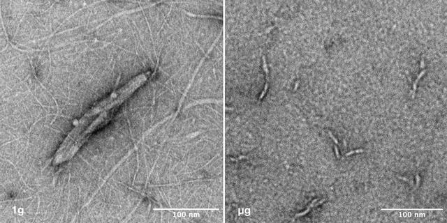
jsc2025e036194 (4/4/2025) --- Image of nanoparticles synthesized under 1g vs microgravity. Left: This transmission electron microscope image depicts the nano-scale structure of Janus Base Nanoparticles encapsulated with mRNA on ground. Right: This is a transmission electron microscope image of mRNA-encapsulated JBNp that was produced on ISS during the SpaceX CRS-31 mission. Here, you can see that the space-made JBNp is smaller and more uniform in size and shape with less background material, demonstrating the stark advantage that in-space manufacturing can provide JBNp: improved uniformity and drug loading. Image courtesy of University of Connecticut.
Martian Meteorite (ALH84001): This high resolution transmission electron microscope image is of a cast, or replica, from a chip of a Martian meteorite, labeled ALH84001, that shows the outline of what are believed to be possible microscopic fossils of bacteria-like organisms that may have lived on Mars more than 3.6 billion years ago. The tubular features in this image are less than a micrometer in size, or about 1/500th the diameter of a human hair. (JSC ref: S96-12637)
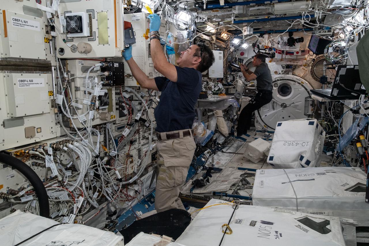
iss065e242201 (Aug. 13, 2021) --- Expedition 65 Commander Akihiko Hoshide of the Japan Aerospace Exploration Agency (JAXA) inserts cell samples into the Kibo laboratory module's Cell Biology Experiment Facility. At the rear of Kibo, NASA Flight Engineer Shane Kimbrough trains to use a miniature electron microscope, called Mochii, to support spectroscopic investigations and analyses of microscopic particles aboard the International Space Station.
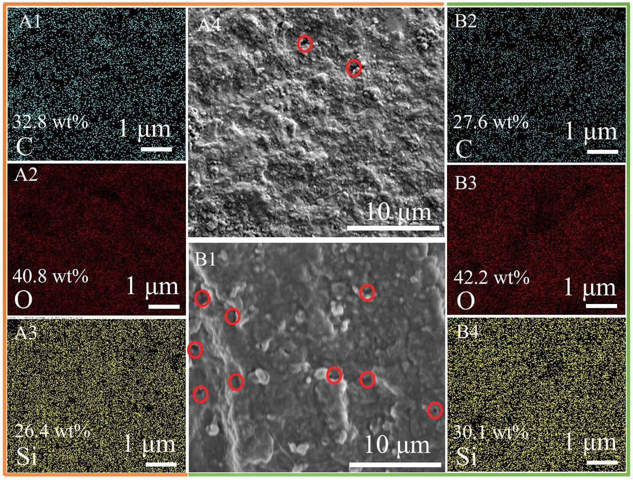
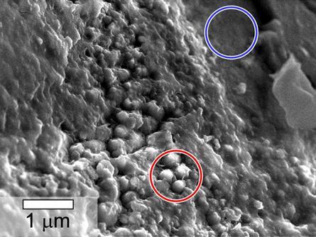
jsc2025e064333 (5/31/2024) --- Scanning electron microscope (SEM) image graphs and elemental mappings of the samples pyrolyzed at 1200 oC in the Ar (A1-A4) and microgravity (B1-B4). (The red circles in the SEM images represent obvious pores).
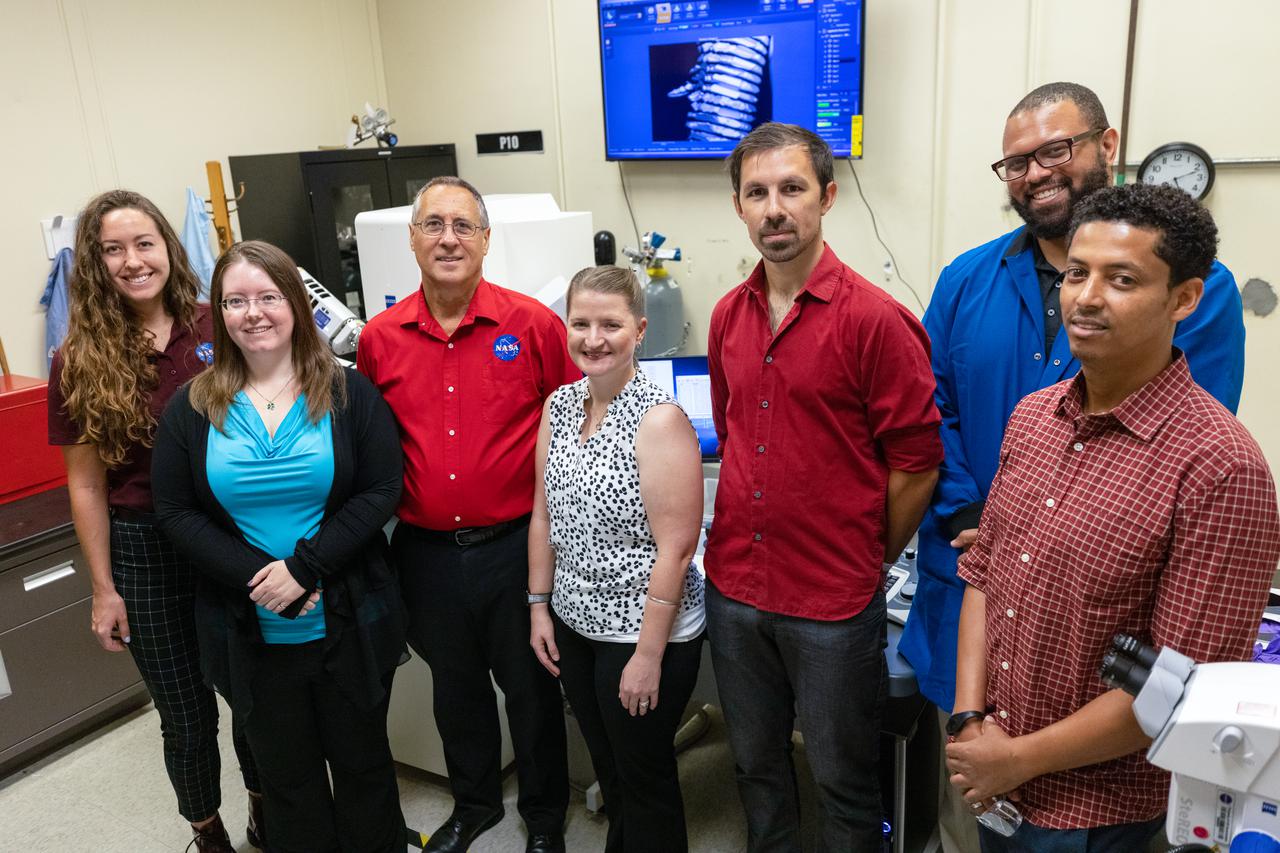
Chemists from NASA Engineering’s Analytical Laboratories at Kennedy Space Center in Florida pose for a photo near a scanning electron microscope on July 7, 2021. From left to right is Macy Mullen, structural materials; Athela Frandsen, structural materials; Philip Howard, lead, structural materials; Janelle Coutts, gas and fluid systems; David Rinderknecht, structural materials; Trey Barnes, Earth biosphere studies; and Misle Tessema, Earth biosphere studies. One of seven branches in the NASA Laboratories, Development, and Testing Division, the Analytical Laboratories branch provides microscopic imagery and analysis through the use of a wide variety of microscopic techniques to identify contaminants and other urgent problems associated with aerospace flight hardware, ground support equipment, and related facilities.
iss068e016422 (Oct. 12, 2022) --- NASA astronaut and Expedition 68 Flight Engineer Jessica Watkins works with Mochii, a miniature scanning electron microscope (SEM) with spectroscopy to conduct real-time, on-site imaging and compositional measurements of particles on the International Space Station (ISS). Such particles can cause vehicle and equipment malfunctions and threaten crew health, but currently, samples must be returned to Earth for analysis, leaving crew and vehicle at risk. Mochii also provides a powerful new analysis platform to support novel microgravity science and engineering.
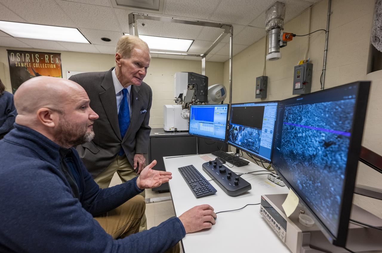
NASA Administrator Bill Nelson uses an electron microscope to view a sample from asteroid Bennu, Friday, Nov. 3, 2023, at the Smithsonian’s National Museum of Natural History in Washington. The sample was collected from the carbon rich near Earth asteroid Bennu in October 2020 by NASA’s OSIRIS-REx spacecraft. Photo Credit: (NASA/Keegan Barber)
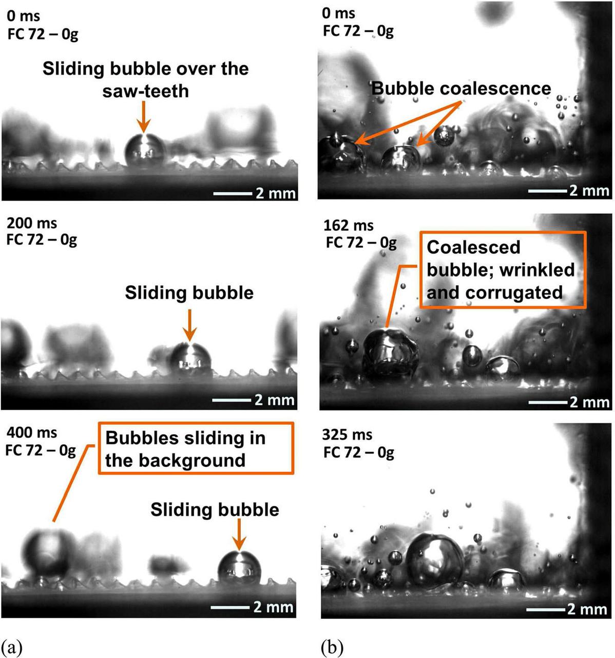
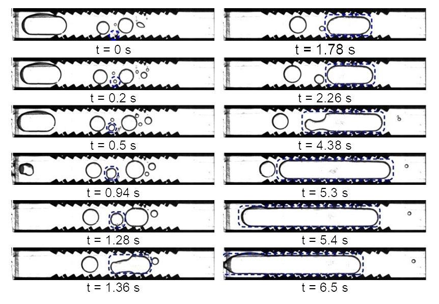
jsc2020e030483 (4/20/2020) --- A preflight image sequence from parabolic flight experiments indicating motion of vapor bubble on heated ratchet surface. Asymmetric Sawtooth and Cavity-Enhanced Nucleation-Driven Transport (PFMI-ASCENT) demonstrates a passive cooling system for electronic devices in microgravity using a microstructured surface. When fluids boil over flat heated surfaces in microgravity, vapor bubbles grow larger in size, causing poor heat transfer that can lead to damage of devices. Adding microscopic rachets on the surface may passively enable mobility of vapor bubbles and prevent this damage. (Image courtesy of: Techshot, Inc.)
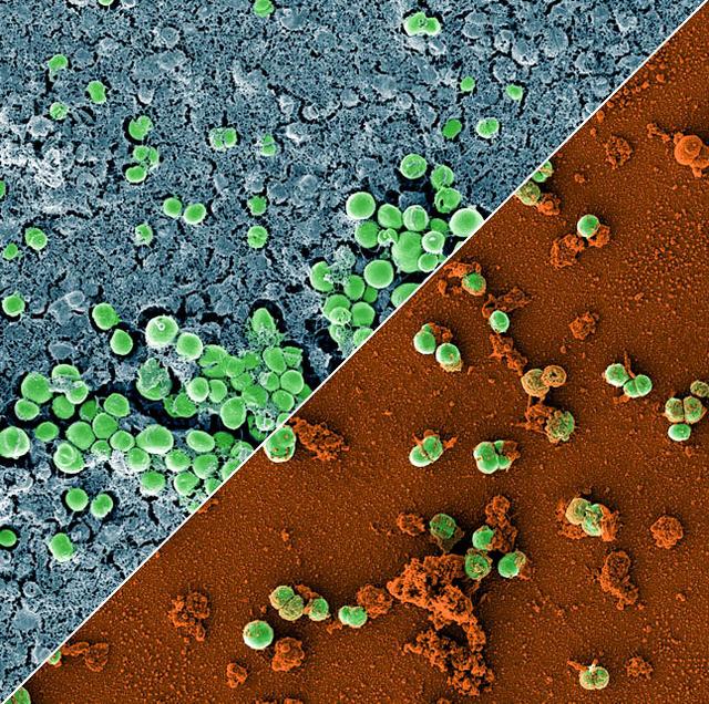
jsc2023e010179 (2/28/2023) --- This image is a composition of two scanning electron microscopic images of the bacterium Staphylococcus capitis on stainless steel versus antimicrobial copper. The image was colored to visualize the bacterial cells (green) either embedded in a biofilm matrix (blue), or covered with copper particles (red/orange). The ESA-Biofilms investigation studies bacterial biofilm formation and antimicrobial properties of different metal surfaces under spaceflight conditions in altered gravity. Both images were taken as part of the preflight experiments for ESA-Biofilms. Image courtesy of DLR, CC BY-NC-ND 3.0.
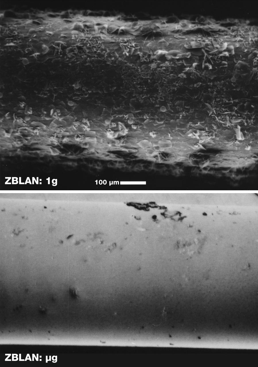
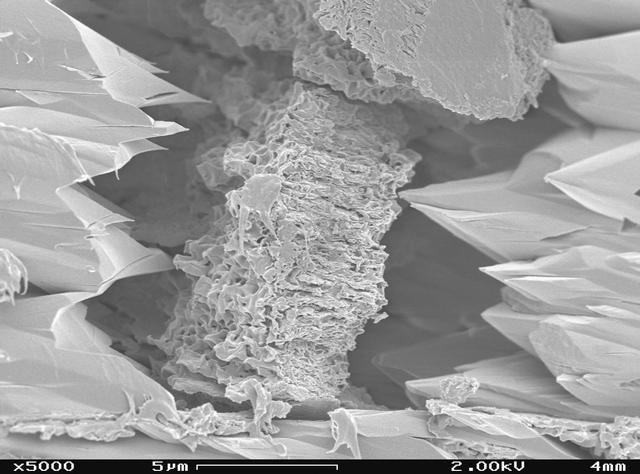
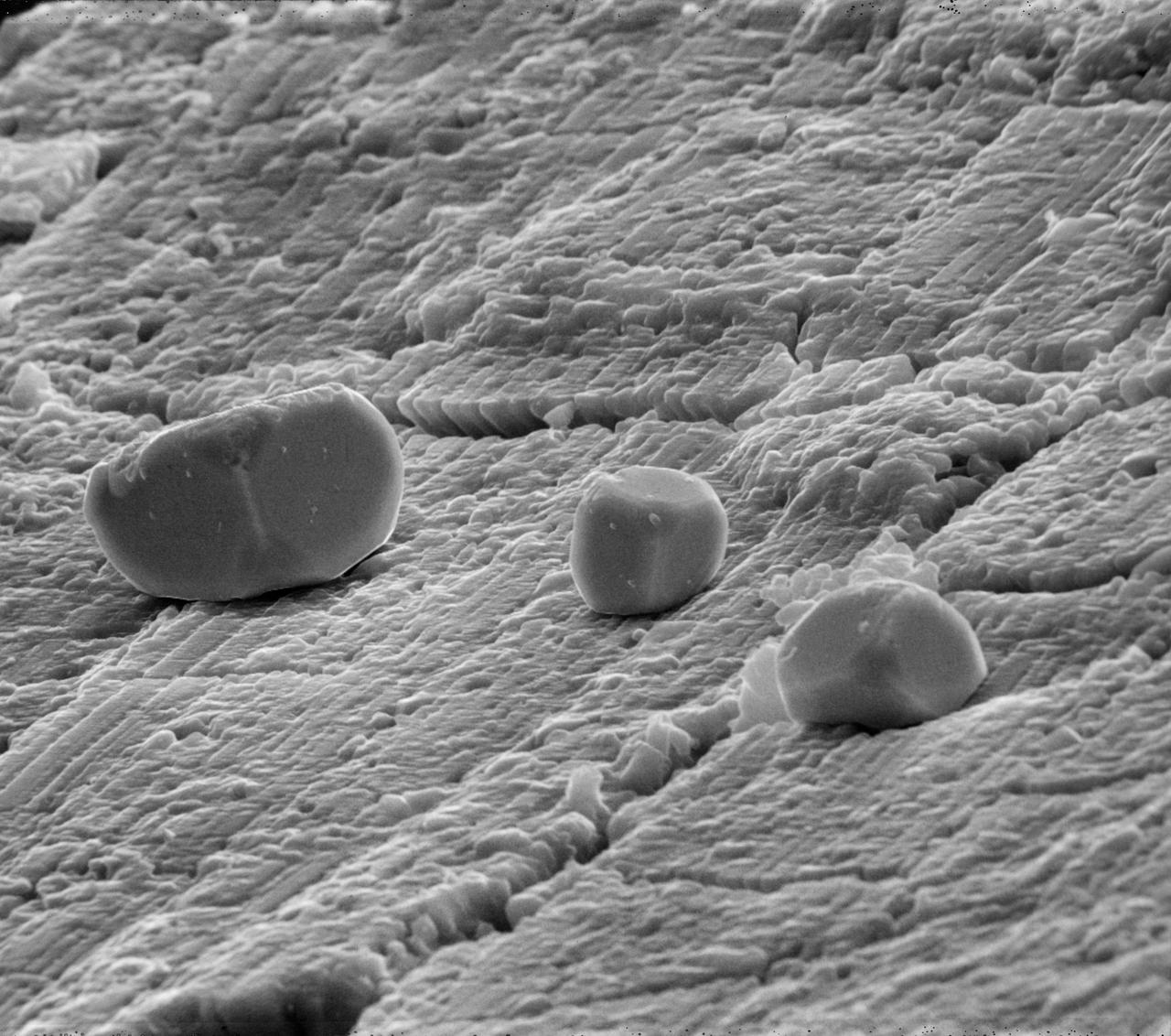
Scarning electron microscope images of the surface of ZBLAN fibers pulled in microgravity (ug) and on Earth (1g) show the crystallization that normally occurs in ground-based processing. The face of each crystal will reflect or refract a portion of the optical signal, thus degrading its quality. NASA is conducting research on pulling ZBLAN fibers in the low-g environment of space to prevent crystallization that limits ZBLAN's usefulness in optical fiber-based communications. ZBLAN is a heavy-metal fluoride glass that shows exdeptional promise for high-throughput communications with infrared lasers. Photo credit: NASA/Marshall Space Flight Center
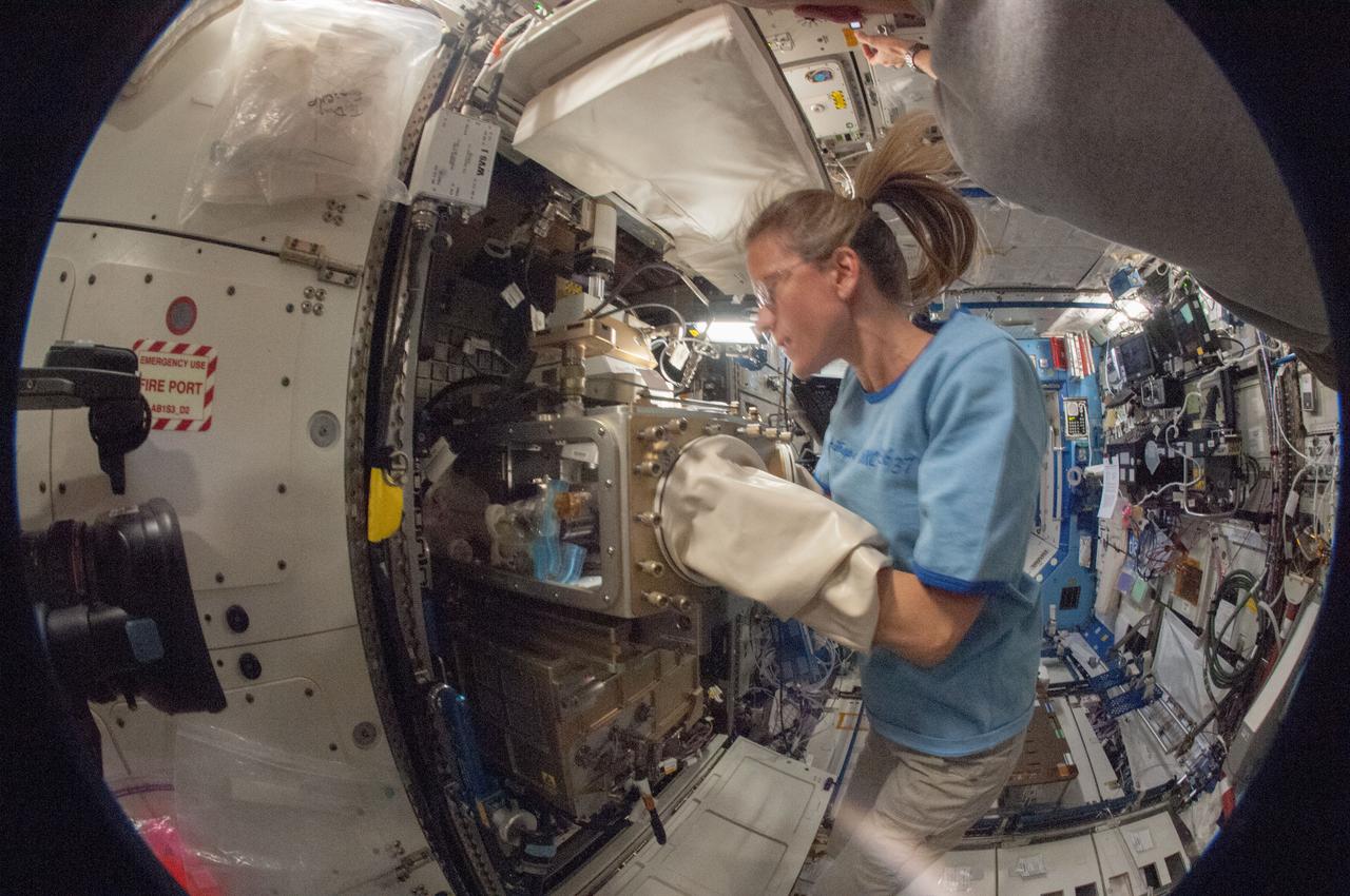
ISS036-E-019783 (24 June 2013) --- In the International Space Station’s Destiny laboratory, a fisheye lens attached to an electronic still camera was used to capture this image of NASA astronaut Karen Nyberg, Expedition 36 flight engineer, as she conducts a session with the Advanced Colloids Experiment (ACE)-1 sample preparation at the Light Microscopy Module (LMM) in the Fluids Integrated Rack / Fluids Combustion Facility (FIR/FCF). ACE-1 is a series of microscopic imaging investigations that uses the microgravity environment to examine flow characteristics and the evolution and ordering effects within a group of colloidal materials.
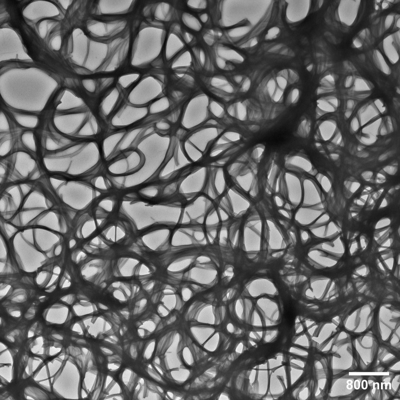
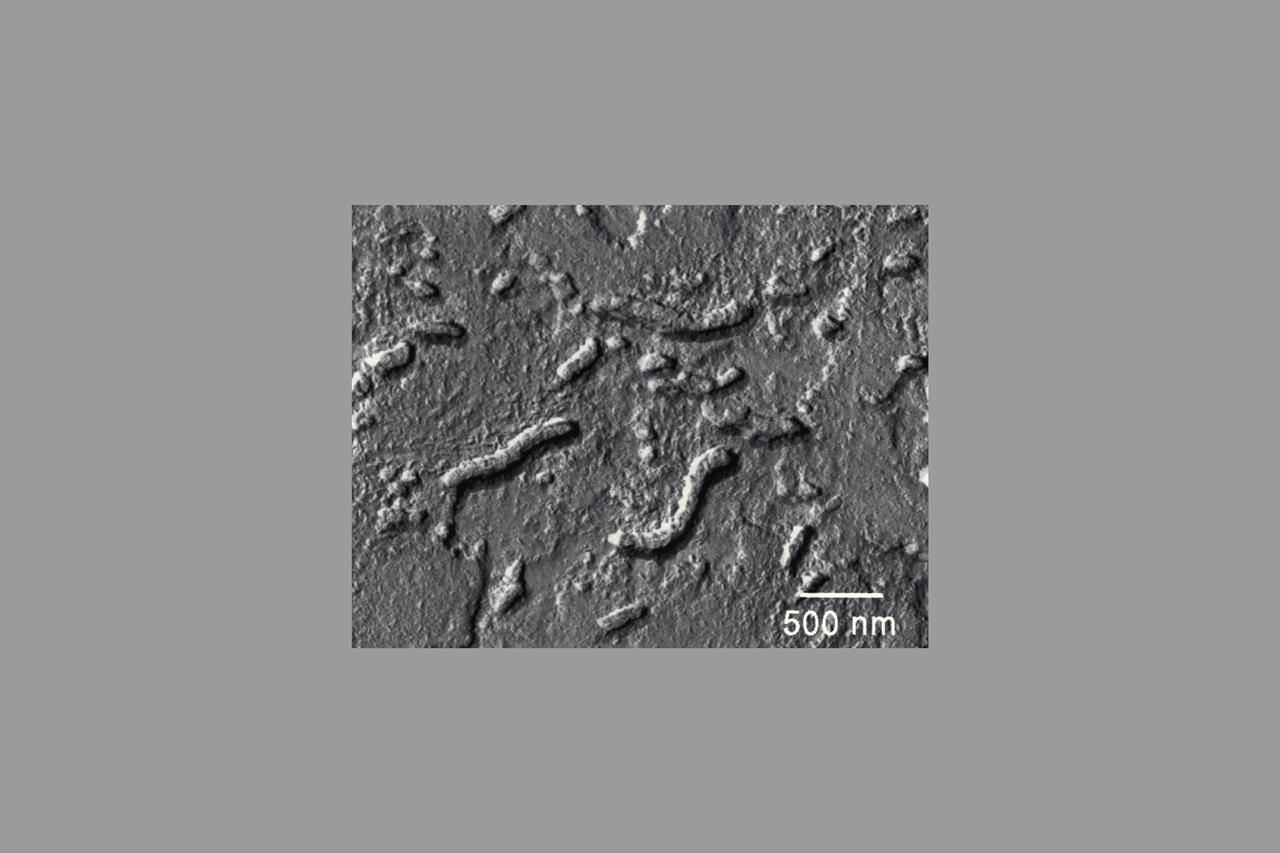
jsc2025e036191 (4/4/2025) --- An electron microscope image taken of a Janus Base Nanomaterial (JBNm) made on the International Space Station. The scale bar on the bottom represents 1/62500 the width of a human hair, making the JBNm bundles thick and interspersed, lead to better outcomes for cells. Biomimetic Fabrication of Multi-Functional DNA-Inspired Nanomaterials via Controlled Self-assembly in Space (DNA Nano Therapeutics-Mission 2) continues prior research on in-space manufacturing of nanomaterials that mimic DNA and have applications for vaccines and regenerative medicine. Image courtesy of University of Connecticut.

jsc2023e010177 (4/7/2022) --- This image is a scanning electron microscopic image of one of the ESA-Biofilms sample plates from the first launch to the ISS. The sample plate in this image is made of copper, which naturally has antimicrobial properties. This surface has a 3 µm laser structure engraved to the surface which improves antimicrobial efficacy. On the surface, only few cells of the bacterial species Staphylococcus capitis are attached. The cells appear small and are not actively dividing. The ESA-Biofilms investigation studies bacterial biofilm formation and antimicrobial properties of different metal surfaces under spaceflight conditions in altered gravity. Image courtesy of DLR, CC BY-NC-ND 3.0.
jsc2020e030484 (4/20/2020) --- A preflight image sequence from terrestrial experiments with two vertically oriented ratchet surfaces; subcooling: 9.5 ℃; heat flux: 1.31 W/cm2. Asymmetric Sawtooth and Cavity-Enhanced Nucleation-Driven Transport (PFMI-ASCENT) demonstrates a passive cooling system for electronic devices in microgravity using a microstructured surface. When fluids boil over flat heated surfaces in microgravity, vapor bubbles grow larger in size, causing poor heat transfer that can lead to damage of devices. Adding microscopic rachets on the surface may passively enable mobility of vapor bubbles and prevent this damage. (Image courtesy of: Techshot, Inc.)

jsc2023e010175 (2/28/2023) --- This image shows a monospecies biofilm through the view of a scanning electron microscope. The image was colored to visualize the bacterial cells (orange) embedded in the biofilm matrix (blue). The biofilm was formed by a strain of the bacterial species Staphylococcus capitis that was isolated from the International Space Station. The ESA-Biofilms investigation studies bacterial biofilm formation and antimicrobial properties of different metal surfaces under spaceflight conditions in altered gravity. The image was taken as part of the preflight experiments for ESA-Biofilms together with the Robert Koch Institute in Berlin, Germany. Image courtesy of DLR, CC BY-NC-ND 3.0.
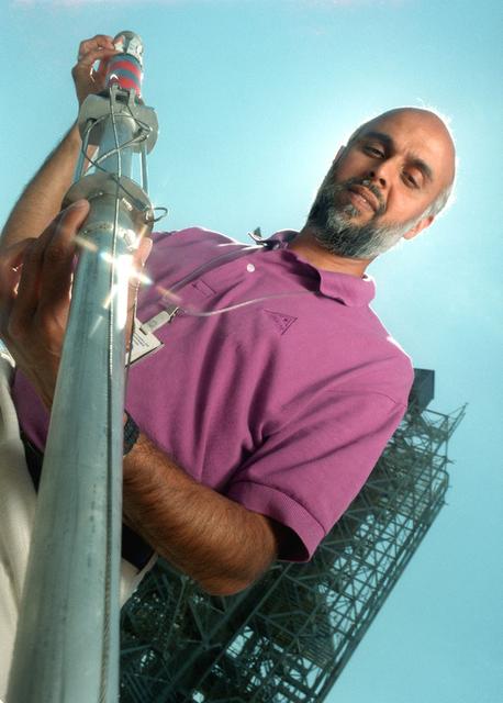
The dart and associated launching system was developed by engineers at MSFC to collect a sample of the aluminum oxide particles during the static fire testing of the Shuttle's solid rocket motor. The dart is launched through the exhaust and recovered post test. The particles are collected on sticky copper tapes affixed to a cylindrical shaft in the dart. A protective sleeve draws over the tape after the sample is collected to prevent contamination. The sample is analyzed under a scarning electron microscope under high magnification and a particle size distribution is determined. This size distribution is input into the analytical model to predict the radiative heating rates from the motor exhaust. Good prediction models are essential to optimizing the development of the thermal protection system for the Shuttle.

S93-E-5003 (23 July 1999) --- Astronaut Jeffrey S. Ashby, pilot, works at the Space Tissue Loss-B experiment on Space Shuttle Columbia's middeck. The experiment is set up to observe cells in culture with a video microscope imaging system to record near-real-time interactions of detecting and inducing cellular responses (macromorphological changes). Just above and to the right of STL-B is the part of the Commercial Generic Bioprocessing Apparatus (CGBA) for the National Institute of Health (NIH-B experiment). It is an experiment designed to investigate the effects of space flight on neural development in Drosophila melanogaster (fruit fly) larvae. This information may help scientists understand how gravity affects nerve growth and development and how neural connections to muscle fibers work. The photo was recorded with an electronic still camera (ESC) on Flight Day 1. Ashby and his four crew mates are scheduled to spend five days aboard Columbia in Earth orbit.
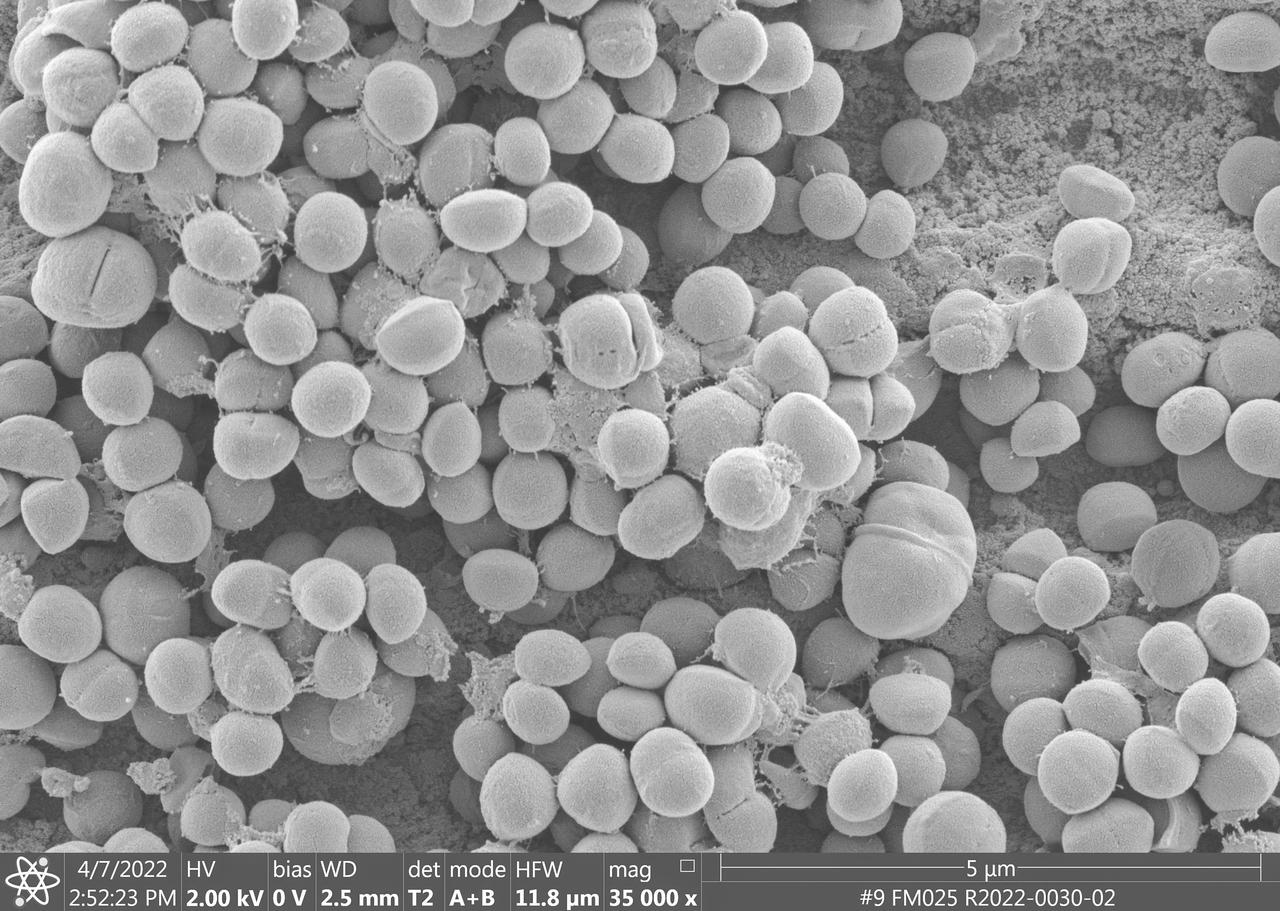
jsc2023e010178 (4/7/2022) --- This image taken by a scanning electron microscope shows one of the ESA-Biofilms sample plates from its first launch to the International Space Station. The sample plate in this image is made of stainless steel, which is the reference surface in the experiment since it has no antimicrobial properties. This surface also has a 3 µm laser structure engraved to the surface as control. In contrast to the copper surface, there are many Staphylococcus capitis cells attached to the steel surface that are actively dividing and starting to from components of a biofilm matrix. The ESA-Biofilms investigation studies bacterial biofilm formation and antimicrobial properties of different metal surfaces under spaceflight conditions in altered gravity. Image courtesy of DLR, CC BY-NC-ND 3.0.
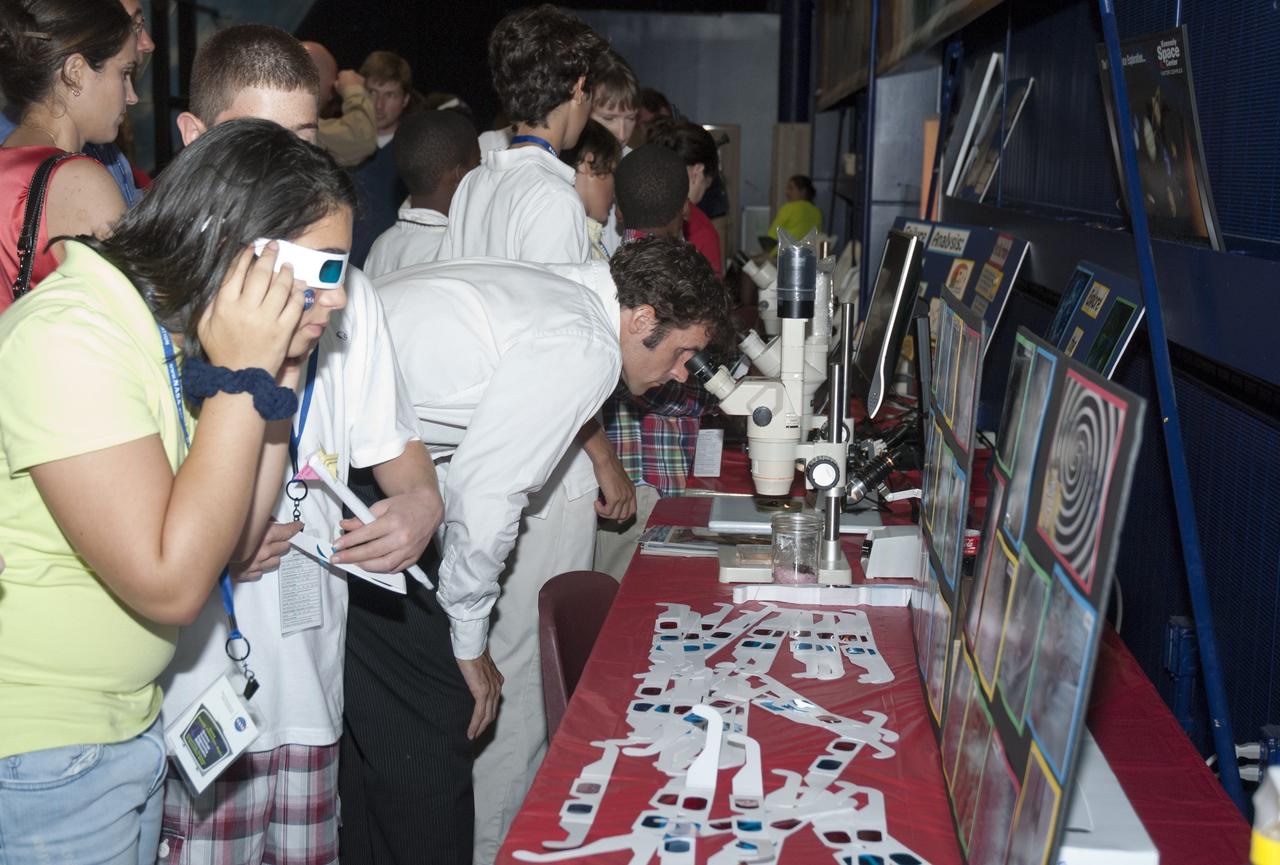
CAPE CANAVERAL, Fla. -- At the Astronaut Hall of Fame near the Kennedy Space Center Visitor Complex in Florida, fifth- through eighth-grade students and their parents participate in the Materials Science Laboratory exhibit by using stereomicroscopes and viewing 3-D scanning electron microscope images during the last NASA family education night event. Other activities included "gee-whiz" presentations, astronaut appearances, a hovercraft, vortex cannon and alternative fuel vehicles, which promote science, technology, engineering and mathematics (STEM) education. The event is part of NASA's Summer of Innovation initiative to provide interactive learning experiences to middle school students nationwide during the summer months. The program is a cornerstone of the Educate to Innovate campaign announced by President Barack Obama in November 2009. Photo credit: NASA/Charisse Nahser
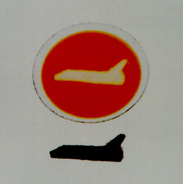
Polydiacetylenes are a unique class of highly conjugated organic polymers that are of interest for both electronic and photonic applications. Photodeposition from solutions is a novel process superior to those grown by conventional techniques. Evidence of this is seen when the films are viewed under a microscope; they exhibit small particles of solid polymer which form in the bulk solution, get transported by convection to the surface of the growing film, and become embedded. Also convection tends to cause the film thickness to be less uniform, and may even affect the molecular orientation of the films. The thrust of the research is to investigate in detail, both in 1-g and low-g, the effects of convection (and lack thereof) on this novel and interesting reaction. In this example, a portion of the substrate was blocked from exposure to the UV light by the mask, which was placed on the opposite side of the glass disk as the film, clearly demonstrating that photodeposition occurs only where the substrate is irradiated directly.
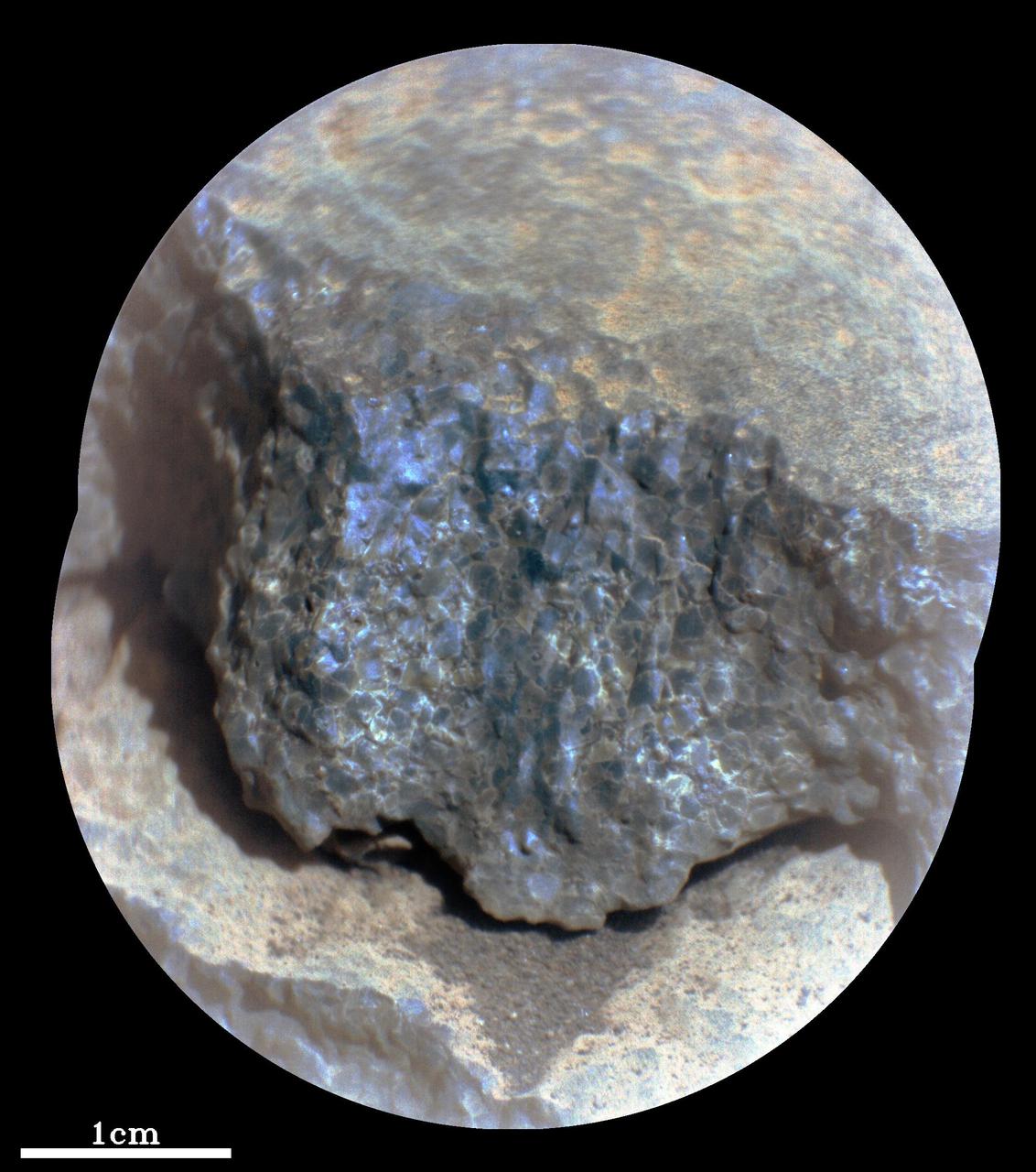
This enhanced-color close-up of a rock target called "Cine" was captured by the SuperCam instrument aboard NASA's Perseverance Mars rover on Sept. 17, 2021, the 206th Martian day, or sol, of rover's mission. SuperCam’s Remote Microscopic Imager took two images that were later combined to form this close-up. The target is 92 inches (2 meters) away, seen from the rover's mast. The image shows a rock layer made up of tightly packed millimeter-size gray, angular grains, or crystals. The image on the right shows a detail of the grain/crystal texture. The composition of this rock target was investigated with SuperCam's laser and spectrometer, along with the Mastcam-Z camera. Using these instruments, scientists can study the chemical composition of rocks from a distance. Analysis of "Cine" showed that it is rich in the mineral olivine. After the image was taken, the mission’s science team debated whether the rock is igneous (volcanic) or consists of fine sedimentary grains of igneous material that were cemented together in a watery environment. SuperCam is led by Los Alamos National Laboratory in New Mexico, where the instrument's body unit was developed. That part of the instrument includes several spectrometers as well as control electronics and software. The mast unit, including the Remote Microscopic Imager used for these images, was developed and built by several laboratories of the CNRS (the French research center) and French universities under the contracting authority of Centre National d'Etudes Spatiales (CNES), the French space agency. A key objective for Perseverance's mission on Mars is astrobiology, including the search for signs of ancient microbial life. The rover will characterize the planet's geology and past climate, pave the way for human exploration of the Red Planet, and be the first mission to collect and cache Martian rock and regolith (broken rock and dust). Subsequent NASA missions, in cooperation with ESA (European Space Agency), would send spacecraft to Mars to collect these sealed samples from the surface and return them to Earth for in-depth analysis. The Mars 2020 Perseverance mission is part of NASA's Moon to Mars exploration approach, which includes Artemis missions to the Moon that will help prepare for human exploration of the Red Planet. https://photojournal.jpl.nasa.gov/catalog/PIA24936
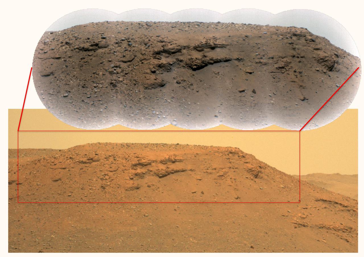
This composite image of the "Delta Scarp" in Mars' Jezero Crater was generated using data from two imagers aboard NASA's Perseverance rover. Taken by the rover's Mastcam-Z, the bottom image shows both the base and plateau of the escarpment. The inset above, created from a mosaic of five Remote Microscopic Imager (RMI) pictures, zooms in on a 377-foot-wide (115-meter-wide) portion of the scarp, allowing closer inspection of some of its intriguing geologic features. Part of the rover's SuperCam instrument, the RMI is able to spot an object the size of a softball from nearly a mile away, allowing scientists to take images of details from a long distance. It also provides fine details of nearby targets zapped by SuperCam's laser. SuperCam is led by Los Alamos National Laboratory in New Mexico, where the instrument's Body Unit was developed. That part of the instrument includes several spectrometers, control electronics and software. The Mast Unit was developed and built by several laboratories of the CNRS (French National Centre for Scientific Research) and French universities under the contracting authority of CNES. Arizona State University in Tempe leads the operations of the Mastcam-Z instrument, working in collaboration with Malin Space Science Systems in San Diego. A key objective for Perseverance's mission on Mars is astrobiology, including the search for signs of ancient microbial life. The rover will characterize the planet's geology and past climate, pave the way for human exploration of the Red Planet, and be the first mission to collect and cache Martian rock and regolith (broken rock and dust). Subsequent NASA missions, in cooperation with ESA (European Space Agency), would send spacecraft to Mars to collect these sealed samples from the surface and return them to Earth for in-depth analysis. The Mars 2020 Perseverance mission is part of NASA's Moon to Mars exploration approach, which includes Artemis missions to the Moon that will help prepare for human exploration of the Red Planet. https://photojournal.jpl.nasa.gov/catalog/PIA24684
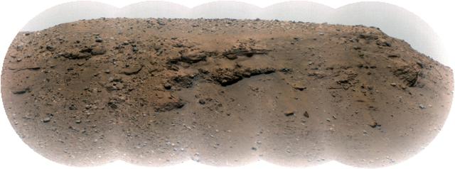
Composed of five images, this mosaic of the Jezero Crater's "Delta Scarp" was taken on March 17, 2021, by the Remote Microscopic Imager (RMI) camera aboard NASA's Perseverance rover from 1.4 miles (2.25 kilometers) away. Scientists believe the 377-foot-wide (115-meter-wide) escarpment is a portion of the remnants of a fan-shaped deposit of sediments that resulted from the confluence between an ancient river and an ancient lake. An annotated version of the same image (Figure 1) reveals location of a conglomerate (rock composed of coarse-grained pebbles mixed with sand) and examples of crossbedding (tilted layers of sedimentary rock that can result from water passing over a loose bed of sediment). Part of the SuperCam instrument, the RMI is able to spot an object the size of a softball from nearly a mile away, allowing scientists to take images of details from a long distance. It also provides fine details of nearby targets zapped by SuperCam's laser. SuperCam is led by Los Alamos National Laboratory in New Mexico, where the instrument's Body Unit was developed. That part of the instrument includes several spectrometers as well as control electronics and software. The Mast Unit was developed and built by several laboratories of the CNRS (the French research center) and French universities under the contracting authority of CNES (the French space agency). A key objective for Perseverance's mission on Mars is astrobiology, including the search for signs of ancient microbial life. The rover will characterize the planet's geology and past climate, pave the way for human exploration of the Red Planet, and be the first mission to collect and cache Martian rock and regolith (broken rock and dust). Subsequent NASA missions, in cooperation with ESA (European Space Agency), would send spacecraft to Mars to collect these sealed samples from the surface and return them to Earth for in-depth analysis. The Mars 2020 Perseverance mission is part of NASA's Moon to Mars exploration approach, which includes Artemis missions to the Moon that will help prepare for human exploration of the Red Planet. https://photojournal.jpl.nasa.gov/catalog/PIA24683
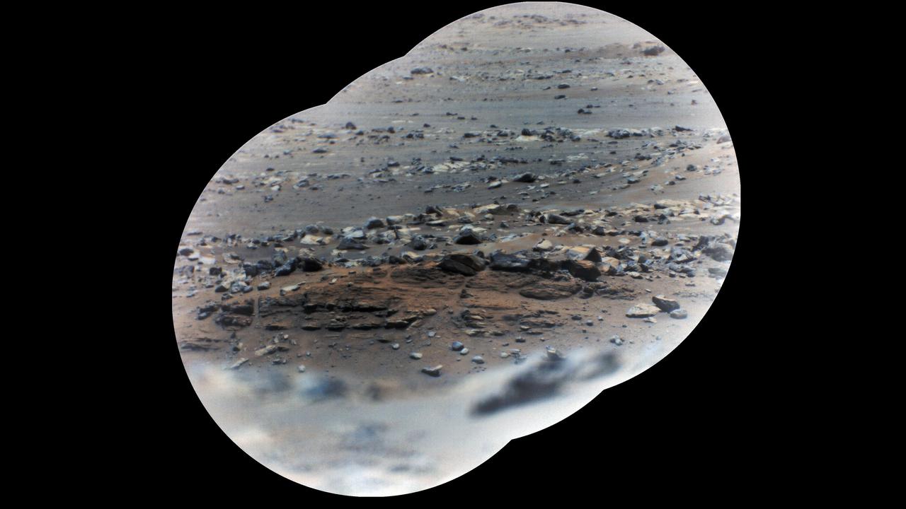
NASA's Perseverance rover took these zoomed-in images of a layered outcrop (just below center of image) nicknamed "Artuby" on June 17, 2021 (the 116th sol, or Martian Day, of its mission), from a little more than a third of a mile (615 meters) away. This mosaic is made up of three images taken by the Remote Microscopic Imager (RMI), part of the rover's SuperCam instrument. Each circular image has a field of view of 37.73 feet (11.50 meters) at this distance. The images were combined using an algorithm that weights the image centers. The outcrop shows evidence of being formed in an ancient lake. The feature is in the 'Verdon' quadrangle of Mars' Jezero Crater, south of the landing site. Artuby is the name of a river in southern France. Perseverance has been exploring the floor of Jezero Crater since it landed on Feb. 18, 2021. SuperCam is led by Los Alamos National Laboratory in New Mexico, where the instrument's Body Unit was developed. That part of the instrument includes several spectrometers as well as control electronics and software. The Mast Unit, including the RMI used for these images, was developed and built by several laboratories of the CNRS (the French research center) and French universities under the contracting authority of Centre National d'Etudes Spatiales (CNES, the French space agency). A key objective for Perseverance's mission on Mars is astrobiology, including the search for signs of ancient microbial life. The rover will characterize the planet's geology and past climate, pave the way for human exploration of the Red Planet, and be the first mission to collect and cache Martian rock and regolith (broken rock and dust). Subsequent NASA missions, in cooperation with ESA (European Space Agency), would send spacecraft to Mars to collect these sealed samples from the surface and return them to Earth for in-depth analysis. The Mars 2020 Perseverance mission is part of NASA's Moon to Mars exploration approach, which includes Artemis missions to the Moon that will help prepare for human exploration of the Red Planet. https://photojournal.jpl.nasa.gov/catalog/PIA24747
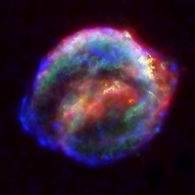
NASA's three Great Observatories -- the Hubble Space Telescope, the SpitzerSpace Telescope, and the Chandra X-ray Observatory -- joined forces to probe theexpanding remains of a supernova, called Kepler's supernova remnant, first seen 400 years ago by sky watchers, including astronomer Johannes Kepler. The combined image unveils a bubble-shaped shroud of gas and dust that is 14light-years wide and is expanding at 4 million miles per hour (2,000 kilometersper second). Observations from each telescope highlight distinct features of thesupernova remnant, a fast-moving shell of iron-rich material from the explodedstar, surrounded by an expanding shock wave that is sweeping up interstellar gasand dust. Each color in this image represents a different region of the electromagneticspectrum, from X-rays to infrared light. These diverse colors are shown in thepanel of photographs below the composite image. The X-ray and infrared datacannot be seen with the human eye. By color-coding those data and combining themwith Hubble's visible-light view, astronomers are presenting a more completepicture of the supernova remnant. Visible-light images from the Hubble telescope (colored yellow) reveal where the supernova shock wave is slamming into the densest regions of surrounding gas.The bright glowing knots are dense clumps from instabilities that form behindthe shock wave. The Hubble data also show thin filaments of gas that look likerippled sheets seen edge-on. These filaments reveal where the shock wave isencountering lower-density, more uniform interstellar material. The Spitzer telescope shows microscopic dust particles (colored red) that havebeen heated by the supernova shock wave. The dust re-radiates the shock wave'senergy as infrared light. The Spitzer data are brightest in the regionssurrounding those seen in detail by the Hubble telescope. The Chandra X-ray data show regions of very hot gas, and extremely high-energyparticles. The hottest gas (higher-energy X-rays, colored blue) is locatedprimarily in the regions directly behind the shock front. These regions alsoshow up in the Hubble observations, and also align with the faint rim of glowingmaterial seen in the Spitzer data. The X-rays from the region on the lower left(colored blue) may be dominated by extremely high-energy electrons that wereproduced by the shock wave and are radiating at radio through X-ray wavelengthsas they spiral in the intensified magnetic field behind the shock front. CoolerX-ray gas (lower-energy X-rays, colored green) resides in a thick interior shelland marks the location of heated material expelled from the exploded star. Kepler's supernova, the last such object seen to explode in our Milky Waygalaxy, resides about 13,000 light-years away in the constellation Ophiuchus. The Chandra observations were taken in June 2000, the Hubble in August 2003;and the Spitzer in August 2004. http://photojournal.jpl.nasa.gov/catalog/PIA06907
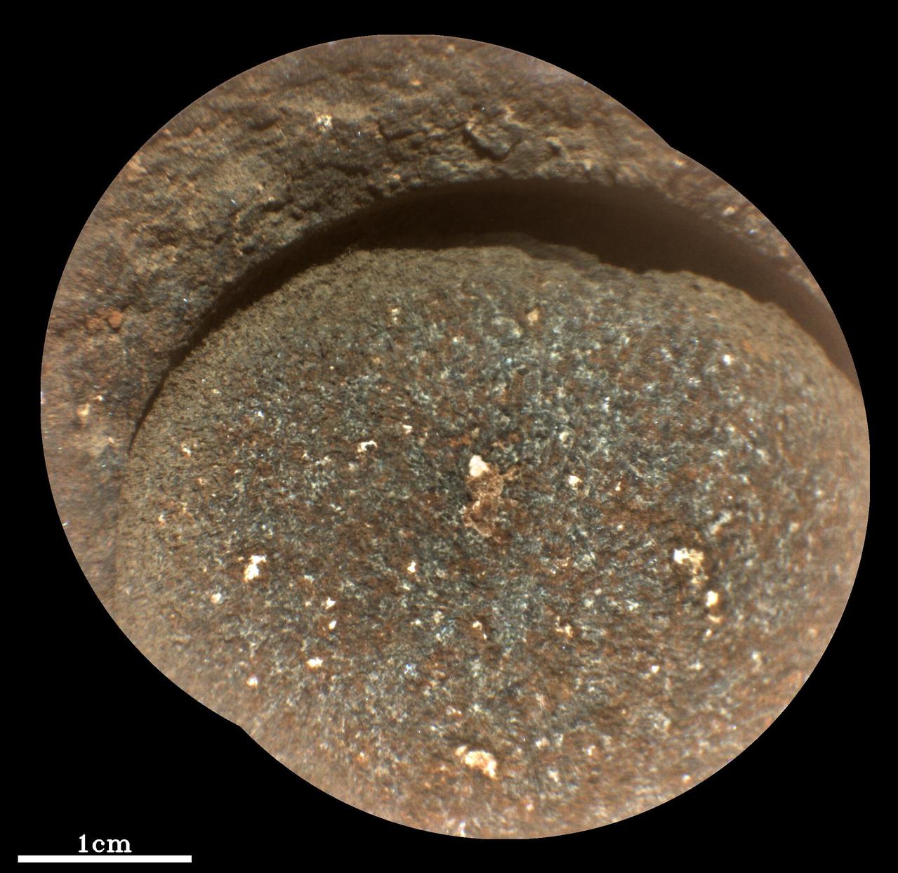
NASA's Perseverance Mars rover used its abrasion tool to grind down the rock surface at this target, nicknamed "Bellegarde," on Aug. 29, 2021, the 188th Martian day, or sol, of the mission. The abraded patch is 0.4 inches (5 centimeters) in diameter. The mission has nicknamed the rock itself "Rochette" and acquired its first two core samples from it. The rover abrades rocks using a tool on its robotic arm before drilling them in order to clear away dust and weathering rinds, allowing other instruments to study the rocks and determine if scientists want to grab a sample of them. This close-up image was produced by Perseverance's SuperCam instrument in natural color, as it would appear under daytime lighting conditions. Besides imagery, SuperCam has a rock-vaporizing laser and spectrometer. By studying a rock's vapor after each laser zap, scientists can study the chemical composition of rocks from a distance. Perseverance landed in Mars' Jezero Crater on Feb. 18, 2021, and has been exploring the floor of the crater since. At the time these images were taken, Perseverance was in an area nicknamed the "Crater Floor Fractured Rough" area. SuperCam is led by Los Alamos National Laboratory in New Mexico, where the instrument's Body Unit was developed. That part of the instrument includes several spectrometers as well as control electronics and software. The Mast Unit, including the Remote Microscopic Imager used for these images, was developed and built by several laboratories of the CNRS (the French research center) and French universities under the contracting authority of Centre National d'Etudes Spatiales (CNES, the French space agency). A key objective for Perseverance's mission on Mars is astrobiology, including the search for signs of ancient microbial life. The rover will characterize the planet's geology and past climate, pave the way for human exploration of the Red Planet, and be the first mission to collect and cache Martian rock and regolith (broken rock and dust). Subsequent NASA missions, in cooperation with ESA (European Space Agency), would send spacecraft to Mars to collect these sealed samples from the surface and return them to Earth for in-depth analysis. The Mars 2020 Perseverance mission is part of NASA's Moon to Mars exploration approach, which includes Artemis missions to the Moon that will help prepare for human exploration of the Red Planet. https://photojournal.jpl.nasa.gov/catalog/PIA24768
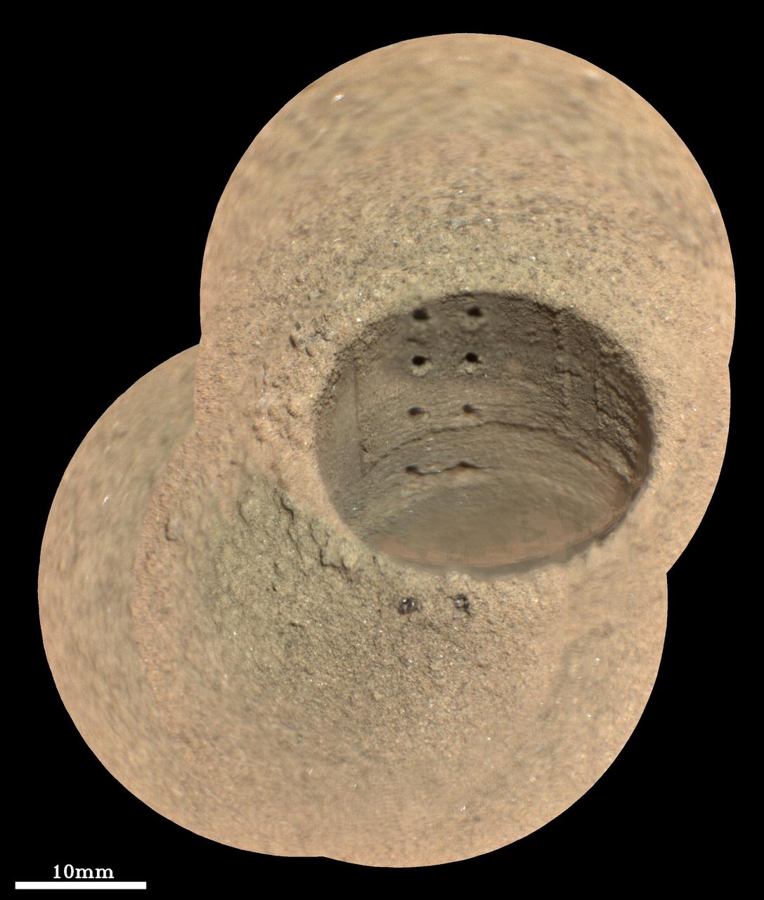
This composite image, made from four taken by the SuperCam instrument aboard NASA's Perseverance rover on August 8, 2021, shows the hole in a Martian rock where the rover attempted to collect its first sample; the small pits within it were created by laser zaps from SuperCam during subsequent efforts to analyze the rock's composition. The rover science team has nicknamed the drill hole "Roubion." The team believes that because of this rock's unusual composition, the process of extracting a core created a significant pile of tailings (or cuttings) around the coring hole. Eight pits produced by 30 laser shots each are seen in two columns inside the drill hole. The SuperCam team's analysis suggests that the top six pits penetrated the compacted mound of tailings around the hole, while the bottom two pits in the hole interrogated material below the rock surface. Two additional laser pits can be seen in the tailings at the near side of the hole. Two vertical ridges inside the hole – one on each side of the laser pits – were produced as the drill was removed, prior to laser analysis. Some bright mineral grains can be seen as glints in the tailings and in the drill hole. A few clumps or larger pieces of material are seen at the top of the tailings pile just to the left of the hole. The SuperCam images were taken from a distance of 7.32 feet (2.23 meters). A scale bar is included in this image. Perseverance landed in Mars' Jezero Crater on February 18, 2021, and has been exploring the floor of the crater since. At the time these images were taken, Perseverance was in an area nicknamed the "Crater Floor Fractured Rough" area. SuperCam is led by Los Alamos National Laboratory in New Mexico, where the instrument's Body Unit was developed. That part of the instrument includes several spectrometers as well as control electronics and software. The Mast Unit, including the Remote Microscopic Imager used for these images, was developed and built by several laboratories of the CNRS (the French research center) and French universities under the contracting authority of Centre National d'Etudes Spatiales (CNES, the French space agency). A key objective for Perseverance's mission on Mars is astrobiology, including the search for signs of ancient microbial life. The rover will characterize the planet's geology and past climate, pave the way for human exploration of the Red Planet, and be the first mission to collect and cache Martian rock and regolith (broken rock and dust). Subsequent NASA missions, in cooperation with ESA (European Space Agency), would send spacecraft to Mars to collect these sealed samples from the surface and return them to Earth for in-depth analysis. The Mars 2020 Perseverance mission is part of NASA's Moon to Mars exploration approach, which includes Artemis missions to the Moon that will help prepare for human exploration of the Red Planet. Movie available at https://photojournal.jpl.nasa.gov/catalog/PIA24749
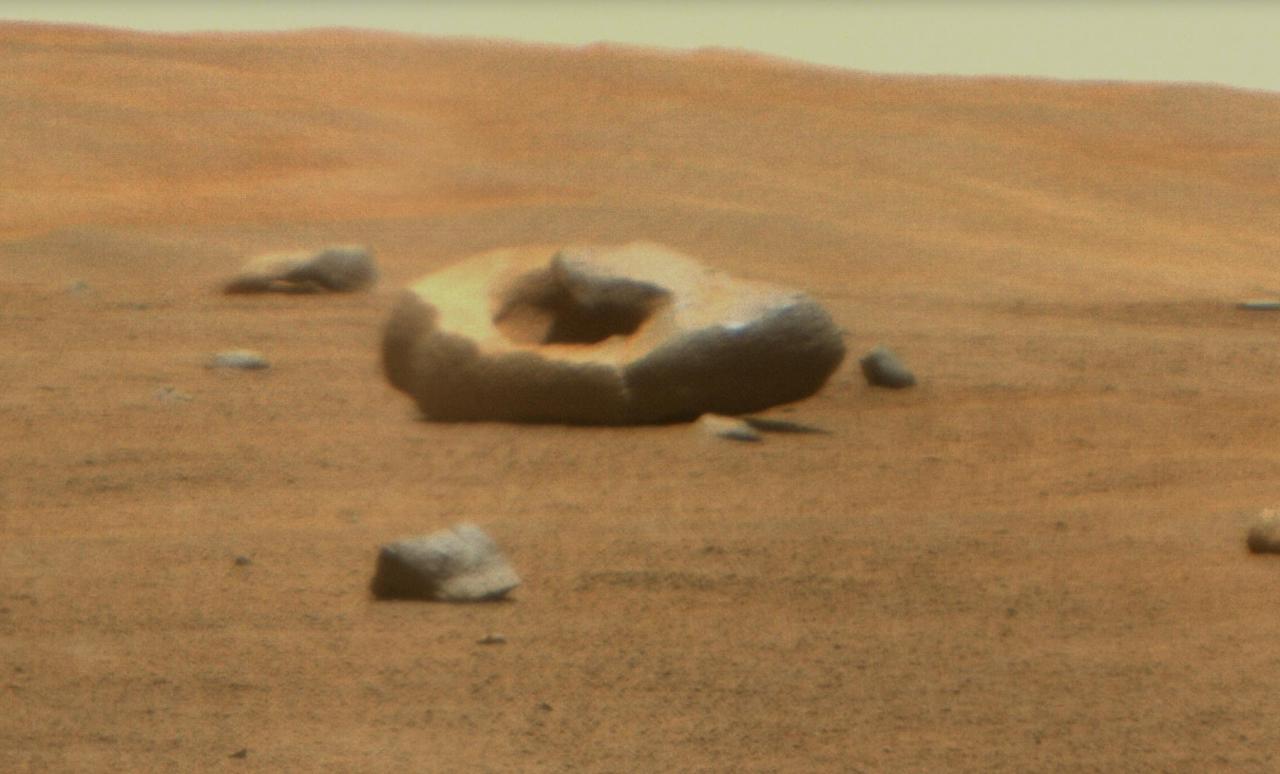
NASA's Perseverance Mars rover captured this doughnut-shaped rock in Jezero Crater from about 328 feet (100 meters) away using its Remote Microscopic Imager (RMI), part of the SuperCam instrument, on June 22, 2023, the 832nd Martian day, or sol, of the mission. Oddly shaped rocks aren't uncommon, either on Earth or Mars; they're often formed over eons as winds sandblast rock faces. This particular rock may have formed after a smaller rock (or multiple rocks) eroded near its center. That left behind a cavity that was later enlarged by the wind. Figure A shows the same rock in its broader context, when it was first spotted by the rover's Mastcam-Z instrument from about 1,312 feet (400 meters away) on April 15, 2023, the 765th Martian day, or sol, of the mission. SuperCam is led by Los Alamos National Laboratory in New Mexico, where the instrument's body unit was developed. That part of the instrument includes several spectrometers as well as control electronics and software. The mast unit, including RMI, was developed and built by several laboratories of the CNRS (the French research center) and French universities under the contracting authority of Centre National d'Études Spatiales (CNES), the French space agency. Arizona State University leads the operations of the Mastcam-Z instrument, working in collaboration with Malin Space Science Systems in San Diego, on the design, fabrication, testing, and operation of the cameras, and in collaboration with the Niels Bohr Institute of the University of Copenhagen on the design, fabrication, and testing of the calibration targets. A key objective for Perseverance's mission on Mars is astrobiology, including the search for signs of ancient microbial life. The rover will characterize the planet's geology and past climate, pave the way for human exploration of the Red Planet, and be the first mission to collect and cache Martian rock and regolith (broken rock and dust). Subsequent NASA missions, in cooperation with ESA (European Space Agency), would send spacecraft to Mars to collect these sealed samples from the surface and return them to Earth for in-depth analysis. The Mars 2020 Perseverance mission is part of NASA's Moon to Mars exploration approach, which includes Artemis missions to the Moon that will help prepare for human exploration of the Red Planet. https://photojournal.jpl.nasa.gov/catalog/PIA25916