Ground testing for the first confocal Light Microscopy Microscope (LMM)  Experiment. Procter and Gamble is working with NASA Glenn scientists to prepare for a study that examines product stabilizers in a microgravity environment. The particles in the tube glow orange because they have been fluorescently tagged with a dye that reacts to green laser lights to allow construction of a 3D image point by point. The experiment, which will be sent to the ISS later this year, will help P&G develop improved product stabilizers to extend shelf life and develop more environmentally friendly packaging.
Light Microscopy Microscope Experiment
Martian Microscope
Martian Microscope
Microscope on Mars
Microscope on Mars
Martian Magnets Under the Microscope
The Biggest Microscopic Image Ever
Vanilla Under the Microscope
Mosaic of Commemorative Microscope Substrate
Mars Under the Microscope color
Punaluu Under the Microscope
Microscope Image of Scavenged Particles
Lanikai Under the Microscope
Mars Under the Microscope
Adirondack Under the Microscope
Microscopic Materials on a Magnet
Mars Under the Microscope stretched
Microscopic Image Inside Endurance
Cookies and Cream Under the Microscope
First Sample Delivery to Mars Microscope
NASA astronaut Karen Nyberg, Expedition 36 flight engineer, works to setup the Multi-Purpose Small Payload Rack (MSPR) fluorescence microscope in the Kibo laboratory of the International Space Station.
Nyberg in JEM (Kibo) with MSPR fluorescence microscope.
Microscopic Image of Martian Surface Material on a Silicone Substrate
Spirit Examines Light-Toned Halley Microscopic Image
Microscopic Comparison of Airfall Dust to Martian Soil
Microscope Image of a Martian Soil Surface Sample
Microscopic View of Soil on a Micromachined Silicone Substrate
In the center of this electron microscope image of a small chip from a meteorite are several tiny structures that are possible microscopic fossils of primitive, bacteria-like organisms that may have lived on Mars more than 3.6 billion years ago.  http://photojournal.jpl.nasa.gov/catalog/PIA00283
Mars Life? - Microscopic Structures
This electron microscope image shows extremely tiny tubular structures that are possible microscopic fossils of bacteria-like organisms that may have lived on Mars more than 3.6 billion years ago.  http://photojournal.jpl.nasa.gov/catalog/PIA00285
Mars Life? - Microscopic Tubular Structures
jsc2022e083574 (8/17/2022) --- A preflight image of the miniature microscope developed for the Moon Microscope investigation. Image courtesy of NASA’s Johnson Space Center Immunology/Virology Laboratory.
Moon Microscope
jsc2022e031227 (5/4/2016) --- A preflight view of the BioServe Microscope. The BioServe Microscope facility allows astronauts to capture digital, full-color, high-definition microscopy images and videos of scientific investigations.
BioServe Microscope
ELLEN RABENBERG,  EM31, FAILURE ANALYSIS AND METALLURGY BRANCH, DIAGNOSTICS TEAM MEMBER WITH 3D MICROSCOPE
1300928
This electron microscope image shows egg-shaped structures, some of which may be possible microscopic fossils of Martian origin as discussed by NASA research published in the Aug. 16, 1996.  http://photojournal.jpl.nasa.gov/catalog/PIA00286
Mars Life? - Microscopic Egg-shaped Structures
This calibration image presents three-dimensional data from the atomic force microscope on NASA Phoenix Mars Lander, showing surface details of a substrate on the microscope station sample wheel.
First Atomic Force Microscope Image from Mars
This image shows the workings of the microscope station of the Microscopy, Electrochemistry and Conductivity Analyzer MECA instrument suite of NASA Phoenix Mars Lander.
Schematic Animation of Phoenix Microscope Station
A scientific illustration of the operation of NASA Phoenix Mars Lander Atomic Force Microscope, or AFM. The AFM is part of Phoenix Microscopy, Electrochemistry, and Conductivity Analyzer, or MECA.
Atomic Force Microscope Operation
A scanning electron microscope photograph of iron crystals which grow in a small vug or cavity in a recrystallized breccia (fragmented rock) from the Apollo 15 Hadley-Apennino lunar landing site. The largest crystal is three microns across. Perfectly developed crystals such as these indicate slow formation from a hot vapor as the rock was cooling. The crystals are resting on an interlocking lattice of pyroxene (calsium-magnesium-iron silicate).
Scanning electron microscope view of iron crystal
Dressed in a cleanroom suit to prevent contamination, Optics Technician Jeff Gum aligns a replacement Focal Plane Assembly (FPA) with a powerful three-dimensional microscope at NASA's Goddard Space Flight Center in Greenbelt, Md. This FPA will be installed on the Near Infrared Camera (NIRCam) instrument, which has unique components that are individually tailored to see in a particular infrared wavelength range. By using the microscope, Gum ensures the FPA detectors are characterized and ready for installation onto NIRCam, the James Webb Space Telescope's primary imager that will see the light from the earliest stars and galaxies that formed in the universe.  Credit: NASA/Goddard/Chris Gunn  <b><a href="http://www.nasa.gov/audience/formedia/features/MP_Photo_Guidelines.html" rel="nofollow">NASA image use policy.</a></b>  <b><a href="http://www.nasa.gov/centers/goddard/home/index.html" rel="nofollow">NASA Goddard Space Flight Center</a></b> enables NASA’s mission through four scientific endeavors: Earth Science, Heliophysics, Solar System Exploration, and Astrophysics. Goddard plays a leading role in NASA’s accomplishments by contributing compelling scientific knowledge to advance the Agency’s mission.  <b>Follow us on <a href="http://twitter.com/NASAGoddardPix" rel="nofollow">Twitter</a></b>  <b>Like us on <a href="http://www.facebook.com/pages/Greenbelt-MD/NASA-Goddard/395013845897?ref=tsd" rel="nofollow">Facebook</a></b>  <b>Find us on <a href="http://instagram.com/nasagoddard?vm=grid" rel="nofollow">Instagram</a></b>
Webb Instruments Perfected to Microscopic Levels
This image taken by the NASA Mars rover Opportunity shows locations of the microscopic imager observations on a rock informally named Tisdale 2.
Locations of Microscopic Imager Observations on Tisdale 2
This electron microscope image is a close-up of the center part of photo number S96-12301.  http://photojournal.jpl.nasa.gov/catalog/PIA00284
Mars Life? - Microscopic Tube-like Structures
Marshall's 1992 Inventor of the Year demonstrates his multi-layer water window imaging x-ray microscope.
Around Marshall
jsc2022e083573 (9/2/2022) --- A preflight image of staining reagents, casing, and staining device developed for the Moon Microscope investigation. Image courtesy of NASA’s Johnson Space Center Immunology/Virology Laboratory.
Moon Microscope
jsc2022e083575 (10/4/2022) --- A preflight image of the slide staining device developed for the Moon Microscope investigation. Image courtesy of NASA’s Johnson Space Center Immunology/Virology Laboratory.
Moon Microscope
This electron microscope image shows tubular structures of likely Martian origin. These structures are very similar in size and shape to extremely tiny microfossils found in some Earth rocks.  http://photojournal.jpl.nasa.gov/catalog/PIA00287
Mars Life? - Microscopic Tubular Structures
This image shows the eight sharp tips of the NASA Phoenix Mars Lander Atomic Force Microscope, or AFM. The AFM is part of Phoenix Microscopy, Electrochemistry, and Conductivity Analyzer, or MECA.
Sharp Tips on the Atomic Force Microscope
A scanning electron microscope captured this image of terresterial soil containing a phyllosilicate mineral from Koua Bocca, Ivory Coast, West Africa. This soil shares some similarities with Martian soil scooped by NASA Phoenix Lander.
Terrestrial Clay under Microscope
iss058e001945 (Jan. 3, 2019) --- Expedition 58 Flight Engineer and astronaut Anne McClain of NASA peers into a microscope and takes photographs for the Protein Crystal Growth-16 experiment that is exploring therapies for Parkinson's disease.
Astronaut Anne McClain of NASA peers into a microscope
Mars Particle and Terrestrial Soil, Compared Microscopically
This high-resolution scanning electron microscope image shows an unusual tube-like structural form that is less than 1/100th the width of a human hair in size found in meteorite ALH84001, a meteorite believed to be of Martian origin.  http://photojournal.jpl.nasa.gov/catalog/PIA00288
Mars Life? - Microscopic Tube-like Structures
This image of a target called "Private Joseph Field" combines four images from the microscopic imager on the robotic arm of NASA's Mars Exploration Rover Opportunity, with enhanced color information added from the rover's panoramic camera.  This target is within the "Marathon Valley" area of the western rim of Endeavour Crater. The component images were taken on May 29, 2016, during the 4,389th Martian day, or sol, of Opportunity's work on Mars. The mosaic shows an area spanning about 2 inches (5 centimeters).  Geochemical data indicate the presence of magnesium and iron sulfates at this location, most likely corresponding to the white pebble visible near the center of the image. These sulfates may have formed by the interaction of acidic fluids with the rocks along the rim of Endeavour crater.  http://photojournal.jpl.nasa.gov/catalog/PIA21142
Opportunity View of Private Joseph Field on Mars
This relatively bright outcropping of rock, dubbed "Gasconade," was investigated by NASA's Mars Exploration Rover Opportunity while the rover was perched on "Spirit Mound" at the western edge of Mars' Endeavour Crater.  This mosaic combines four frames taken by the microscopic imager on Opportunity's robotic arm on Oct. 2, 2016, during the 4,512st Martian day, or sol, of the rover's work on Mars. Enhanced color information from Opportunity's panoramic camera has been added to emphasize differences in the materials visible in the target. Figure A is a version with no color information added to the microscopic imager mosaic.  The view covers an area about 2 inches (5 centimeters) wide. Opportunity's inspection found Gasconade to be a wind-etched outcrop with angular bits of darker rock within a lighter matrix, which may have been formed from fallout of the impact event that excavated the crater.  This location of Spirit Mound, shown at PIA20854, is the deeper on the western rim of Endeavour Crater than any site visited previously by Opportunity.  http://photojournal.jpl.nasa.gov/catalog/PIA21141
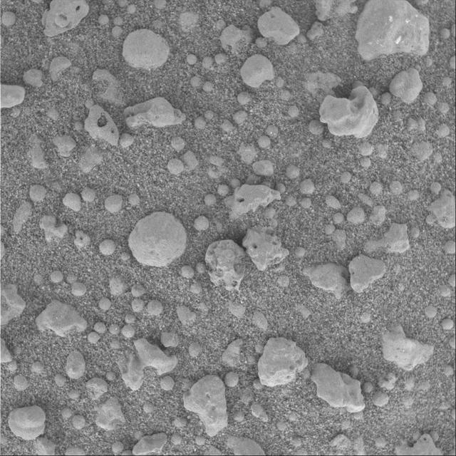
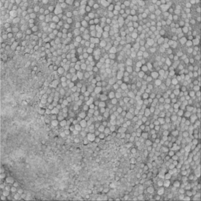
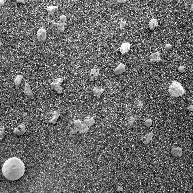
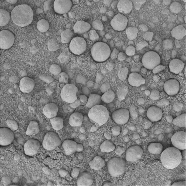
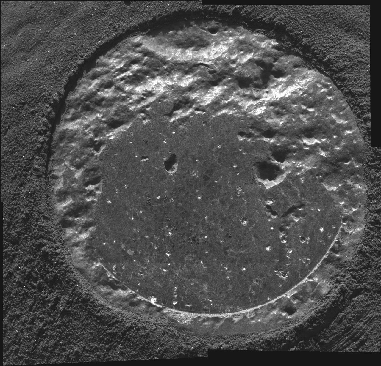
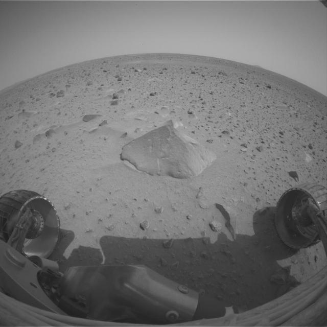
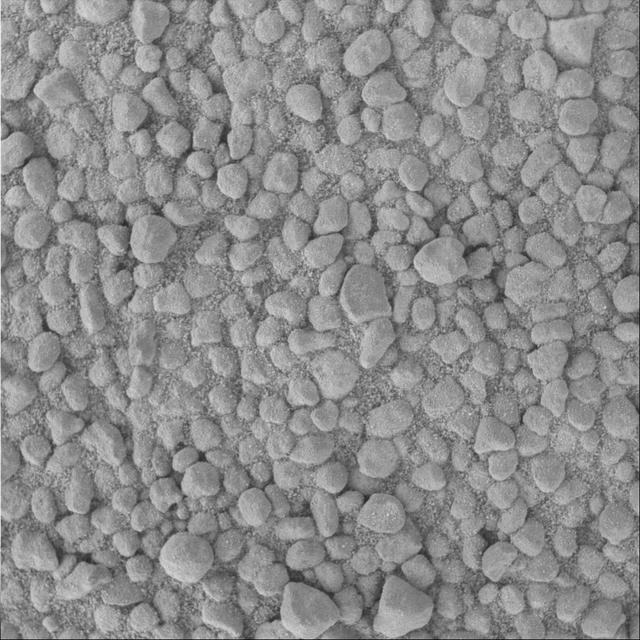
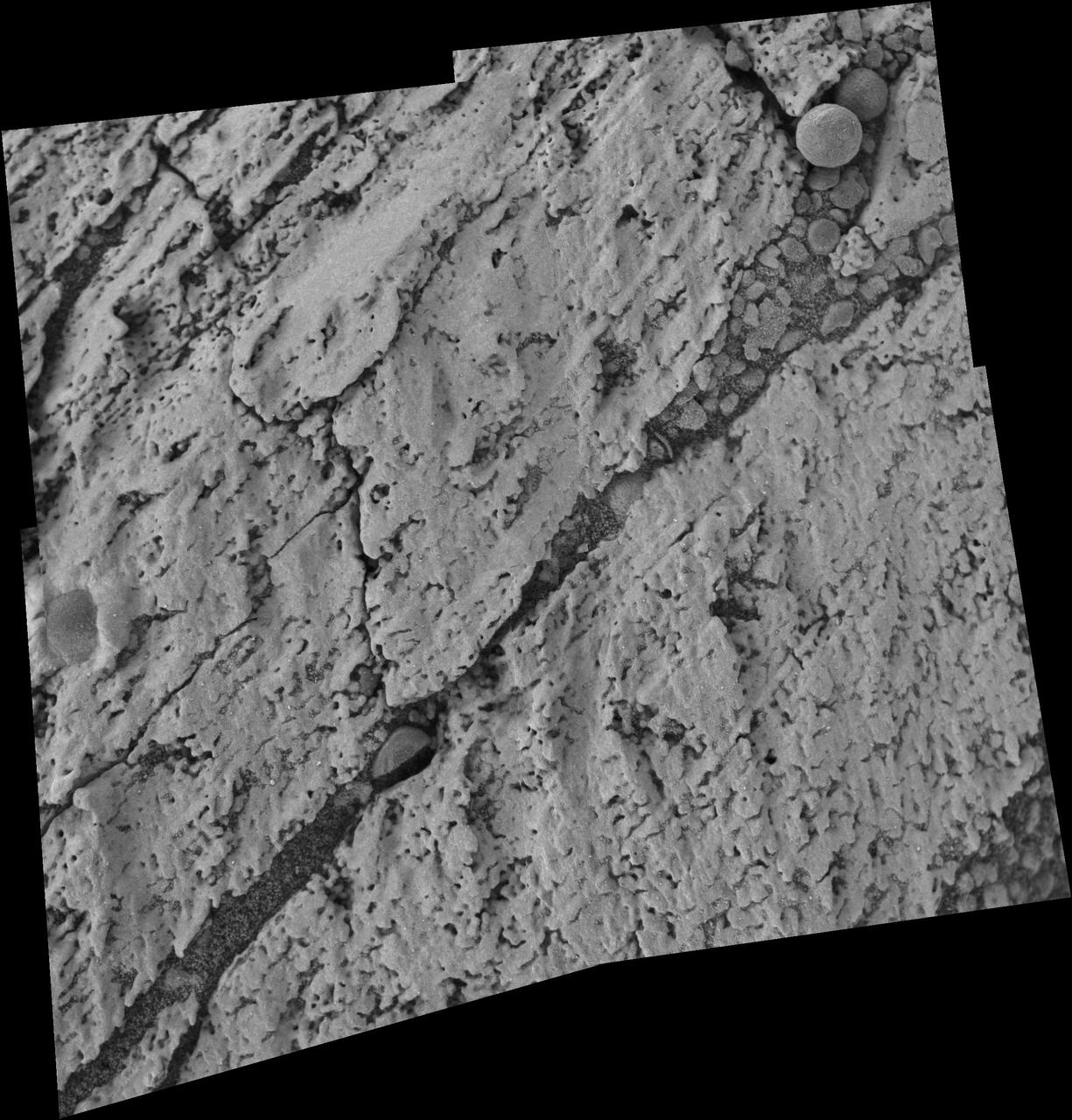
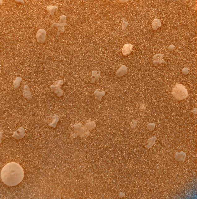
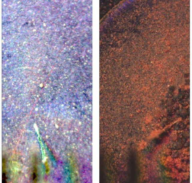
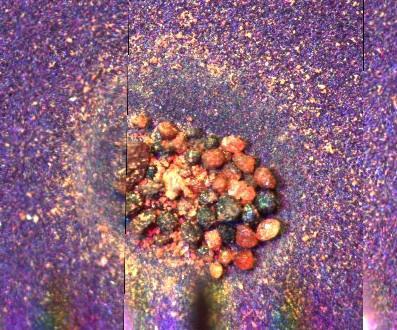
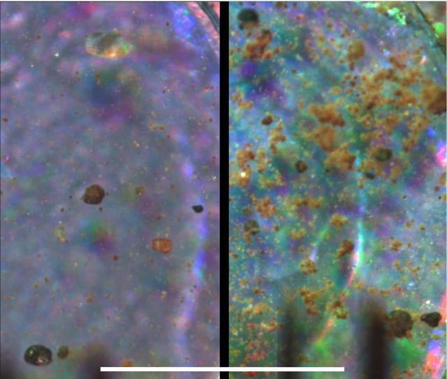
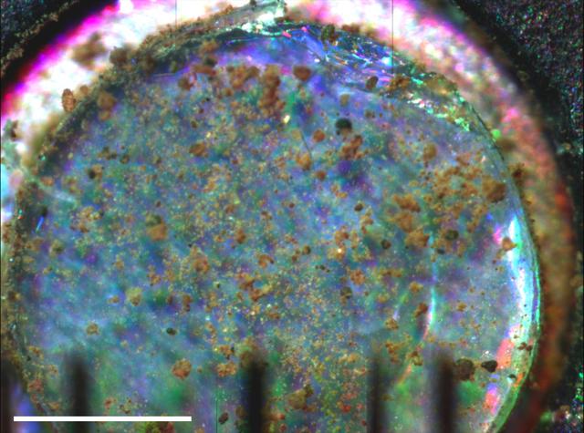
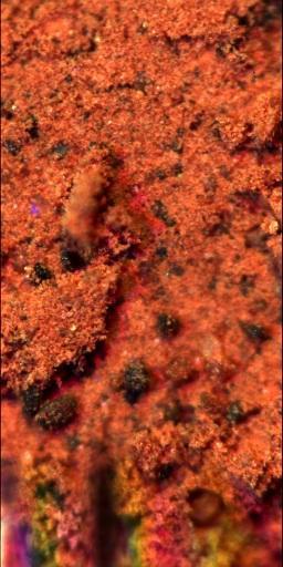
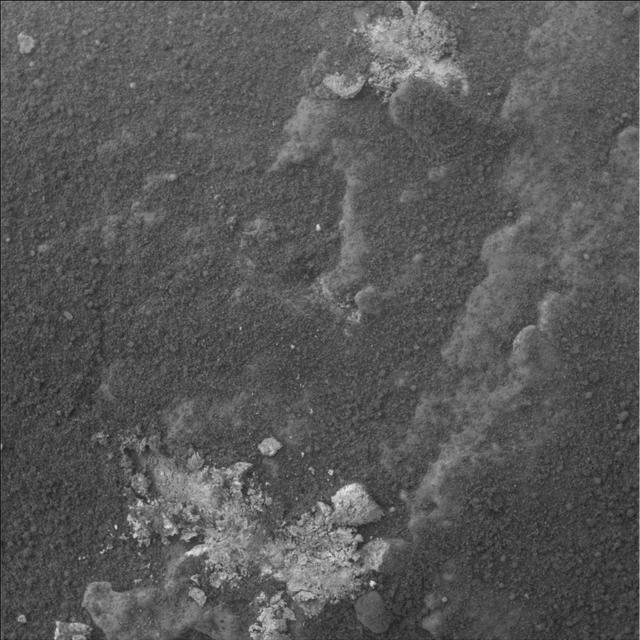
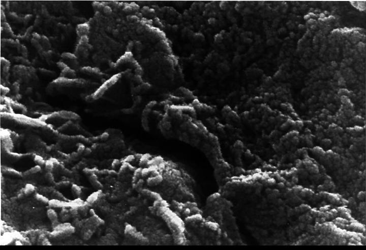
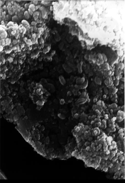
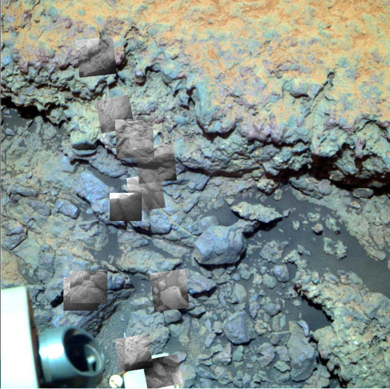
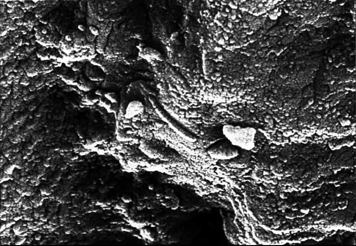
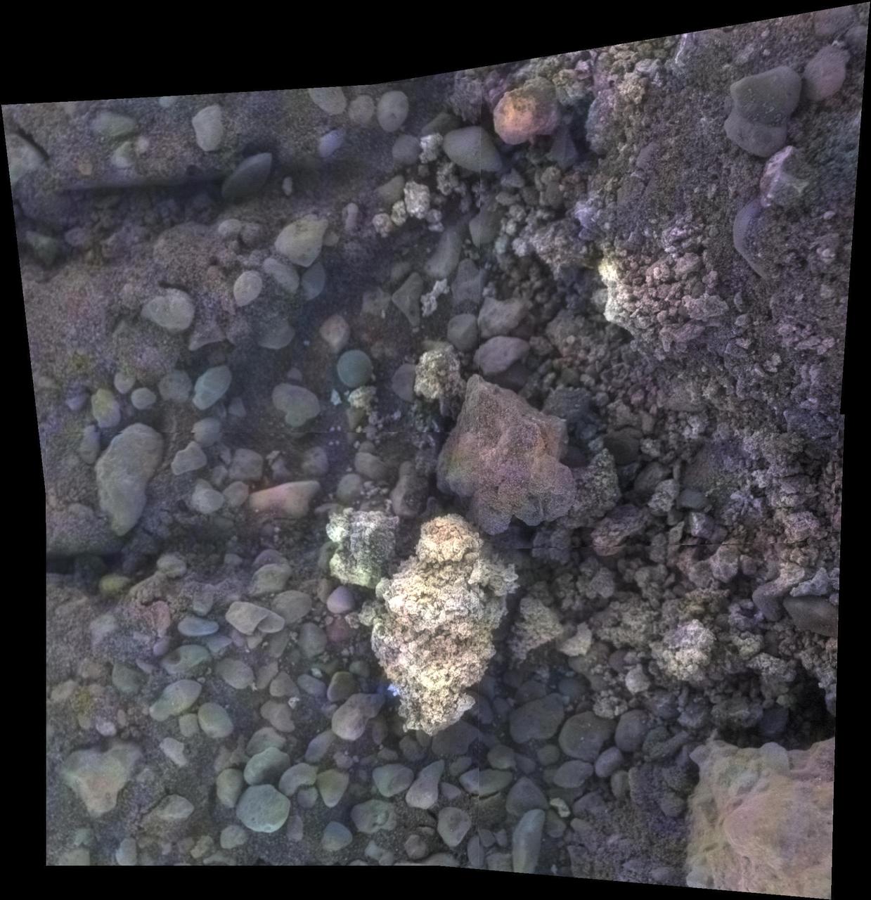
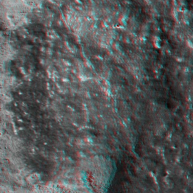
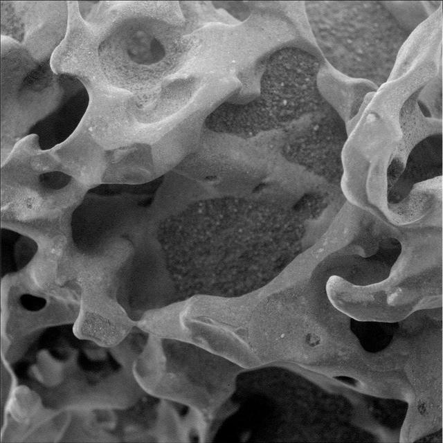
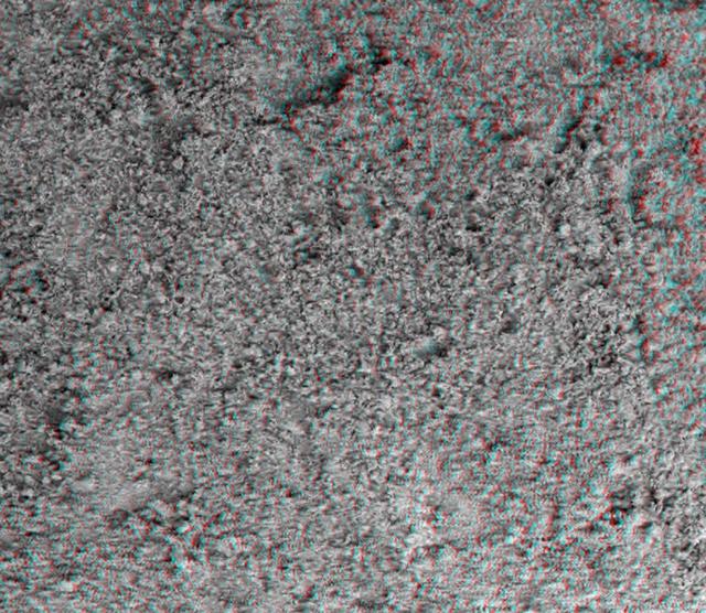
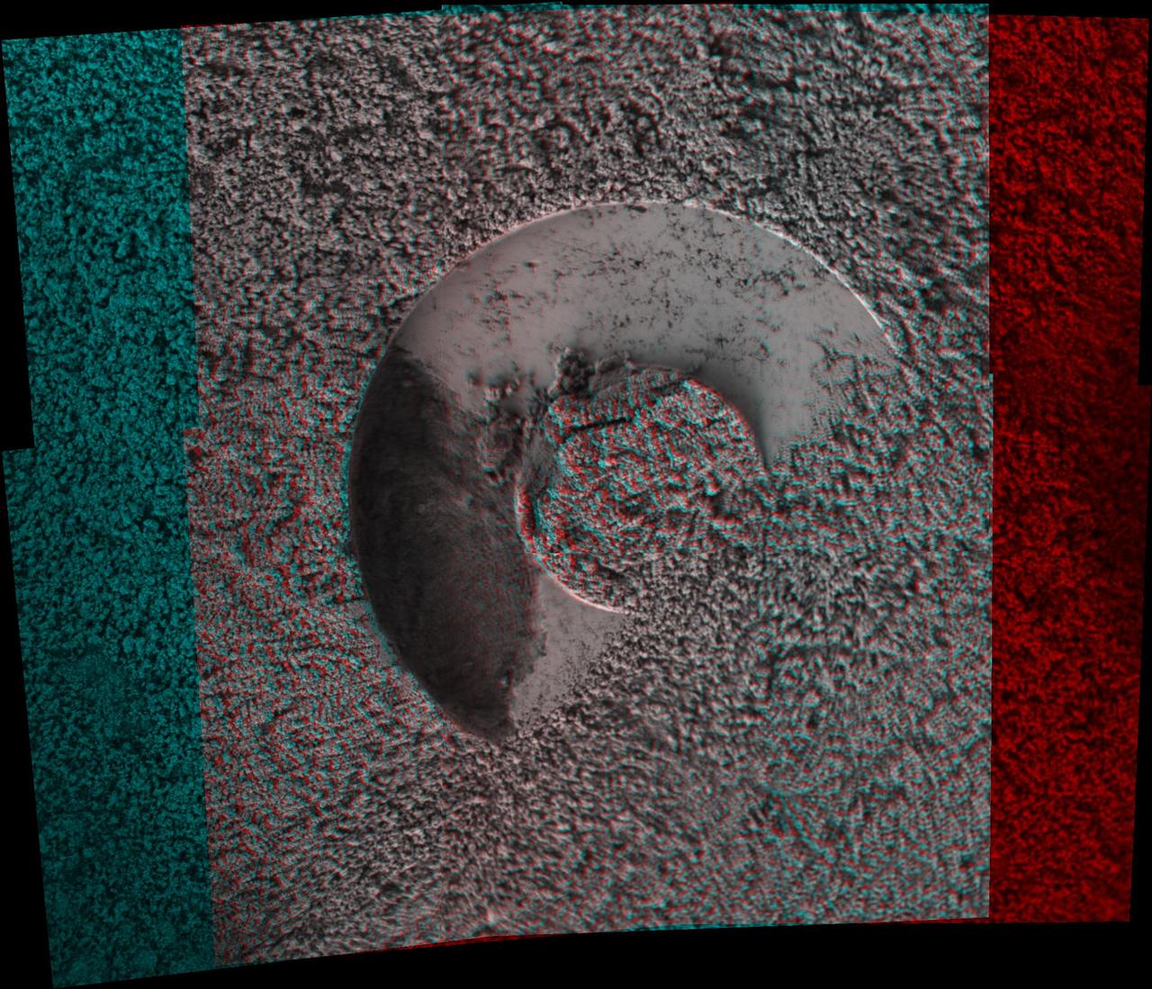
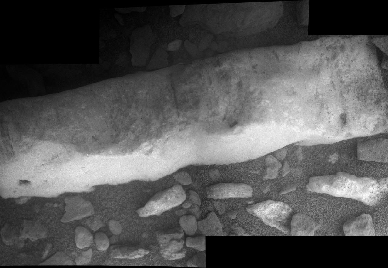
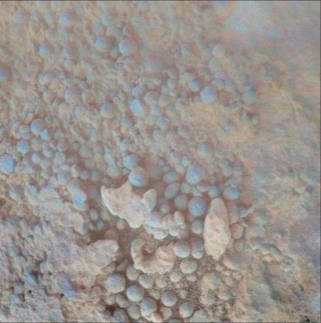
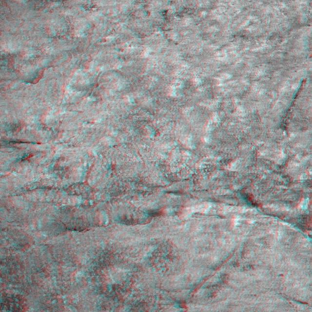
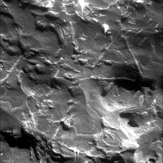
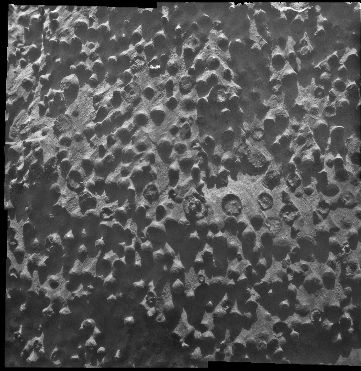
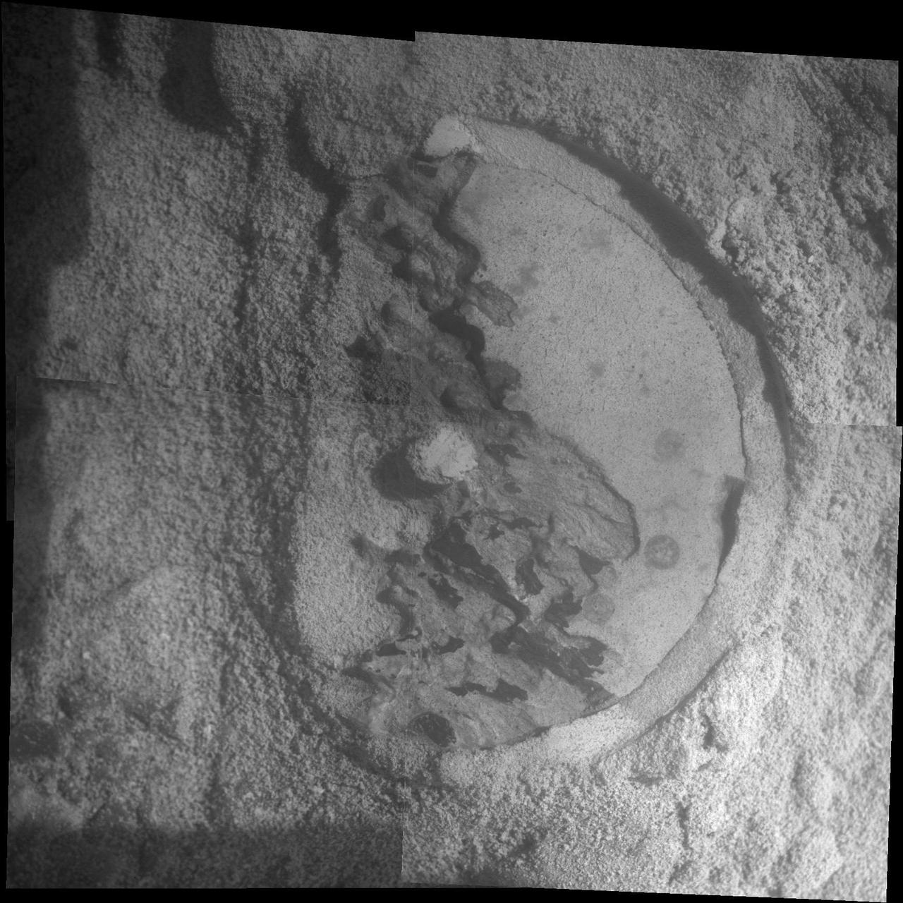
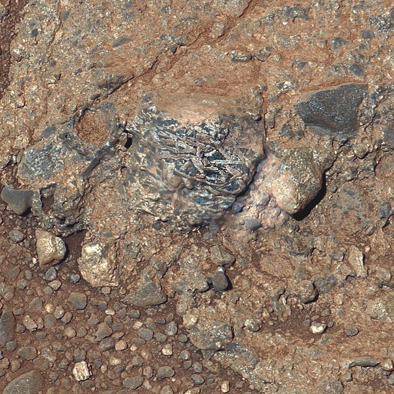
Opportunity Inspects Gasconade on Spirit Mound of Mars
ISS038-E-029082 (12 Jan. 2014) --- Japan Aerospace Exploration Agency astronaut Koichi Wakata, Expedition 38 flight engineer, performs microscopic analysis of the NanoRacks Module-38 Petri Dishes, using Celestron Reflective Microscope, in the Kibo laboratory of the International Space Station. These Module-38 experiments are designed by students as part of a competition sponsored by the International Space School Educational Trust (ISSET). This experiment examines three-dimensional growth of slime mold in petri dishes utilizing the NanoRacks Microscopes Facility.
Wakata performs microscopic analysis of the NanoRacks Module-38 Petri Dishes
iss073e1049692 (Nov. 6, 2025) --- NASA astronaut and Expedition 73 Flight Engineer Jonny Kim poses for a portrait while servicing the KERMIT (Keyence Research Microscope Testbed) fluorescence microscope inside the Materials Science Research Rack aboard the International Space Station’s Destiny laboratory module. KERMIT is a commercial off-the-shelf microscope that provides researchers with essential imaging capabilities for biological, physical, and materials science research in microgravity.
NASA astronaut Jonny Kim poses for a portrait while servicing a fluorescence microscope
iss073e0118086 (May 29, 2025) --- NASA astronaut and Expedition 73 Flight Engineer Anne McClain works inside the Destiny laboratory module's Materials Science Research Rack and swaps filters inside the Kermit microscope. Kermit is an all-in-one fluorescence microscope system used to conduct biological, physical, and materials science research.
NASA astronaut Anne McClain swaps filters inside the Kermit microscope
iss054e067484 (12/21/2017) --- Japan Aerospace Exploration Agency (JAXA) astronaut Norishige Kanai is photographed in the U.S. Lab performing Microscope operations for Synthetic Bone experiment. Synthetic Bone tests the functionality and effectiveness of a new material that can assist in recovery from bone injuries or dental work during long-term space travel.
Synthetic Bone Microscope Operations
S94-E-5001 (5 July 1997) --- Astronaut Donald Thomas, mission specialist, uses a microscope at the glovebox onboard the Space Shuttle Columbia's Spacelab Module during flight day five activities.
BDND - MGBX - Thomas with microscope at glovebox in Spacelab
STS047-05-025 (12 - 20 Sept 1992) --- Payload specialist Mamoru Mohri, representing Japan's National Space Development Agency (NASDA), uses a microscope to produce photomicrographs of mammalian cells.  The mammal cell structure experiment is one of a large number of tests that were performed during the eight-day Spacelab-J mission.  On his back, Dr. Mohri totes a health monitoring experiment.  The primary objective of the physiological monitoring system is to observe the health condition of the Japanese payload specialist so that good health can be maintained during and after the spaceflight.
STS-47 PS Mohri uses Spacelab Japan microscope to study cells aboard OV-105
iss072e034672 (Oct. 9, 2024) --- Roscosmos cosmonaut and Expedition 72 Flight Engineer Alexander Grebenkin peers into a microscope during research activities aboard the International Space Station's Destiny laboratory module.
Cosmonaut Alexander Grebenkin peers into a microscope during research activities
ISS036-E-020935 (17 July 2013) --- NASA astronaut Karen Nyberg, Expedition 36 flight engineer, works to setup the Multi-Purpose Small Payload Rack (MSPR)  fluorescence microscope in the Kibo laboratory of the International Space Station. This configuration, along with the leak checks that the crew performed in June, is in preparation for the Aniso Tubule experiment launching on HTV-4 in August. Aniso Tubule will investigate the role of cortical microtubules and microtubule-associated proteins in plant growth while in microgravity.
Nyberg in JEM (Kibo) with MSPR fluorescence microscope.
ISS036-E-020938 (17 July 2013) --- NASA astronaut Karen Nyberg, Expedition 36 flight engineer, works to setup the Multi-Purpose Small Payload Rack (MSPR)  fluorescence microscope in the Kibo laboratory of the International Space Station. This configuration, along with the leak checks that the crew performed in June, is in preparation for the Aniso Tubule experiment launching on HTV-4 in August. Aniso Tubule will investigate the role of cortical microtubules and microtubule-associated proteins in plant growth while in microgravity.
Nyberg in JEM (Kibo) with MSPR fluorescence microscope.
ISS057E106426 - European Space Agency (ESA) astronaut Alex Gerst uses a microscope with the Space Automated Bioproduct Laboratory (SABL) Camera attached to document a Protein Crystal Growth (PCG) MicroG Card. The photo was taken in the Destiny U.S. Laboratory aboard the International Space Station (ISS) for the Crystallization of LRRK2 Under Microgravity Conditions-2 (CASIS PCG 16) investigation.
PCG 16 Microscope Card Imagery OPS
European Space Agency astronaut Alexander Gerst,Expedition 40 flight engineer,installs a microscope for the Cell Mechanosensing-2 experiment in the Kibo laboratory of the International Space Station. The Japanese experiment,which is conducted in Kibos Kobairo rack,seeks to identify gravity sensors in cells that may change the expression of key proteins and genes and allowing muscles to atrophy in microgravity.
Gerst installs CMS-2 in KIBO rack
ISS040-E-130233 (9 Sept. 2014) --- European Space Agency astronaut Alexander Gerst, Expedition 40 flight engineer, installs a microscope for the Cell Mechanosensing-2 experiment in the Kibo laboratory of the International Space Station. The Japanese experiment, which is conducted in Kibo’s Kobairo rack, seeks to identify gravity sensors in cells that may change the expression of key proteins and genes and allowing muscles to atrophy in microgravity.
Gerst installs CMS-2 in KIBO rack
iss073e0134904 (June 5, 2025) --- NASA astronaut and Expedition 73 Flight Engineer Nichole Ayers works inside the Kibo laboratory module to test imaging operations of a 3D research microscope, also known as the Extant Life Volumetric Imaging System, or ELVIS. The specialized 3D imaging device, located in Kibo's Life Science Glovebox, could be used to monitor water quality, detect potentially infectious organisms, and study liquid mixtures and microorganisms in space and on Earth.
NASA astronaut Nichole Ayers tests imaging operations of a 3D research microscope
NASA Mars 2020 rover would have capabilities for nested-scale observations and localized composition identification down to microscopic scale.
The Importance of Nested Scales of Observations, Fine Scales
Saturn bright ringlets seen here are populated with microscopic icy particles and are among the brightest features in the rings at high phase angles
Bright in the Night
This mosaic image was taken with the microscopic imager on NASA Mars Exploration Rover Spirit to get a look underneath the rover.
Spirit View of Own Underbelly, Sol 1990
This scanning electron microscope image of a polished thin section of a meteorite from Mars shows tunnels and curved microtunnels.
Microtunnels in Yamato Meteorite From Mars
This self-portrait of NASA's Opportunity Mars rover shows the vehicle at a site called "Perseverance Valley" on the slopes of Endeavour Crater. It was taken with the rover's Microscopic Imager to celebrate the 5000th Martian Day, or sol, of the rover's mission.  The Microscopic Imager is a fixed-focus camera mounted at the end of the rover's robotic arm. Because it was designed for close inspection of rocks, soils and other targets at a distance of around 2.7 inches (7 cm), the rover is out of focus.  The rover's self-portrait view is made by stitching together multiple images take on Sol 5,000 and 5,006 of the mission. Wrist motions and turret rotations on the arm allowed the Microscopic Imager to acquire the mosaic's component images. The resulting mosaic does not include the rover's arm.  This simulation from planning software used to write commands for the rover shows the motion of the robotic arm, and an inset view of the Microscopic Imager.  https://photojournal.jpl.nasa.gov/catalog/PIA22222
Opportunity's First Selfie
This close-up image of a penny shows the degree to which the microscopic imager on NASA Mars Exploration Rover Spirit can zoom in on a target.
Penny for Your Reference
This image taken by the microscopic imager on NASA Mars Exploration Rover Spirit shows the powdery soil of Mars in 3-D. 3D glasses are necessary to view this image.
Martian Soil in 3-D
This 3D anaglyph, from NASA Mars Exploration Rover Spirit, shows a microscopic image taken of the rock called Adirondack. 3D glasses are necessary to view this image.
Adirondack Post-Drill 3-D
This scanning electron microscope image shows speroidal features embedded in a layer of iddingsite, a mineral formed by action of water, in a meteorite that came from Mars.
Spheroidal Features in Yamato Meteorite From Mars
NASA Mars Exploration Rover Spirit used its microscopic imager to capture this spectacular, jagged mini-landscape on a rock called GongGong.
Hardened Lava Meets Wind on Mars
This 3D anaglyph, from NASA Mars Exploration Rover Spirit, shows a microscopic image taken of the rock called Adirondack. 3D glasses are necessary to view this image.
A Clean Adirondack 3-D
One part of the MECA instrument for NASA Phoenix Mars Lander is a pair of telescopes with a special wheel on the right in this photograph for presenting samples to be inspected with the microscopes.
Phoenix Mars Lander Chemistry Lab in a Box
iss063e113776 (10/20/2020) --- A view of the CubeLab Microscope Imagery Tech Demo aboard the International Space Station (ISS). The CubeLab Microscope Imagery Technology Demonstration (CubeLab Microscope Imagery Tech Demo) tests enhanced microscope imagery capabilities for experiments aboard the space station. Images provide a primary way to document and analyze many microgravity investigations, and better quality images could lead to better results.
iss063e113776
STS055-233-025 (26 April-6 May 1993) --- German payload specialist Hans Schlegel uses a microscope at the Biolabor workstation in the Spacelab D-2 science module.  The Biolabor facility is a life sciences and biotechnology research device developed by Germany (MBB/ERNO) for use aboard Spacelab.  Schlegel was joined by five NASA astronauts and a fellow German payload specialist for the 10-day mission aboard the Space Shuttle Columbia.
STS-55 German Payload Specialist Schlegel works at SL-D2 Biolabor microscope
iss073e0025978 (May 9, 2025) --- NASA astronaut and Expedition 73 Flight Engineer Nichole Ayers works in the Kibo laboratory module's Life Sciences Glovebox processing bacteria samples before viewing them inside a 3D imaging microscope called Extant Life Volumetric Imaging System, or ELVIS. The technology demonstration may enable applications for monitoring water quality, detecting infectious organisms on spacecraft, and researching colloids (suspensions of particles in a liquid) and microorganisms in microgravity.
NASA astronaut Nichole Ayers processes bacteria samples for viewing inside a 3D imaging microscope
iss073e0027806 (May 10, 2025) --- NASA astronaut and Expedition 73 Flight Engineer Anne McClain works in the Kibo laboratory module's Life Sciences Glovebox processing bacteria samples before viewing them inside a 3D imaging microscope called Extant Life Volumetric Imaging System, or ELVIS. The technology demonstration may enable applications for monitoring water quality, detecting infectious organisms on spacecraft, and researching colloids (suspensions of particles in a liquid) and microorganisms in microgravity.
NASA astronaut Anne McClain processes bacteria samples for viewing inside a 3D imaging microscope
TRANSMISSION ELECTRON MICROSCOPE / TEM
GRC-2002-C-00955
TRANSMISSION ELECTRON MICROSCOPE / TEM
GRC-2002-C-00956
The ELVIS microscope plugged in and running.
jsc2025e007237
TRANSMISSION ELECTRON MICROSCOPE / TEM
GRC-2002-C-00953
Examination of cells in Microscope
GRC-2005-C-01221
Researchers using microscope in the Biophotonics Laboratory
GRC-2005-C-01237
Researchers using microscope in Biophotonics Lab
GRC-2005-C-01228
This 3-D image taken by the microscopic imager on NASA Mars Exploration Rover Opportunity shows a close-up of the center of the rock abrasion tool hole, ground into Bounce. 3D glasses are necessary to view this image.
A Crack Runs Through It
This microscopic image shows dozens of individual bacterial cells of the recently discovered species, Tersicoccus phoenicis, found in only two places: clean rooms in Florida and South America where spacecraft are assembled for launch.
Novel Bacterial Genus Found Only in Spacecraft Assembly Clean Rooms
NASA Mars Exploration Rover Opportunity found this image of a meteorite. The science team used two tools on Opportunity arm, the microscopic imager and the alpha particle X-ray spectrometer, to inspect the rock texture and composition.
Opportunity Close-up of a Meteorite: Oile&aacute;n Ruaidh False Color
NASA Mars Exploration Rover microscopic imager onboard Spirit revealed a gap less than half an inch in the imprint left behind in the soil. 3D glasses are necessary to view this image.
Sampling Martian Soil 3-D
NASA Mars Exploration Rover Opportunity used its microscopic imager to get this view of the surface of a rock called Block Island during the 1,963rd Martian day, or sol, of the rover mission on Mars Aug. 1, 2009.
Magnified Look at a Meteorite on Mars
This artist concept shows microscopic crystals in the dusty disk surrounding a brown dwarf, or failed star. The crystals, made up of a green mineral found on Earth called olivine, are thought to help seed the formation of planets.
Sowing the Seeds of Planets? Artist Concept
This image from the microscopic imager on NASA Mars Exploration Rover Opportunity shows details of the coating on a rock called Chocolate Hills, which the rover found and examined at the edge of a young crater called Concepción.
Coating on Rock Beside a Young Martian Crater
This view of a Martian rock target called /Harrison merges images from two cameras onboard NASA Curiosity Mars rover to provide both color and microscopic detail. The elongated crystals are likely feldspars, and the matrix is pyroxene-dominated.
Martian Rock Harrison in Color, Showing Crystals
Small spherical objects fill the field in this mosaic combining four images from the Microscopic Imager on NASA Mars Exploration Rover Opportunity at an outcrop called Kirkwood in the Cape York segment of the western rim of Endeavour Crater.
Puzzling Little Martian Spheres That Dont Taste Like Blueberries
This panorama image of NASA’s Phoenix Mars Lander’s solar panel and the lander’s Robotic Arm with a sample in the scoop. The image was taken just before the sample was delivered to the Optical Microscope.
Panorama of Phoenix Solar Panel and Robotic Arm
The BICEP2 telescope at the South Pole used a specialized array of superconducting detectors to capture polarized light from billions of years ago. The detector array is shown here, under a microscope.
Superconducting Detectors for Study of Infant Universe
NASA Mars Exploration Rover Opportunity found and examined this meteorite. The science team used two tools on Opportunity arm, the microscopic imager and the alpha particle X-ray spectrometer, to inspect the rock texture and composition.
Opportunity Close-up of a Meteorite: Oile&aacute;n Ruaidh True Color
This stereo view combines a pair of images taken two months apart by the microscopic imager on NASA Mars Exploration Rover Spirit. 3D glasses are necessary to view this image.
Spirit View of Own Underbelly, Stereo from Two Sols
This view from the microscopic imager on NASA Mars Exploration Rover Opportunity shows a type of light-colored, rough-textured spherules scientists call popcorn in contrast to the darker, smoother spherules called blueberries.
Blueberries Inside Popcorn
This close-up view of a mineral vein called Homestake comes from the microscopic imager on NASA Mars Exploration Rover Opportunity; the vein is found to be rich in calcium and sulfur, possibly the calcium-sulfate mineral gypsum.
Close-up View of Homestake Vein