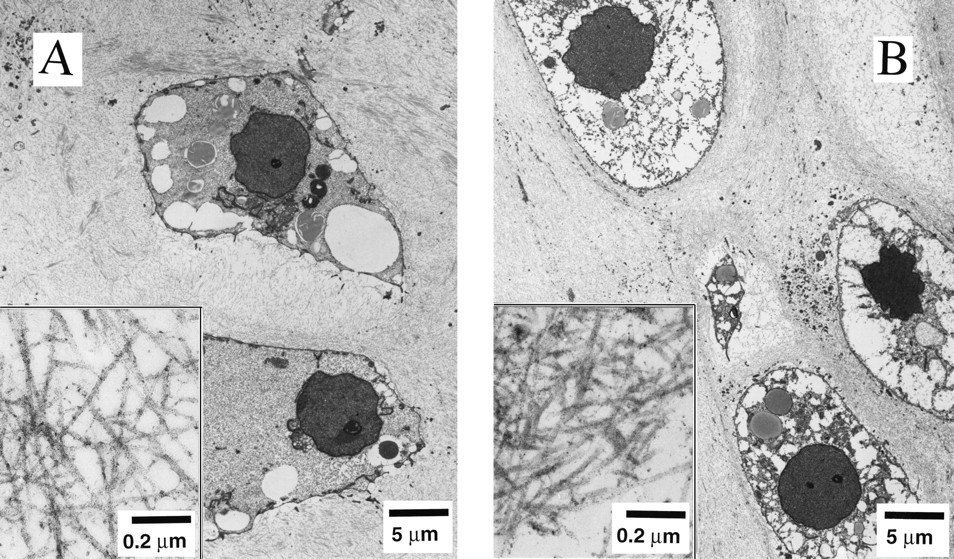
Dr. Lisa E. Freed of the Massachusetts Institute of Technology and her colleagues have reported that initially disc-like specimens tend to become spherical in space, demonstrating that tissues can grow and differentiate into distinct structures in microgravity. The Mir Increment 3 (Sept. 16, 1996 - Jan. 22, 1997) samples were smaller, more spherical, and mechanically weaker than Earth-grown control samples. These results demonstrate the feasibility of microgravity tissue engineering and may have implications for long human space voyages and for treating musculoskeletal disorders on earth. Final samples from Mir and Earth appeared histologically cartilaginous throughout their entire cross sections (5-8 mm thick), with the exception of fibrous outer capsules. Constructs grown on Earth (A) appeared to have a more organized extracellular matrix with more uniform collagen orientation as compared with constructs grown on Mir (B), but the average collagen fiber diameter was similar in the two groups (22 +- 2 nm) and comparable to that previously reported for developing articular cartilage. Randomly oriented collagen in Mir samples would be consistent with previous reports that microgravity disrupts fibrillogenesis. These are transmission electron micrographs of constructs from Mir (A) and Earth (B) groups at magnifications of x3,500 and x120,000 (Inset). The work is sponsored by NASA's Office of Biological and Physical Research. The bioreactor is managed by the Biotechnology Cell Science Program at NASA's Johnson Space Center (JSC). NASA-sponsored bioreactor research has been instrumental in helping scientists to better understand normal and cancerous tissue development. In cooperation with the medical community, the bioreactor design is being used to prepare better models of human colon, prostate, breast and ovarian tumors. Credit: Proceedings of the National Academy of Sciences.