
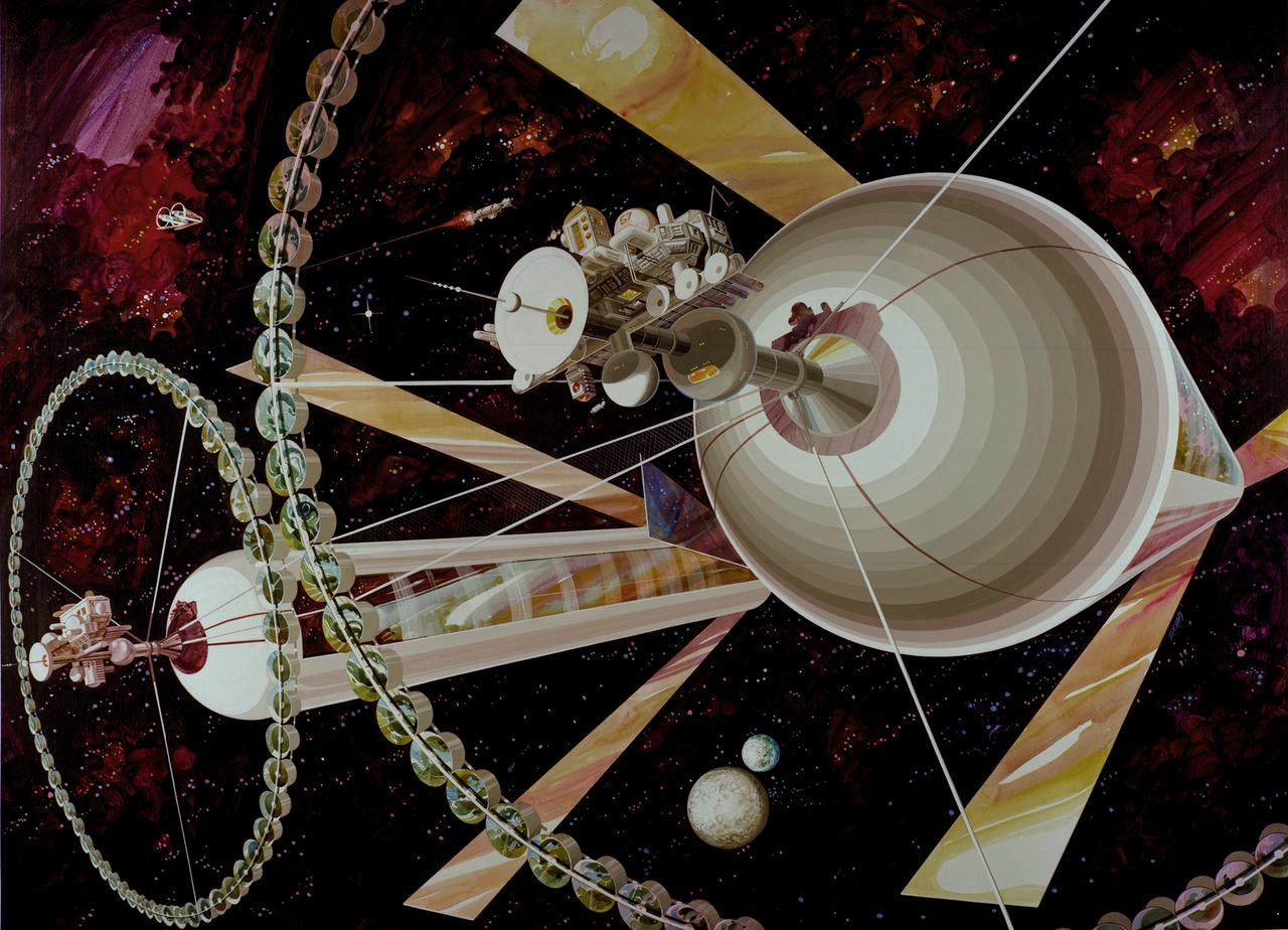
Exterior View. Space Colonization - Artwork.
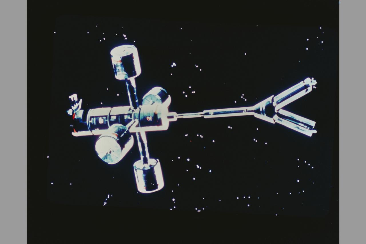
Exterior View of Colony. Space Colonization - Artwork.

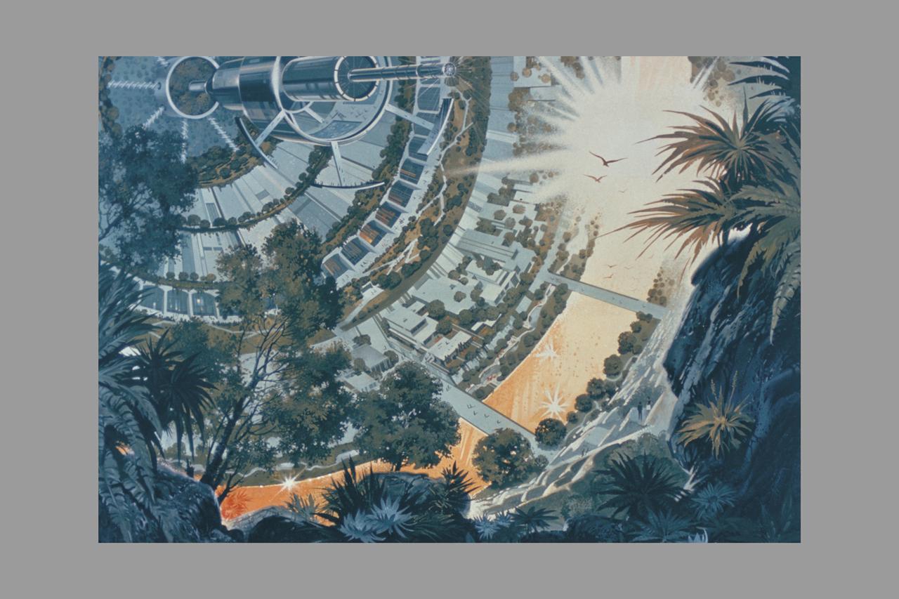
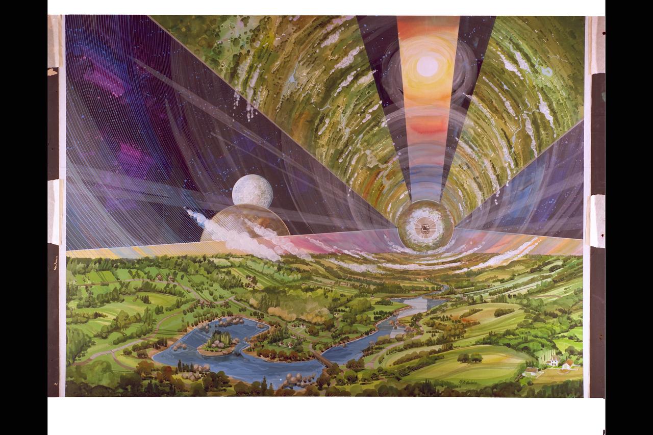
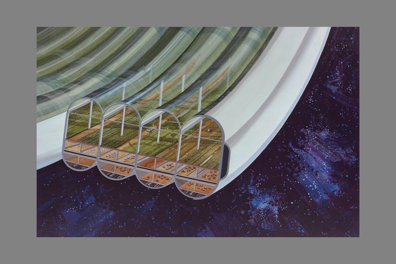
Interior View of L-5 Torus Sphere Colony. Space Colonization. Artwork by Don Davis
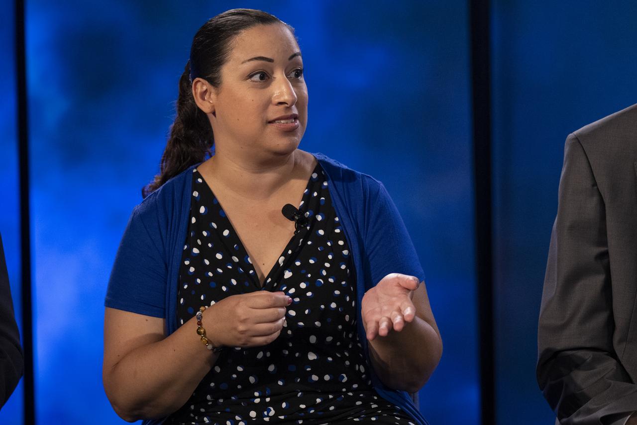
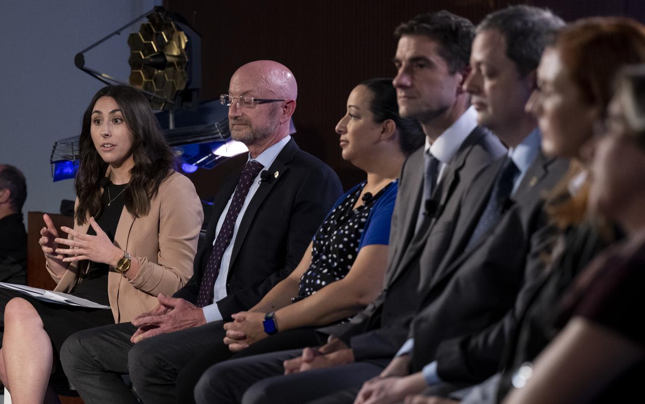
NASA James Webb Space Telescope Deputy Project Scientist for Exoplanet Science Knicole Colón answers a question from a member of the media during a briefing following the release of the first full-color images from NASA’s James Webb Space Telescope, Tuesday, July 12, 2022, at NASA’s Goddard Space Flight Center in Greenbelt, Md. The first full-color images and spectroscopic data from the James Webb Space Telescope, a partnership with ESA (European Space Agency) and the Canadian Space Agency (CSA), are a demonstration of the power of Webb as the telescope begins its science mission to unfold the infrared universe. Photo Credit: (NASA/Bill Ingalls)
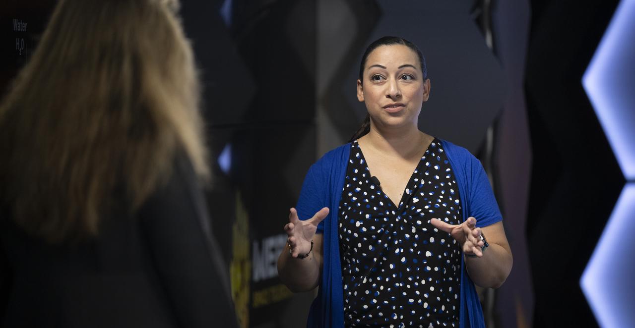
NASA James Webb Space Telescope Deputy Project Scientist for Exoplanet Science Knicole Colón speaks about the measurements of exoplanet WASP-96 b taken by the Near-Infrared Imager and Slitless Spectrograph as it is shown on screen during a broadcast releasing the telescope’s first full-color images, Tuesday, July 12, 2022, at NASA’s Goddard Space Flight Center in Greenbelt, Md. The first full-color images and spectroscopic data from the James Webb Space Telescope, a partnership with ESA (European Space Agency) and the Canadian Space Agency (CSA), are a demonstration of the power of Webb as the telescope begins its science mission to unfold the infrared universe. Photo Credit: (NASA/Bill Ingalls)
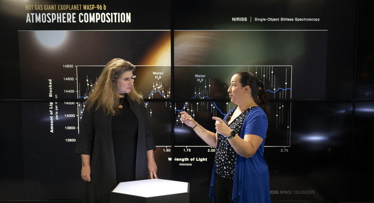
NASA James Webb Space Telescope Deputy Project Scientist for Exoplanet Science Knicole Colón speaks about the measurements of exoplanet WASP-96 b taken by the Near-Infrared Imager and Slitless Spectrograph as it is shown on screen during a broadcast releasing the telescope’s first full-color images, Tuesday, July 12, 2022, at NASA’s Goddard Space Flight Center in Greenbelt, Md. The first full-color images and spectroscopic data from the James Webb Space Telescope, a partnership with ESA (European Space Agency) and the Canadian Space Agency (CSA), are a demonstration of the power of Webb as the telescope begins its science mission to unfold the infrared universe. Photo Credit: (NASA/Bill Ingalls)
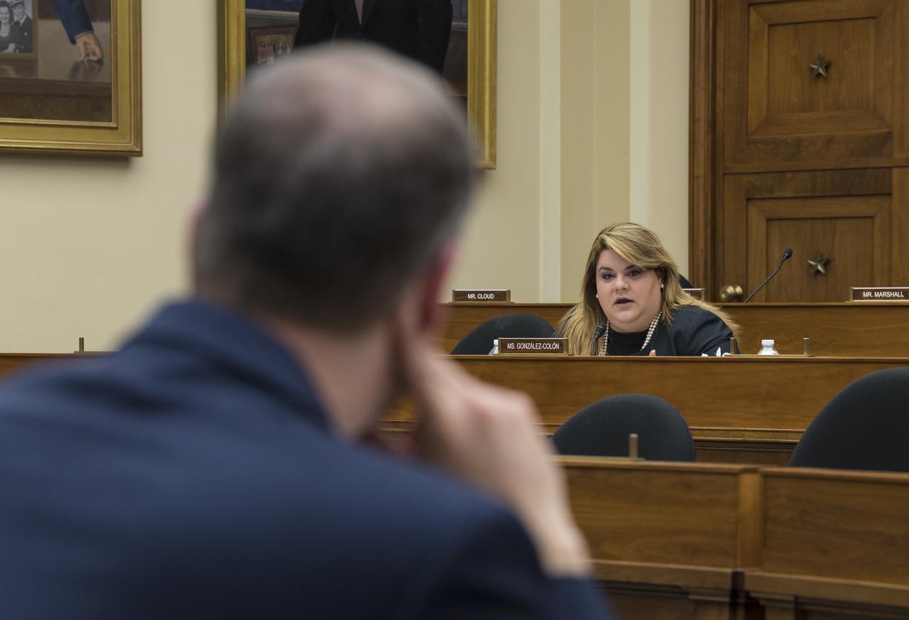
Resident Commissioner Jenniffer González-Colón, R-Puerto Rico, asks NASA Administrator Jim Bridenstine a question during a House Committee on Science, Space, and Technology hearing to review the Fiscal Year 2020 budget request for the National Aeronautics and Space Administration, Tuesday, April 2, 2019 at the Rayburn House Office Building in Washington. Photo Credit: (NASA/Aubrey Gemignani)
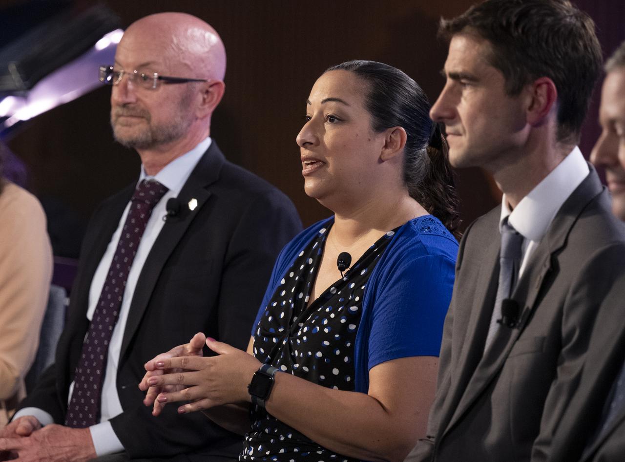
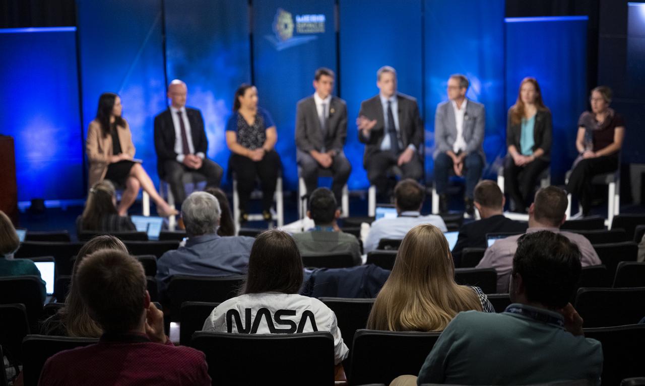
NASA James Webb Space Telescope Deputy Project Scientist for Exoplanet Science Knicole Colón, center, answers a question from a member of the media alongside NASA James Webb Space Telescope Program Scientist and Astrophysics Division Chief Scientist Eric Smith, left, and NASA James Webb Space Telescope Project Scientist at ESA (European Space Agency) Christopher Evans, during a briefing following the release of the first full-color images from NASA’s James Webb Space Telescope, Tuesday, July 12, 2022, at NASA’s Goddard Space Flight Center in Greenbelt, Md. The first full-color images and spectroscopic data from the James Webb Space Telescope, a partnership with ESA (European Space Agency) and the Canadian Space Agency (CSA), are a demonstration of the power of Webb as the telescope begins its science mission to unfold the infrared universe. Photo Credit: (NASA/Bill Ingalls)
Artwork Space Colonization - Bernal sphere
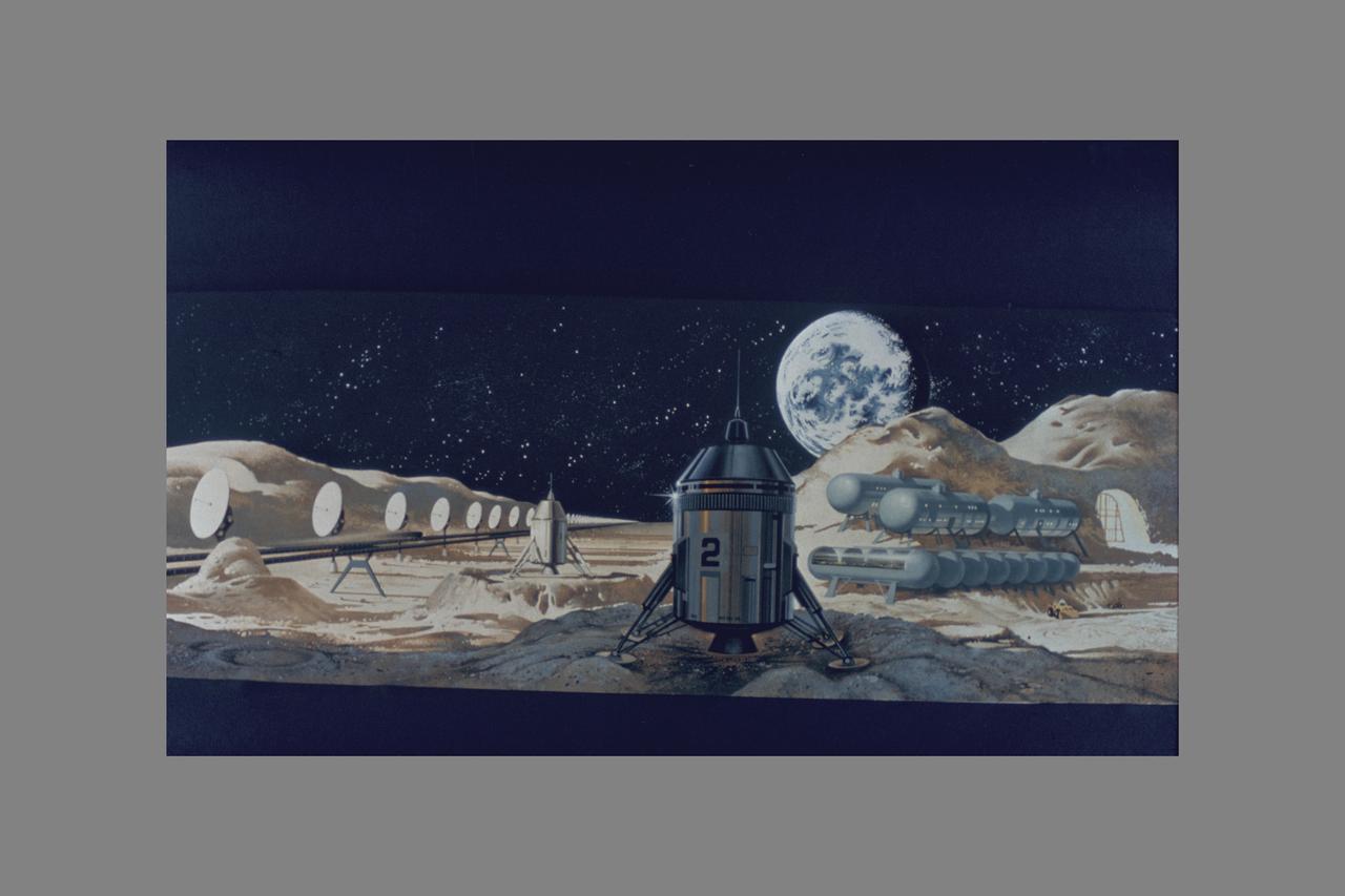
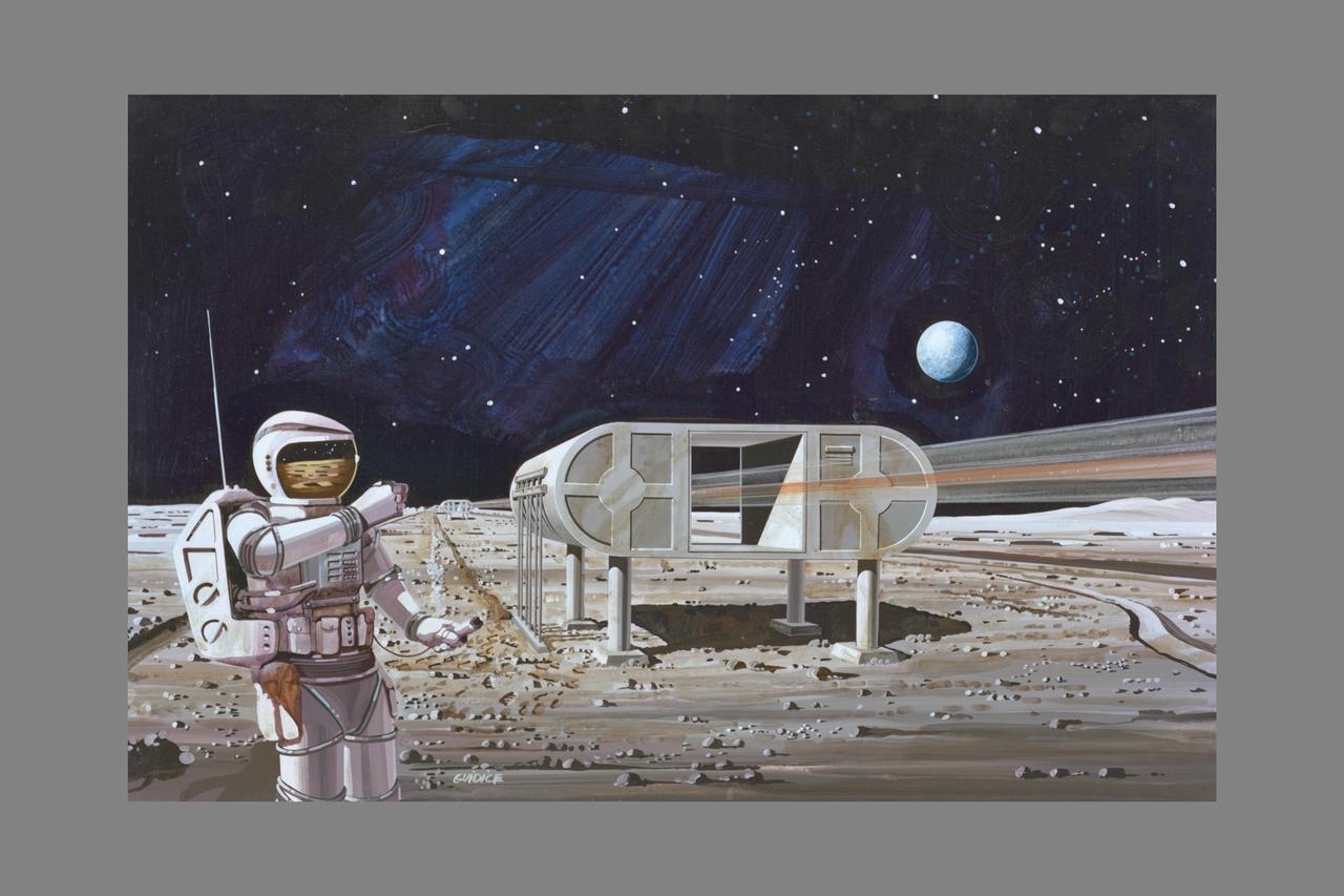
View of Moon of Equipment (transmitters). Space Colonization - Artwork.
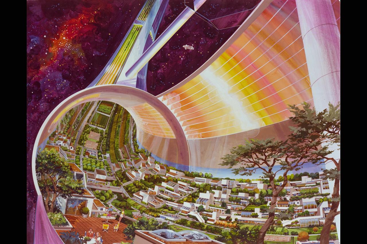
Space Colonization Modules. Artist: Rick Guidice
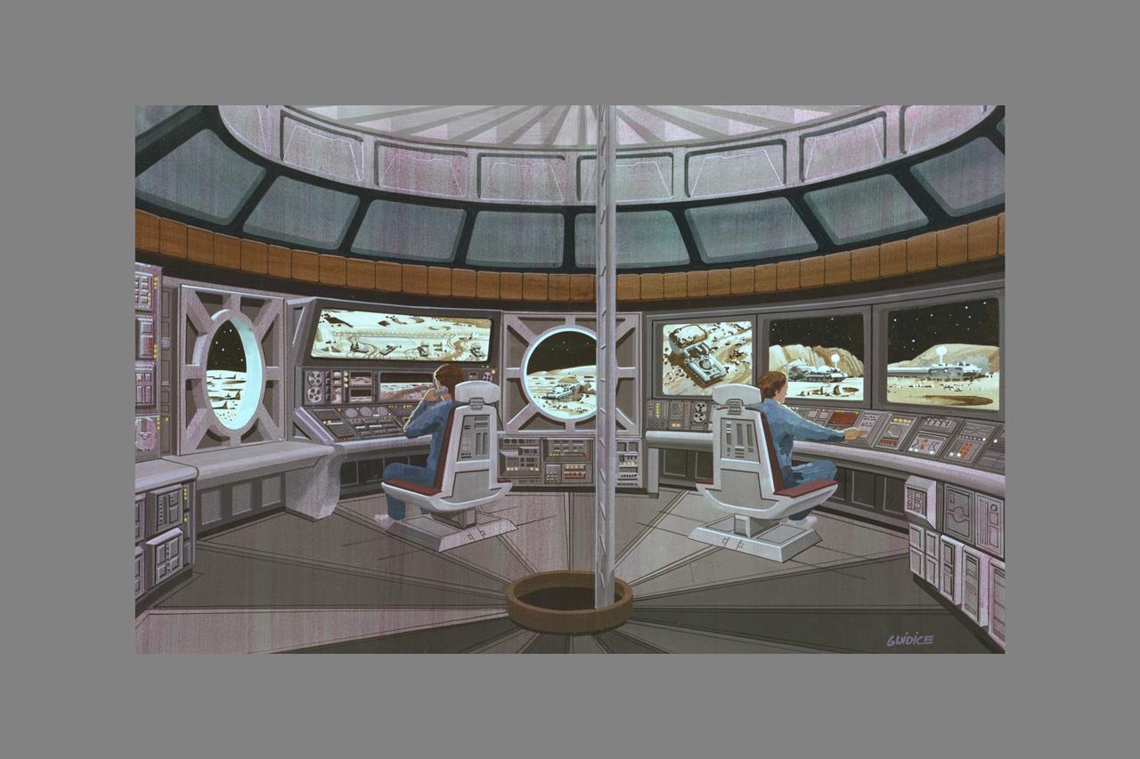
Artist: Rick Guidice Space Colonization, Habitat on Lunar base (Artwork)
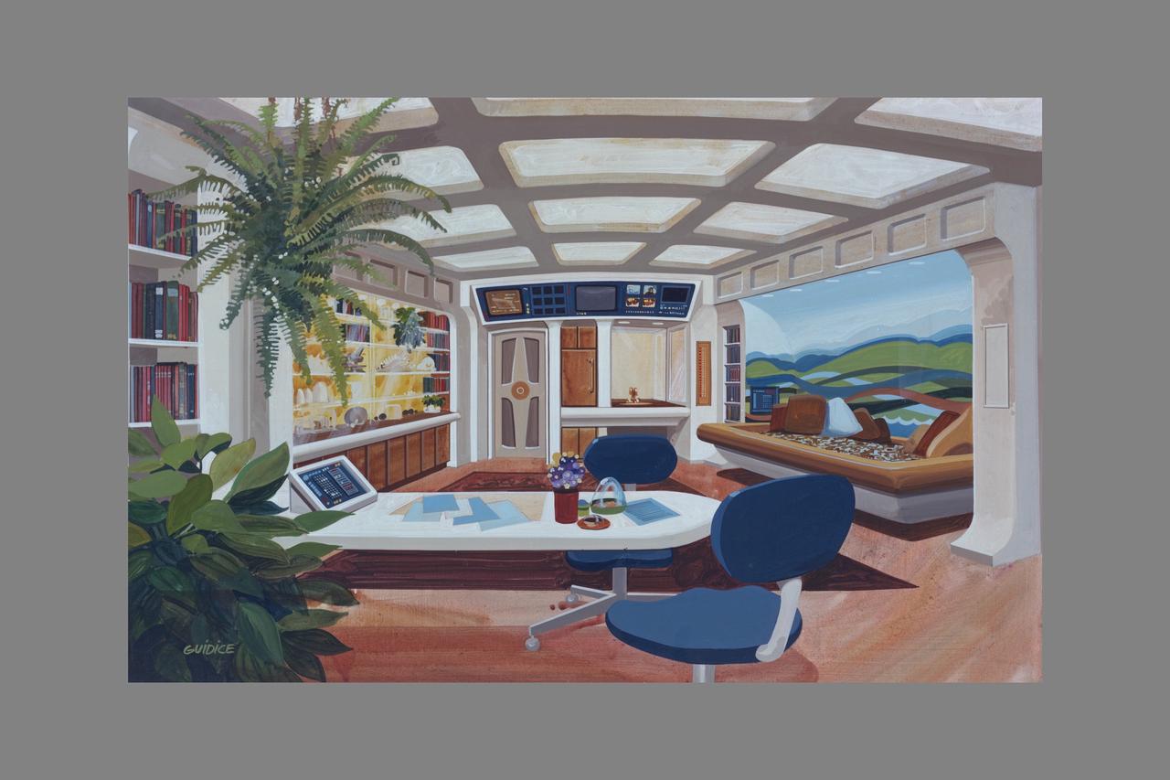
Artist: Rick Guidice Space Colonization, Habitat on Lunar base (Artwork)
ARTIST: RICK GUIDICE SPACE COLONIZATION, MANUFACTURING, HABITAT AND LUNAR BASE (ARTWORK)
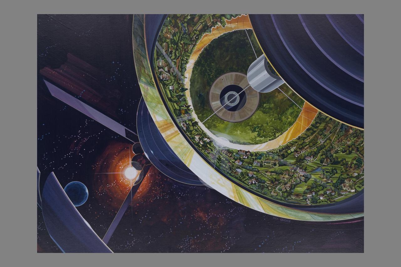
Artwork by: Rick Guidice Space Colonization - Bernal Sphere. EXTERIOR VIEW LOOKING INTO SPHERE.
Artist: Rick Guidice interior of Torus wheel (L-5) Space Colonization Module
NASA Art by Artist Rick Guidice Space Colonization - Torus Wheel Toroidal Colonies
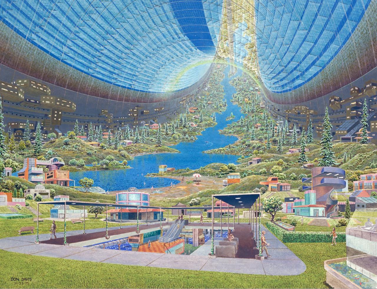
Torus sphere Interior view. Space Colonization. Artist: Don Davis ref: NASA SP-413; Space Settlements: A Design Study

“I liked every subject in school, but I think I always liked math and science the most. At the time, math seemed to always have just one answer, versus English or history where you always had to interpret things. The funny part is, once you get more into research, there’s never really one answer. You are always interpreting your data. “Data are almost tangible, even though you can’t literally touch them. You can measure the brightness of a star. You can measure the radius of a planet. You can physically see what a telescope collected and the data that come out. You can see how a star is changing over time. You can say, oh look, there’s a periodic dip! There’s a planet! And then you can say, okay, let’s take this dip and look at it across the whole spectrum of light and then you realize: this planet has an atmosphere! It has clouds, just like Earth, or Jupiter! So what does that all mean? So now, I appreciate the interpretive aspect of astronomy. “I love that there’s just so much to learn from all different-sized planets. The giant ones, down to the little ones.” Knicole Colon, research astrophysicist in the Exoplanets and Stellar Astrophysics Laboratory at NASA’s Goddard Space Flight Center, Friday, Feb. 21, 2020, in Greenbelt, Md. Photo Credit: (NASA/Joel Kowsky)
Artist: Rick Guidice Space Colonization regenerative life support systems. This concept from a summer study done in 1977 depicts a closed loop life support system for long duration space settlements or space industrialization.

Angel Colon-Rios, NASA Commercial Crew Program electrical power systems engineer, monitors the countdown during a dress rehearsal in preparation for the launch of a SpaceX Falcon 9 rocket carrying the company's Dragon spacecraft on NASA’s SpaceX Crew-10 mission with NASA astronauts Anne McClain and Nichole Ayers, JAXA (Japan Aerospace Exploration Agency) astronaut Takuya Onishi, and Roscosmos cosmonaut Kirill Peskov onboard, Sunday, March 9, 2025, in the control room of SpaceX’s HangarX at NASA’s Kennedy Space Center in Florida. NASA’s SpaceX Crew-10 mission is the tenth crew rotation mission of the SpaceX Dragon spacecraft and Falcon 9 rocket to the International Space Station as part of the agency’s Commercial Crew Program. McClain, Ayers, Onishi, and Peskov are scheduled to launch at 7:48 p.m. EDT on Wednesday, March 12, from Launch Complex 39A at the NASA's Kennedy Space Center. Photo Credit: (NASA/Aubrey Gemignani)
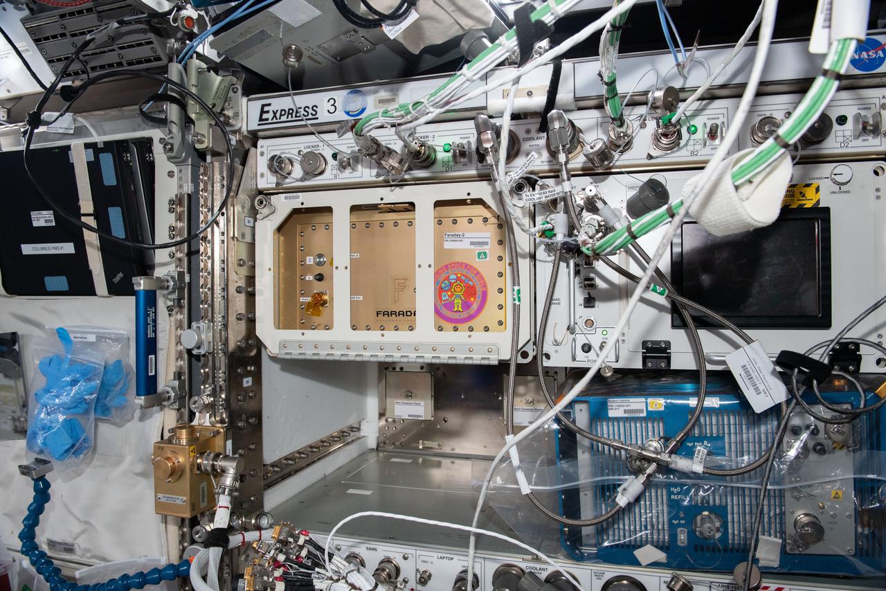
iss065e335906 (Aug. 31, 2021) --- A view of the Faraday-2 facility inside the International Space Station's U.S. Destiny laboratory module. The payload gives the Girl Scouts on Earth the opportunity to conduct a control experiment while observing space station experiments on plant growth, ant colonization, and brine shrimp lifecycle aboard the orbiting lab.
iss065e335891 (Aug. 31, 2021) --- A view of the Faraday-2 facility inside the International Space Station's U.S. Destiny laboratory module. The payload gives the Girl Scouts on Earth the opportunity to conduct a control experiment while observing space station experiments on plant growth, ant colonization, and brine shrimp lifecycle aboard the orbiting lab.
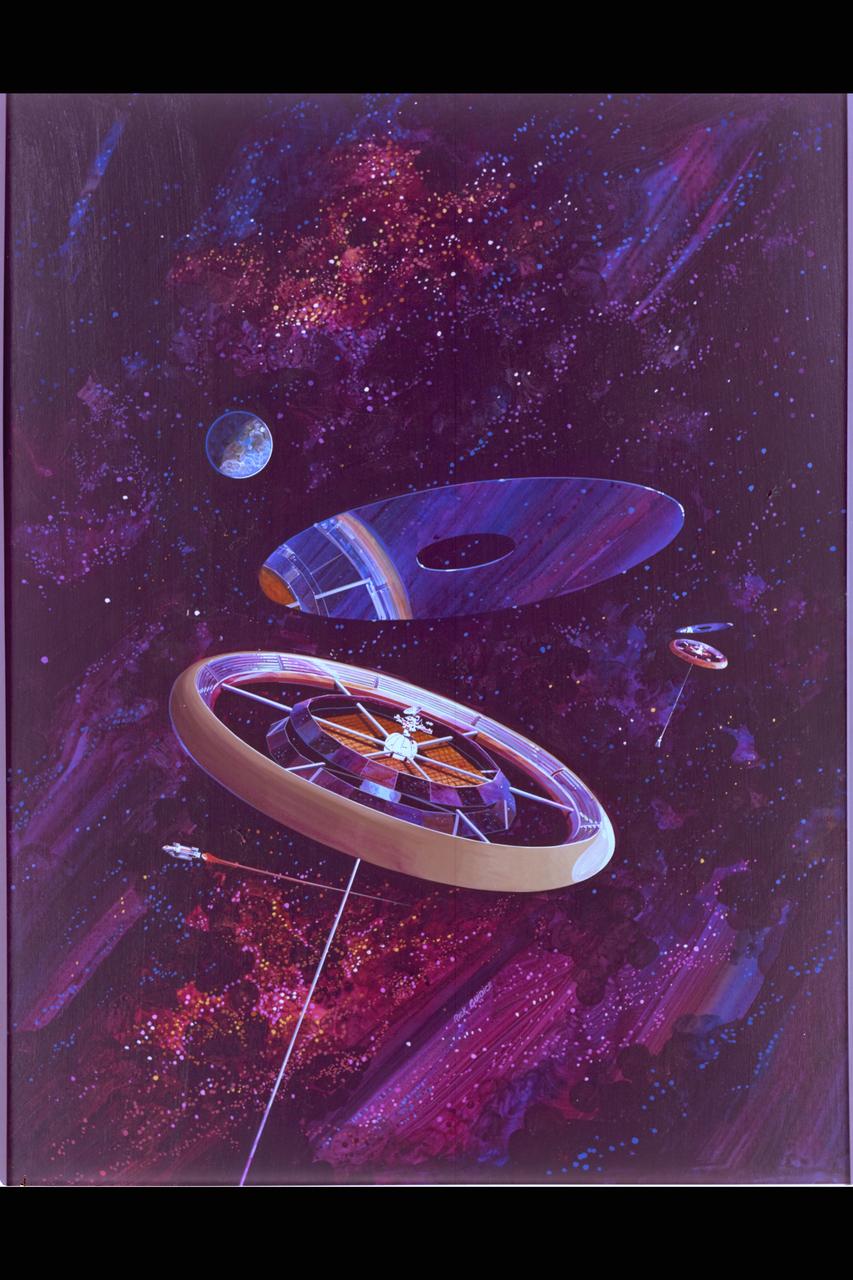
NASA Art by Rick Guidice The Torus Wheel from 'Space Settlements; A Design Study' in colonization sponsored by NASA Ames, ASEE and Stanford University in the summer of 1975 to look at all aspects of sustained life in space. (ref: NASA SP-413, library of congress catalog card number 76-600068)
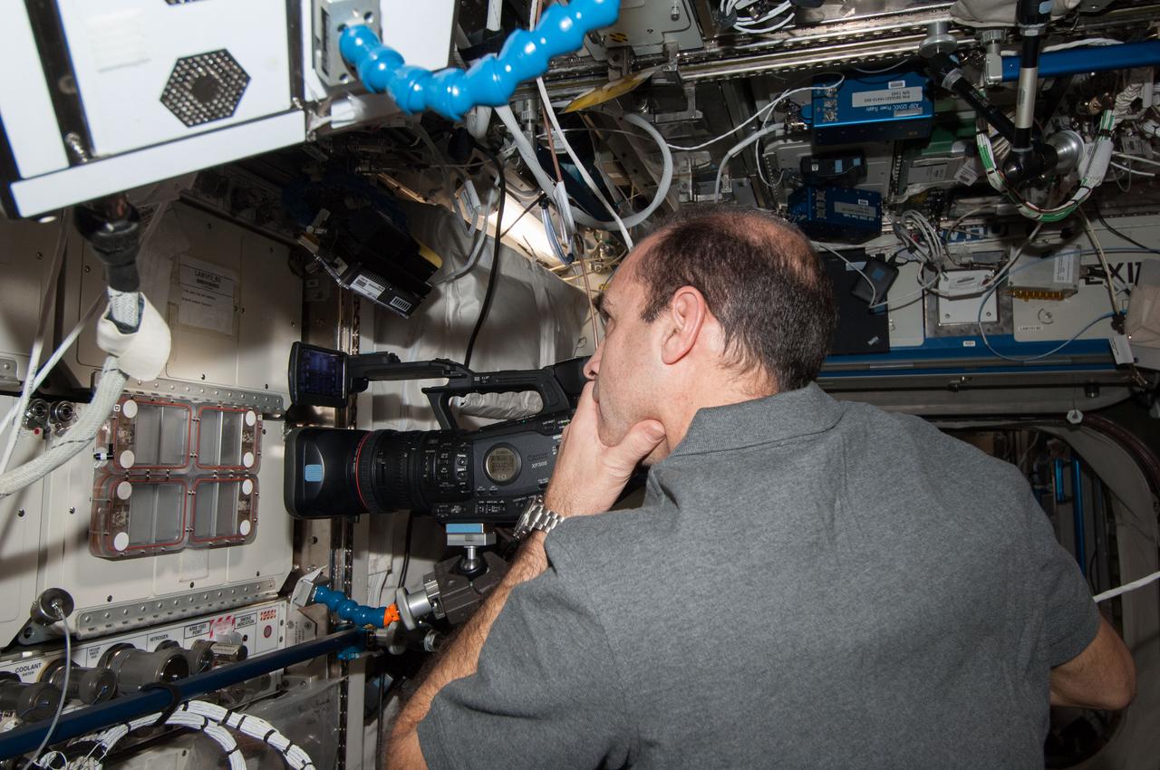
ISS038-E-029059 (12 Jan. 2014) --- In the International Space Station's Destiny laboratory, NASA astronaut Rick Mastracchio, Expedition 38 flight engineer, uses a video camera to photograph the Ant Forage Habitat Facility which will study ant behavior and colonization in microgravity.
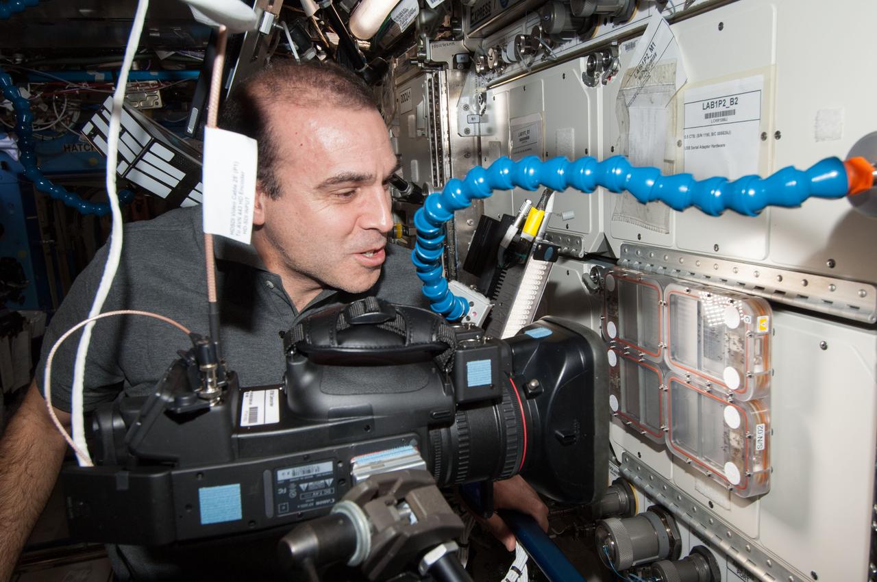
ISS038-E-029065 (12 Jan. 2014) --- In the International Space Station's Destiny laboratory, NASA astronaut Rick Mastracchio, Expedition 38 flight engineer, uses a video camera to photograph the Ant Forage Habitat Facility which will study ant behavior and colonization in microgravity.
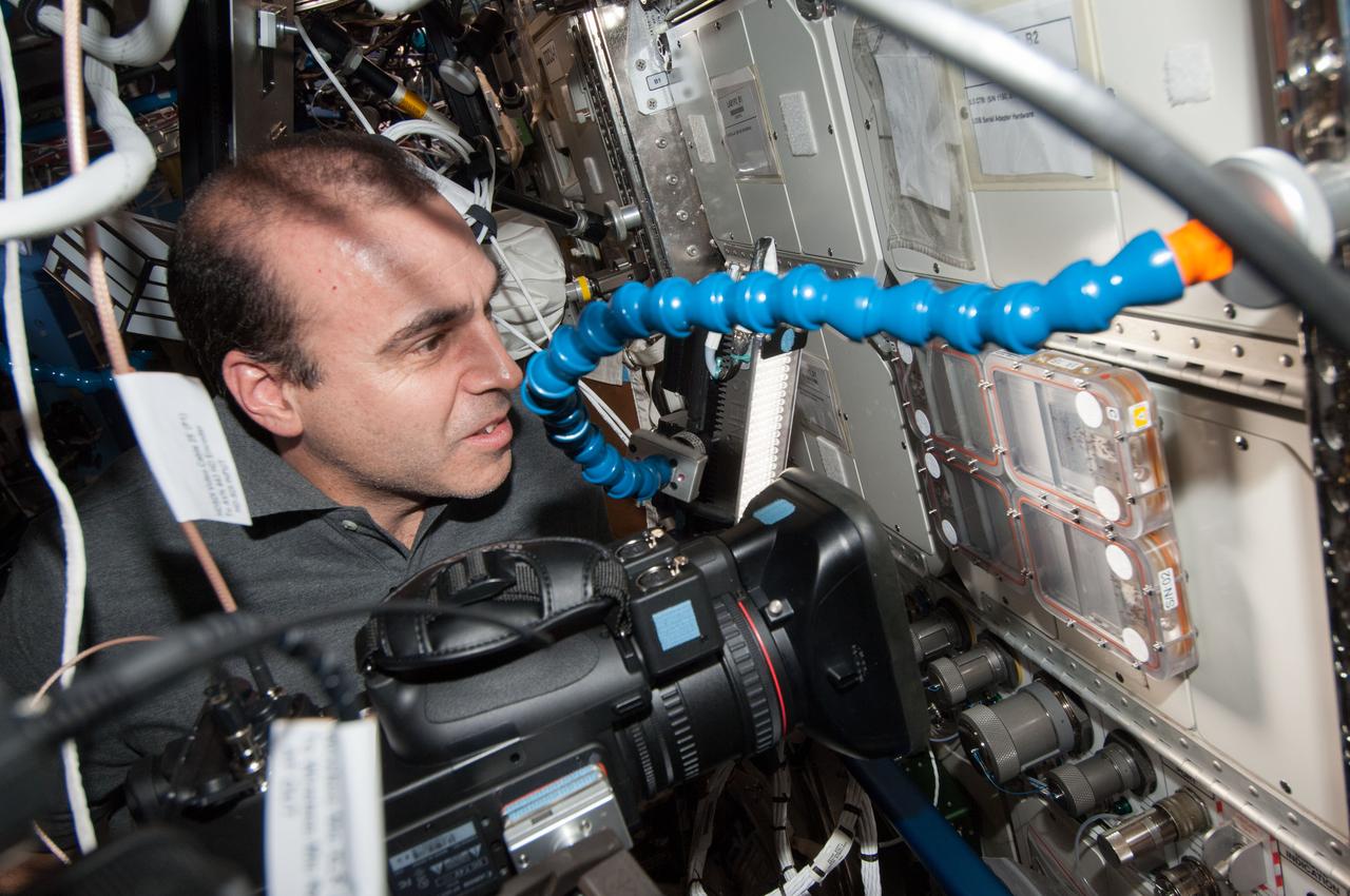
ISS038-E-029062 (12 Jan. 2014) --- In the International Space Station's Destiny laboratory, NASA astronaut Rick Mastracchio, Expedition 38 flight engineer, uses a video camera to photograph the Ant Forage Habitat Facility which will study ant behavior and colonization in microgravity.
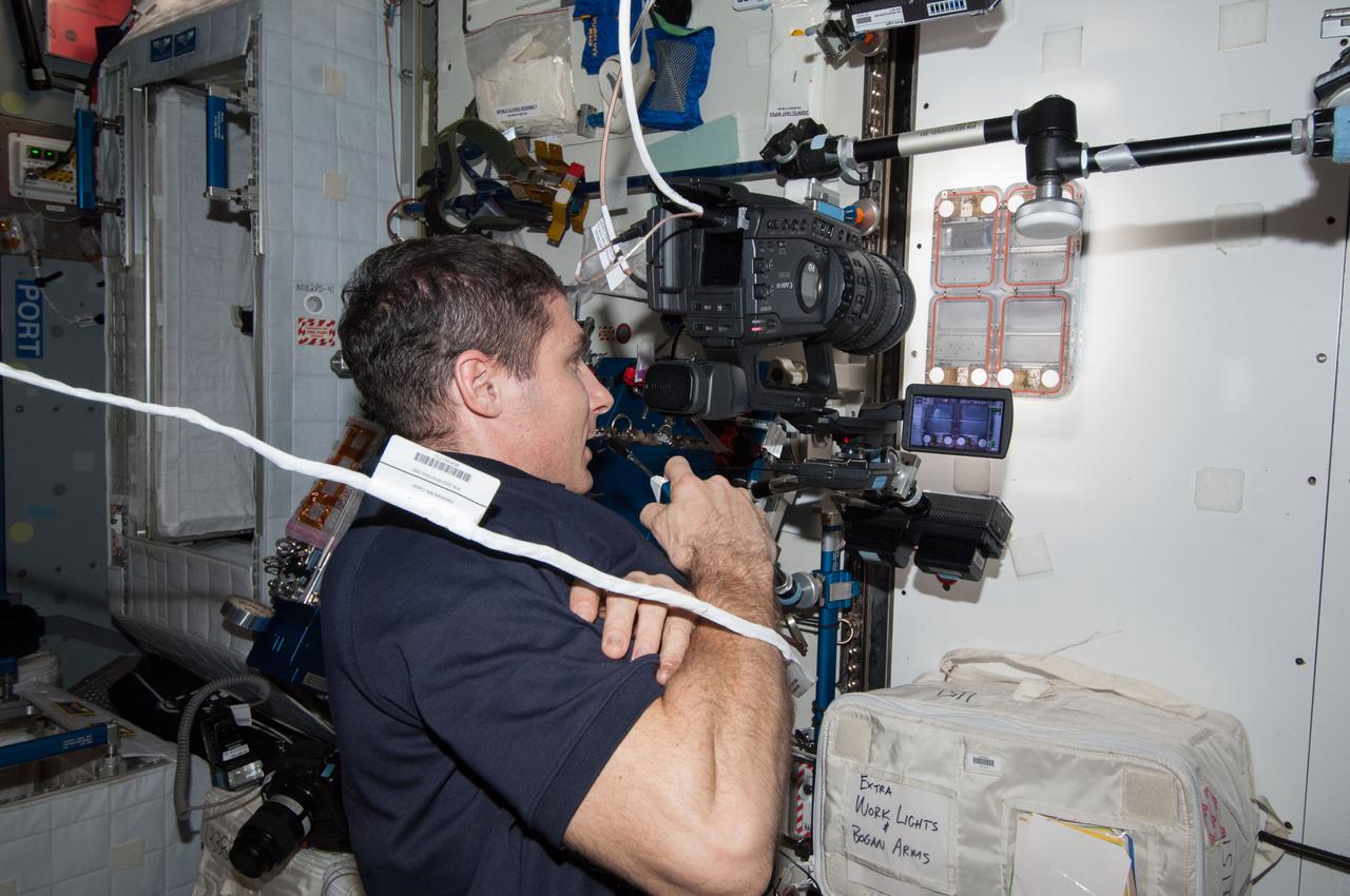
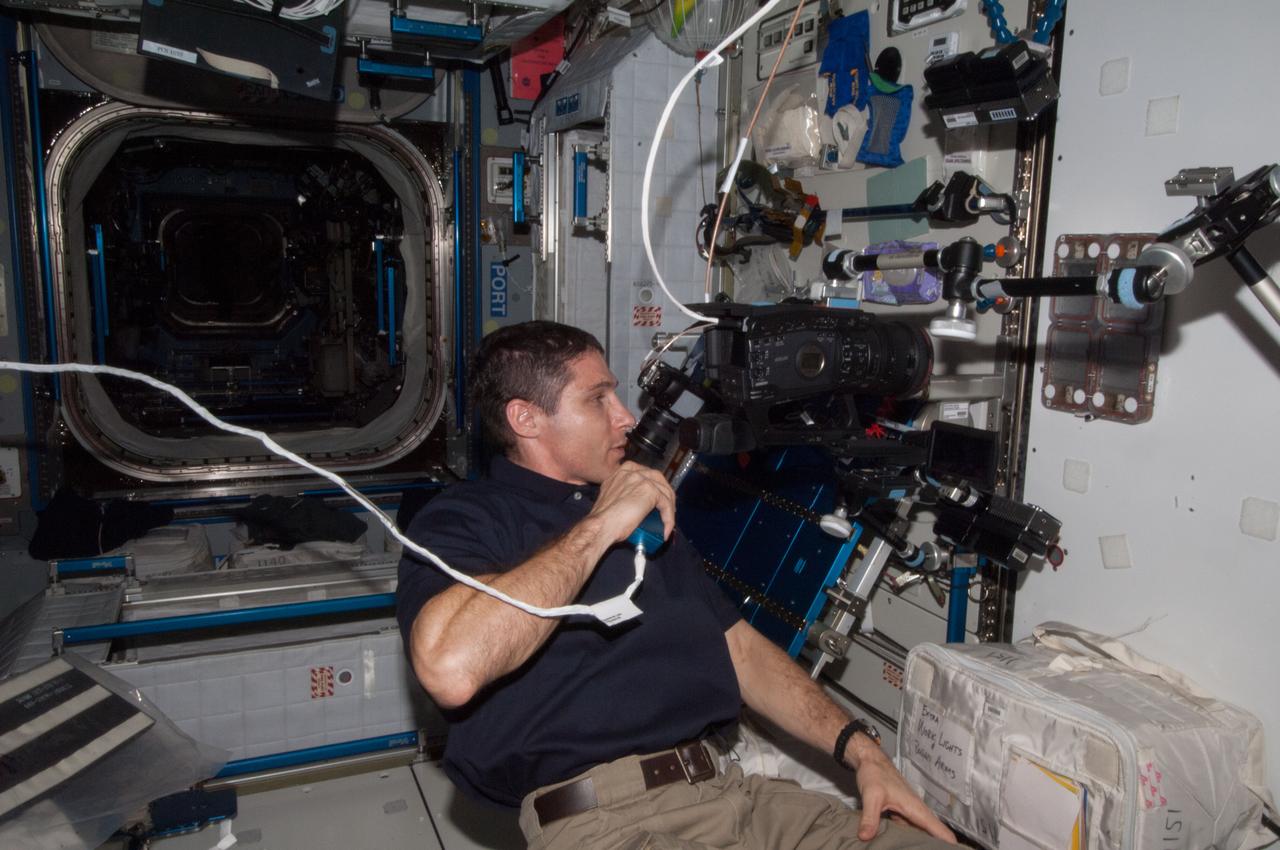
ISS038-E-029077 (12 Jan. 2014) --- In the International Space Station's Harmony node, NASA astronaut Mike Hopkins, Expedition 38 flight engineer, uses a video camera to photograph the Ant Forage Habitat Facility which will study ant behavior and colonization in microgravity.
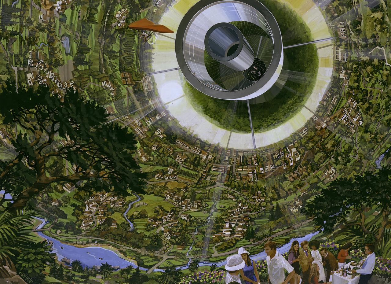
ARTIST: Rick Guidice Space Colonization; inside the sphere gravity is strongest along the equator. as on moves toward the center gravity lessens and one could fly easily. Sunlight enters as shown by the large fuzzy ring. The central tube connects to other sections of the colony.
ISS038-E-029065 (12 Jan. 2014) --- In the International Space Station's Destiny laboratory, NASA astronaut Rick Mastracchio, Expedition 38 flight engineer, uses a video camera to photograph the Ant Forage Habitat Facility which will study ant behavior and colonization in microgravity.
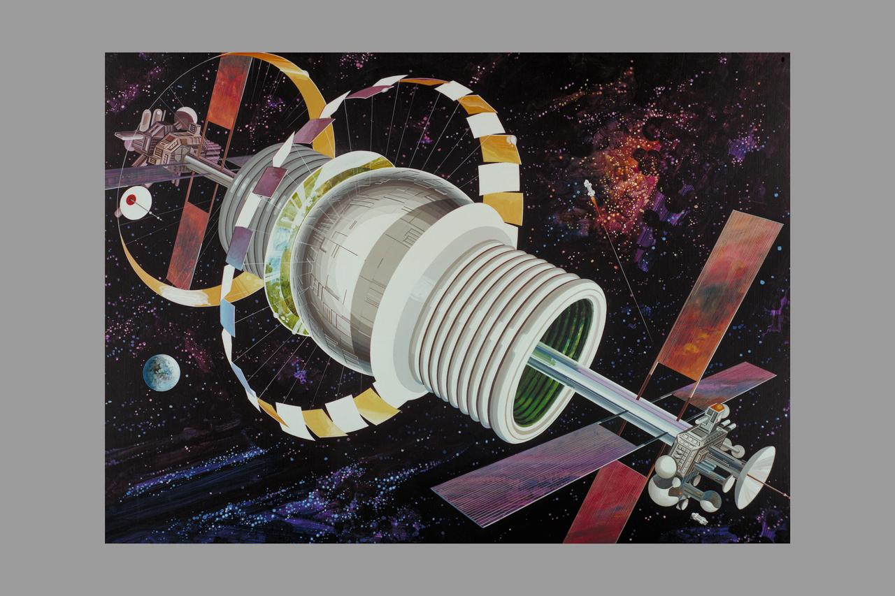
Artist: Rick Guidice Space Colonization - Bernal Sphere - The residential area is in the central sphere. Farming regions are in the 'tires.' Mirrors reflect sunlight into the habitat and farms. The large flat panels radiate away extra heat into space, and panels of solar cells provide electricity. Factories and docks for spaceships are at either end of the long central tube. (NOTE: art printed in Book 'Space Colony - Frontier of the 21st Century by Franklyn M. Branley)
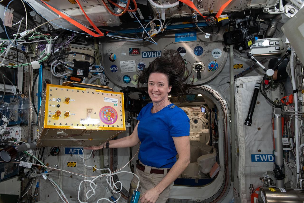
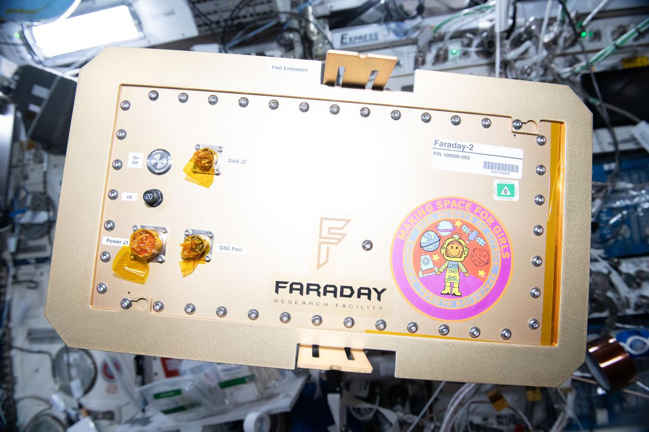
iss065e335890 (Aug. 31, 2021) --- NASA astronaut and Expedition 65 Flight Engineer Megan McArthur installs the Faraday-2 facility inside the International Space Station's U.S. Destiny laboratory module. The payload gives the Girl Scouts on Earth the opportunity to conduct a control experiment while observing space station experiments on plant growth, ant colonization, and brine shrimp lifecycle aboard the orbiting lab.
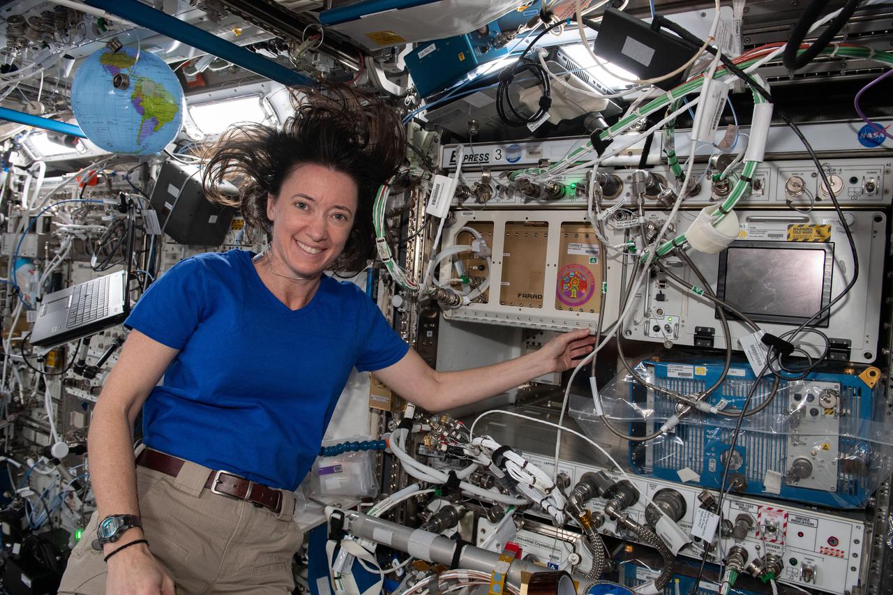
iss065e335909 (Aug. 31, 2021) --- NASA astronaut and Expedition 65 Flight Engineer Megan McArthur installs the Faraday-2 facility inside the International Space Station's U.S. Destiny laboratory module. The payload gives the Girl Scouts on Earth the opportunity to conduct a control experiment while observing space station experiments on plant growth, ant colonization, and brine shrimp lifecycle aboard the orbiting lab.
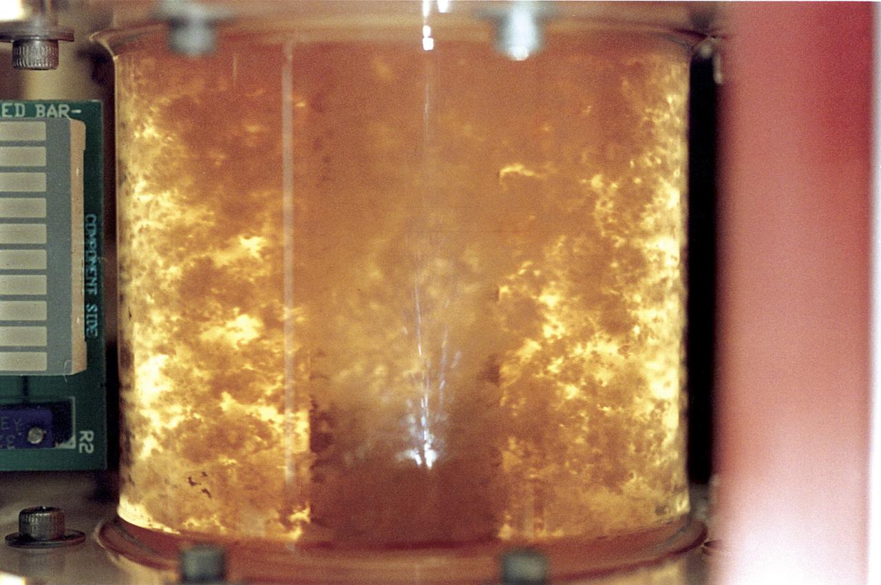
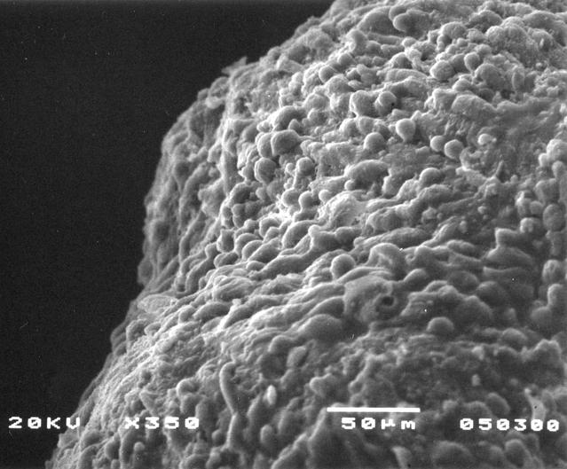
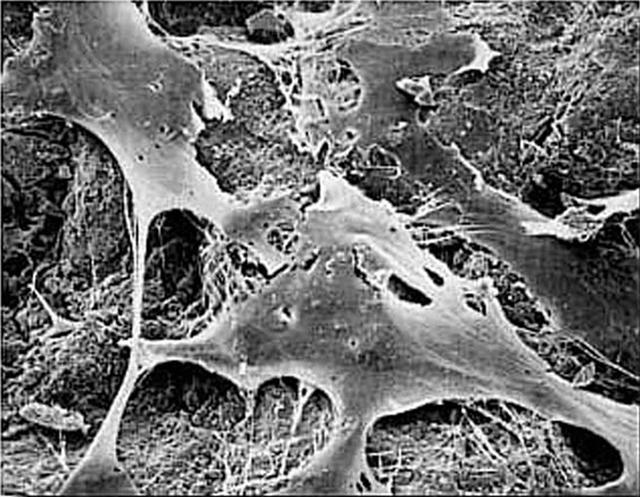
Within five days, bioreactor cultivated human colon cancer cells (shown) grown in Microgravity on the STS-70 mission in 1995, had grown 30 times the volume of the control specimens on Earth. The samples grown in space had a higher level of cellular organization and specialization. Because they more closely resemble tumors found in the body, microgravity grown cell cultures are ideal for research purposes.

ISS038-E-000232 (11 Nov. 2013) --- One of the Expedition 38 crew members aboard the International Space Station used a 180mm lens to photograph this oblique image featuring the Galapagos Islands or Islas Galapagos, distributed on either side of the Equator in the eastern Pacific Ocean. An archipelago of volcanic islands, the group?s official name is Archipielago de Colon.
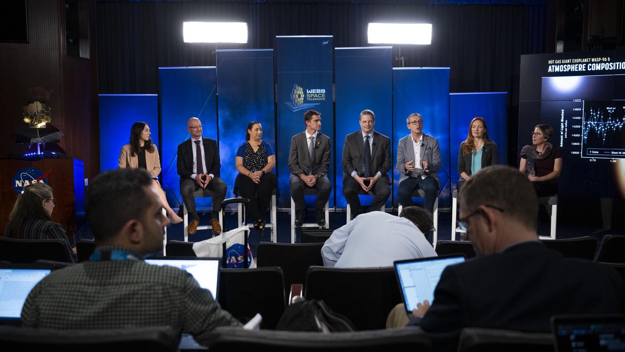
NASA Public Affairs Officer Alise Fisher, left, NASA James Webb Space Telescope Program Scientist and Astrophysics Division Chief Scientist Eric Smith, second from left, NASA James Webb Space Telescope Deputy Project Scientist for Exoplanet Science Knicole Colón, third from left, NASA James Webb Space Telescope Project Scientist at ESA (European Space Agency) Christopher Evans, fourth from left, NASA James Webb Space Telescope Project Scientist, Space Telescope Science Institute, Klaus Pontoppidan, fourth from right, Principal Investigator for the Canadian Near-Infrared Imager and Slitless Spectrograph at the University of Montreal René Doyon, third from right, NASA James Webb Space Telescope Deputy Project Scientist for Communications Amber Straughn, second from right, and NASA James Webb Space Telescope Operations Project Scientist Jane Rigby, right, during a briefing following the release of the first full-color images from NASA’s James Webb Space Telescope, Tuesday, July 12, 2022, at NASA’s Goddard Space Flight Center in Greenbelt, Md. The first full-color images and spectroscopic data from the James Webb Space Telescope, a partnership with ESA (European Space Agency) and the Canadian Space Agency (CSA), are a demonstration of the power of Webb as the telescope begins its science mission to unfold the infrared universe. Photo Credit: (NASA/Bill Ingalls)
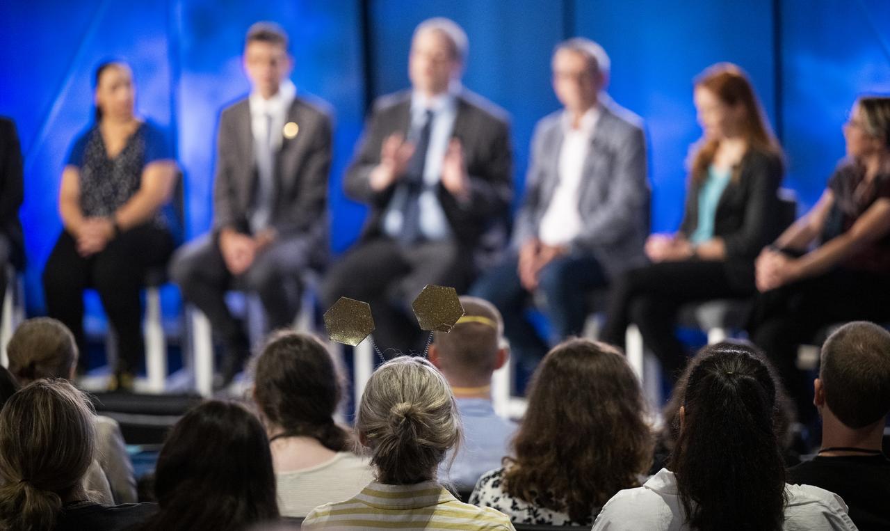
A NASA Social attendee is seen wearing a headband featuring Webb mirrors during a briefing with NASA Public Affairs Officer Alise Fisher, left, NASA James Webb Space Telescope Program Scientist and Astrophysics Division Chief Scientist Eric Smith, second from left, NASA James Webb Space Telescope Deputy Project Scientist for Exoplanet Science Knicole Colón, third from left, NASA James Webb Space Telescope Project Scientist at ESA (European Space Agency) Christopher Evans, fourth from left, NASA James Webb Space Telescope project scietntist, Space Telescope Science Institute, Klaus Pontoppidan, fourth from right, Principal Investigator for the Canadian Near-Infrared Imager and Slitless Spectrograph at the University of Montreal René Doyon, third from right, NASA James Webb Space Telescope Deputy Project Scientist for Communications Amber Straughn, second from right, and NASA James Webb Space Telescope Operations Project Scientist Jane Rigby, right, following the release of the first full-color images from NASA’s James Webb Space Telescope, Tuesday, July 12, 2022, at NASA’s Goddard Space Flight Center in Greenbelt, Md. The first full-color images and spectroscopic data from the James Webb Space Telescope, a partnership with ESA (European Space Agency) and the Canadian Space Agency (CSA), are a demonstration of the power of Webb as the telescope begins its science mission to unfold the infrared universe. Photo Credit: (NASA/Bill Ingalls)
A NASA social attendees and members of the media are seen during a briefing with NASA Public Affairs Officer Alise Fisher, left, NASA James Webb Space Telescope Program Scientist and Astrophysics Division Chief Scientist Eric Smith, second from left, NASA James Webb Space Telescope Deputy Project Scientist for Exoplanet Science Knicole Colón, third from left, NASA James Webb Space Telescope Project Scientist at ESA (European Space Agency) Christopher Evans, fourth from left, NASA James Webb Space Telescope project scietntist, Space Telescope Science Institute, Klaus Pontoppidan, fourth from right, Principal Investigator for the Canadian Near-Infrared Imager and Slitless Spectrograph at the University of Montreal René Doyon, third from right, NASA James Webb Space Telescope Deputy Project Scientist for Communications Amber Straughn, second from right, and NASA James Webb Space Telescope Operations Project Scientist Jane Rigby, right, following the release of the first full-color images from NASA’s James Webb Space Telescope, Tuesday, July 12, 2022, at NASA’s Goddard Space Flight Center in Greenbelt, Md. The first full-color images and spectroscopic data from the James Webb Space Telescope, a partnership with ESA (European Space Agency) and the Canadian Space Agency (CSA), are a demonstration of the power of Webb as the telescope begins its science mission to unfold the infrared universe. Photo Credit: (NASA/Bill Ingalls)
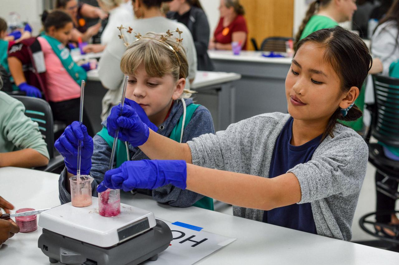
jsc2021e037279 (11/15/2019) --- Girl Scouts prepare experiments as part of a STEM collaboration with the Girl Scouts of Citrus Council. Faraday-Girl Scouts-1 (Faraday-Girls Scouts) offers Girl Scouts the opportunity to conduct a control experiment and observe the actual experiments on plant growth, ant colonization, and brine shrimp lifecycle in Faraday boxes aboard the International Space Station. This is part of a year-long effort by the Girl Scouts of Citrus Council to engage scouts in the study and understanding of space. The program also provides scouts firsthand experience with the concept of an experimental control. Image Credit: Girl Scouts Citrus Council
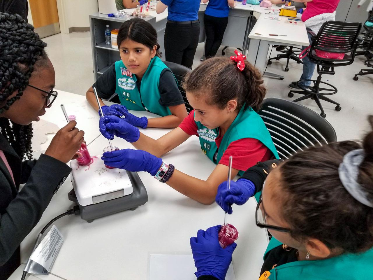
jsc2021e037278 (11/15/2019) --- Girl Scouts prepare experiments as part of a STEM collaboration with the Girl Scouts of Citrus Council. Faraday-Girl Scouts-1 (Faraday-Girls Scouts) offers Girl Scouts the opportunity to conduct a control experiment and observe the actual experiments on plant growth, ant colonization, and brine shrimp lifecycle in Faraday boxes aboard the International Space Station. This is part of a year-long effort by the Girl Scouts of Citrus Council to engage scouts in the study and understanding of space. The program also provides scouts firsthand experience with the concept of an experimental control. Image Credit: Girl Scouts Citrus Council
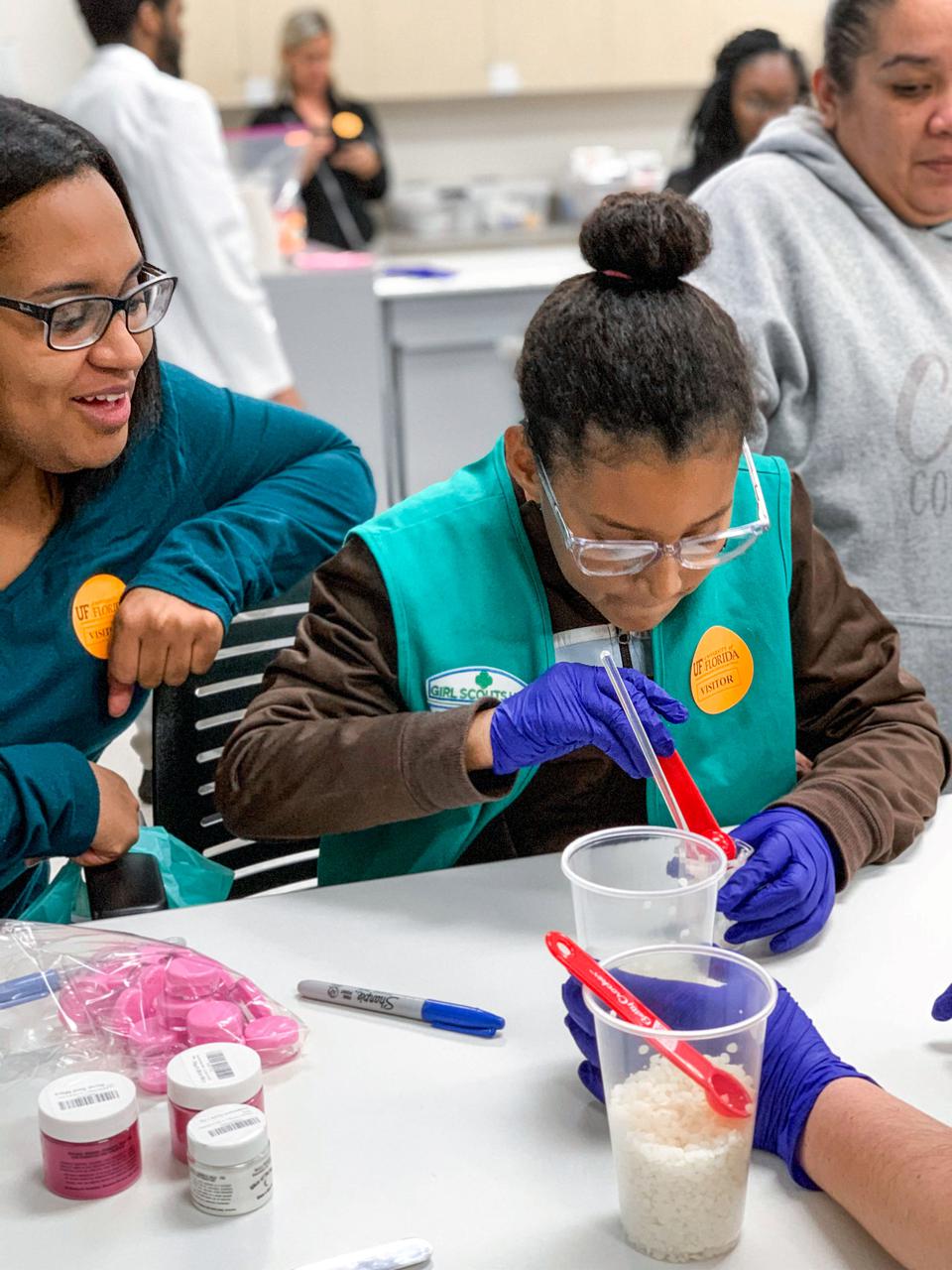
jsc2021e037280 (11/15/2019) --- Girl Scouts prepare experiments as part of a STEM collaboration with the Girl Scouts of Citrus Council. Faraday-Girl Scouts-1 (Faraday-Girls Scouts) offers Girl Scouts the opportunity to conduct a control experiment and observe the actual experiments on plant growth, ant colonization, and brine shrimp lifecycle in Faraday boxes aboard the International Space Station. This is part of a year-long effort by the Girl Scouts of Citrus Council to engage scouts in the study and understanding of space. The program also provides scouts firsthand experience with the concept of an experimental control. Image Credit: Girl Scouts Citrus Council
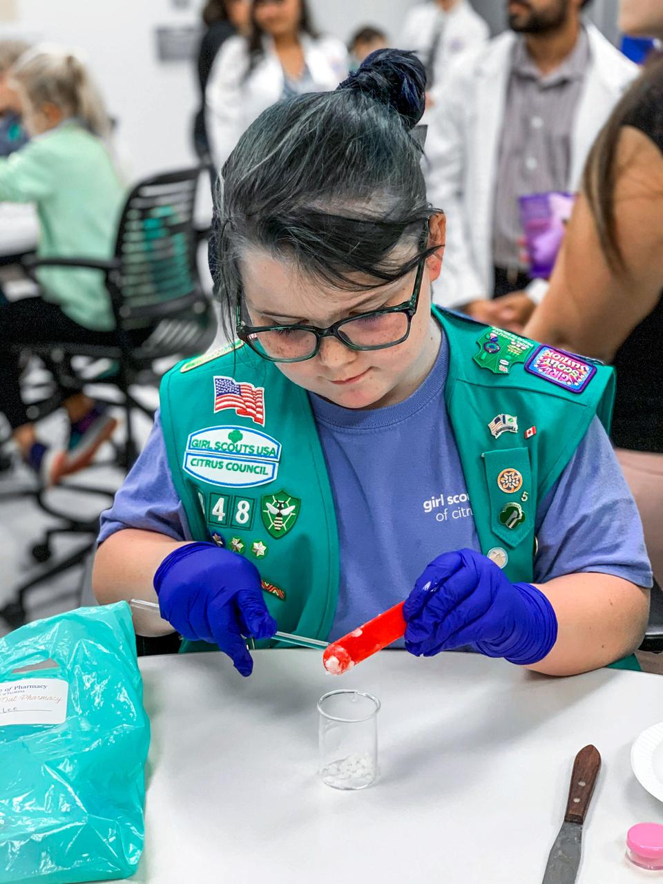
jsc2021e037281 (11/15/2019) --- Girl Scouts prepare experiments as part of a STEM collaboration with the Girl Scouts of Citrus Council. Faraday-Girl Scouts-1 (Faraday-Girls Scouts) offers Girl Scouts the opportunity to conduct a control experiment and observe the actual experiments on plant growth, ant colonization, and brine shrimp lifecycle in Faraday boxes aboard the International Space Station. This is part of a year-long effort by the Girl Scouts of Citrus Council to engage scouts in the study and understanding of space. The program also provides scouts firsthand experience with the concept of an experimental control. Image Credit: Girl Scouts Citrus Council

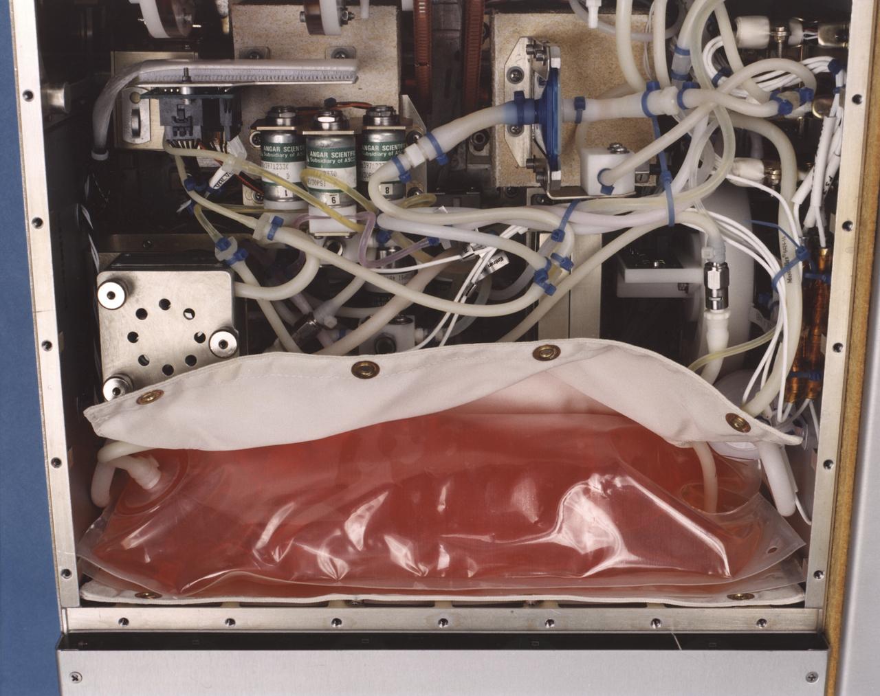
Astronaut John Blaha replaces an exhausted media bag and filled waste bag with fresh bags to continue a bioreactor experiment aboard space station Mir in 1996. NASA-sponsored bioreactor research has been instrumental in helping scientists to better understand normal and cancerous tissue development. In cooperation with the medical community, the bioreactor design is being used to prepare better models of human colon, prostate, breast and ovarian tumors. Cartilage, bone marrow, heart muscle, skeletal muscle, pancreatic islet cells, liver and kidney are just a few of the normal tissues being cultured in rotating bioreactors by investigators. This image is from a video downlink. The work is sponsored by NASA's Office of Biological and Physical Research. The bioreactor is managed by the Biotechnology Cell Science Program at NASA's Johnson Space Center (JSC).
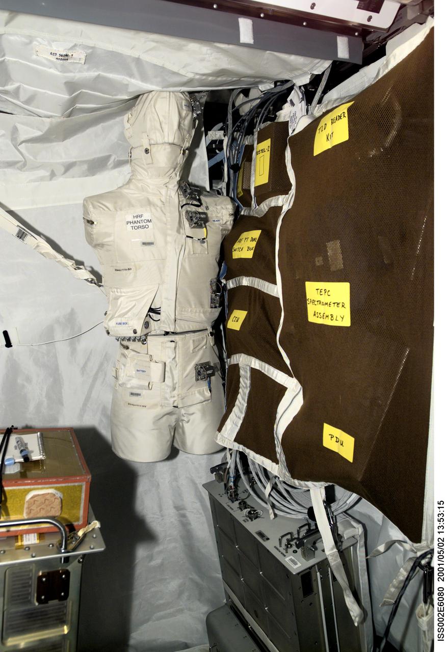
ISS002-E-6080 (2 May 2001) --- The Phantom Torso, seen here in the Human Research Facility (HRF) section of the Destiny/U.S. laboratory on the International Space Station (ISS), is designed to measure the effects of radiation on organs inside the body by using a torso that is similar to those used to train radiologists on Earth. The torso is equivalent in height and weight to an average adult male. It contains radiation detectors that will measure, in real-time, how much radiation the brain, thyroid, stomach, colon, and heart and lung area receive on a daily basis. The data will be used to determine how the body reacts to and shields its internal organs from radiation, which will be important for longer duration space flights. The experiment was delivered to the orbiting outpost during by the STS-100/6A crew in April 2001. Dr. Gautam Badhwar, NASA JSC, Houston, TX, is the principal investigator for this experiment. A digital still camera was used to record this image.
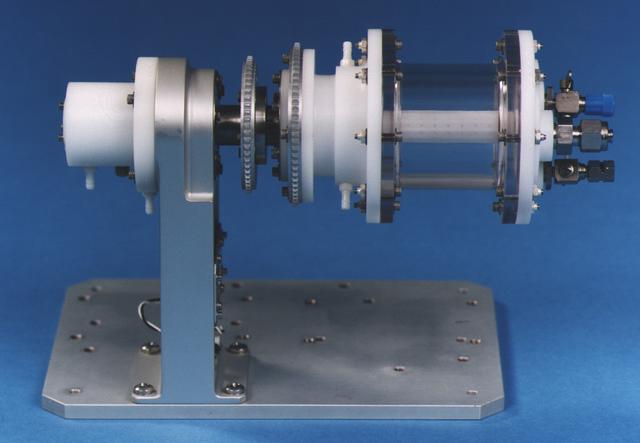
The heart of the bioreactor is the rotating wall vessel, shown without its support equipment. Volume is about 125 mL. The work is sponsored by NASA's Office of Biological and Physical Research. The bioreactor is managed by the Biotechnology Cell Science Program at NASA's Johnson Space Center (JSC). NASA-sponsored bioreactor research has been instrumental in helping scientists to better understand normal and cancerous tissue development. In cooperation with the medical community, the bioreactor design is being used to prepare better models of human colon, prostate, breast and ovarian tumors. Cartilage, bone marrow, heart muscle, skeletal muscle, pancreatic islet cells, liver and kidney are just a few of the normal tissues being cultured in rotating bioreactors by investigators.
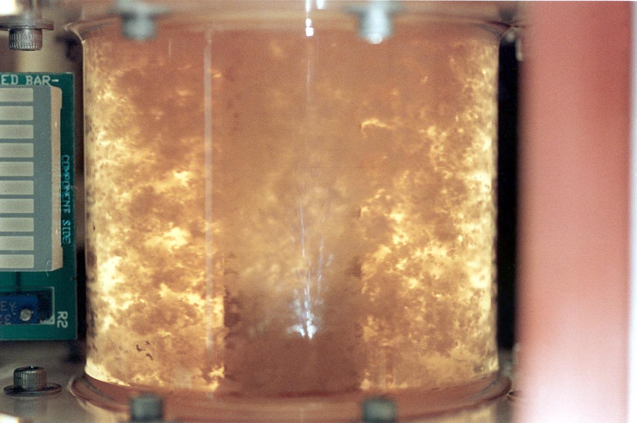
For 5 days on the STS-70 mission, a bioreactor cultivated human colon cancer cells, which grew to 30 times the volume of control specimens grown on Earth. This significant result was reproduced on STS-85 which grew mature structures that more closely match what are found in tumors in humans. Shown here, clusters of cells slowly spin inside a bioreactor. On Earth, the cells continually fall through the buffer medium and never hit bottom. In space, they are naturally suspended. Rotation ensures gentle stirring so waste is removed and fresh nutrient and oxygen are supplied. The NASA Bioreactor provides a low turbulence culture environment which promotes the formation of large, three-dimensional cell clusters. Due to their high level of cellular organization and specialization, samples constructed in the bioreactor more closely resemble the original tumor or tissue found in the body. The Bioreactor is rotated to provide gentle mixing of fresh and spent nutrient without inducing shear forces that would damage the cells. The work is sponsored by NASA's Office of Biological and Physical Research. The bioreactor is managed by the Biotechnology Cell Science Program at NASA's Johnson Space Center (JSC). NASA-sponsored bioreactor research has been instrumental in helping scientists to better understand normal and cancerous tissue development. In cooperation with the medical community, the bioreactor design is being used to prepare better models of human colon, prostate, breast and ovarian tumors. Cartilage, bone marrow, heart muscle, skeletal muscle, pancreatic islet cells, liver and kidney are just a few of the normal tissues being cultured in rotating bioreactors by investigators.
NASA Public Affairs Officer Alise Fisher, left, moderates a briefing with NASA James Webb Space Telescope Program Scientist and Astrophysics Division Chief Scientist Eric Smith, NASA James Webb Space Telescope Deputy Project Scientist for Exoplanet Science Knicole Colón, NASA James Webb Space Telescope Project Scientist at ESA (European Space Agency) Christopher Evans, NASA James Webb Space Telescope project scietntist, Space Telescope Science Institute, Klaus Pontoppidan, Principal Investigator for the Canadian Near-Infrared Imager and Slitless Spectrograph at the University of Montreal René Doyon, NASA James Webb Space Telescope Deputy Project Scientist for Communications Amber Straughn, and NASA James Webb Space Telescope Operations Project Scientist Jane Rigby following the release of the first full-color images from NASA’s James Webb Space Telescope, Tuesday, July 12, 2022, at NASA’s Goddard Space Flight Center in Greenbelt, Md. The first full-color images and spectroscopic data from the James Webb Space Telescope, a partnership with ESA (European Space Agency) and the Canadian Space Agency (CSA), are a demonstration of the power of Webb as the telescope begins its science mission to unfold the infrared universe. Photo Credit: (NASA/Bill Ingalls)
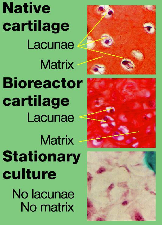
Dr. Lisa E. Freed of the Massachusetts Institute of Technology and her colleagues have reported that initially disc-like specimens tend to become spherical in space, demonstrating that tissues can grow and differentiate into distinct structures in microgravity. The Mir Increment 3 (Sept. 16, 1996 - Jan. 22, 1997) samples were smaller, more spherical, and mechanically weaker than Earth-grown control samples. These results demonstrate the feasibility of microgravity tissue engineering and may have implications for long human space voyages and for treating musculoskeletal disorders on earth. The work is sponsored by NASA's Office of Biological and Physical Research. The bioreactor is managed by the Biotechnology Cell Science Program at NASA's Johnson Space Center (JSC). NASA-sponsored bioreactor research has been instrumental in helping scientists to better understand normal and cancerous tissue development. In cooperation with the medical community, the bioreactor design is being used to prepare better models of human colon, prostate, breast and ovarian tumors. Cartilage, bone marrow, heart muscle, skeletal muscle, pancreatic islet cells, liver and kidney are just a few of the normal tissues being cultured in rotating bioreactors by investigators.
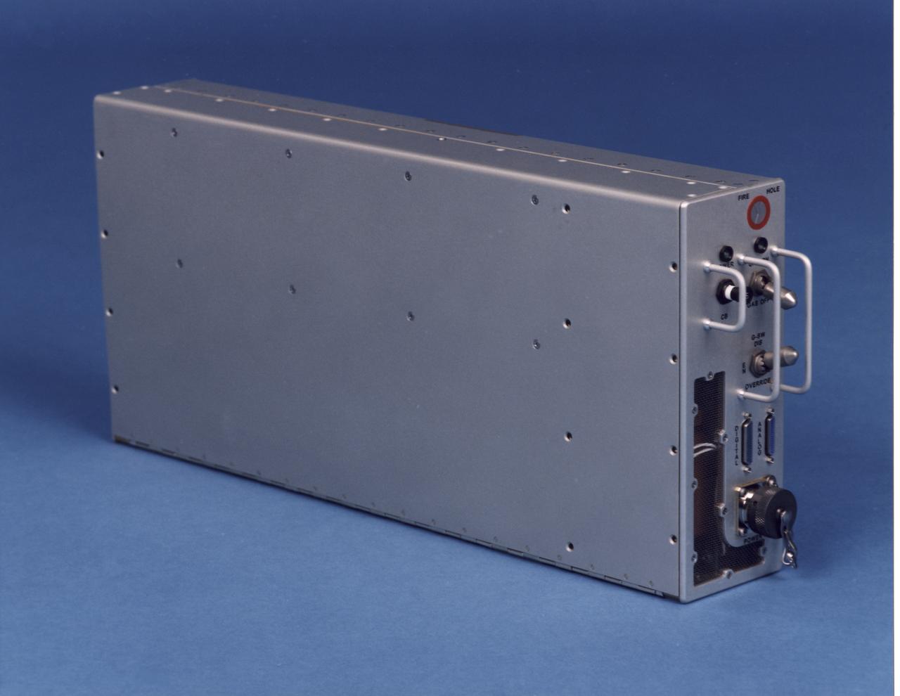
Electronics control module for the NASA Bioreactor. The NASA Bioreactor provides a low turbulence culture environment which promotes the formation of large, three-dimensional cell clusters. The Bioreactor is rotated to provide gentle mixing of fresh and spent nutrient without inducing shear forces that would damage the cells. Due to their high level of cellular organization and specialization, samples constructed in the bioreactor more closely resemble the original tumor or tissue found in the body. The work is sponsored by NASA's Office of Biological and Physical Research. The bioreactor is managed by the Biotechnology Cell Science Program at NASA's Johnson Space Center (JSC). NASA-sponsored bioreactor research has been instrumental in helping scientists to better understand normal and cancerous tissue development. In cooperation with the medical community, the bioreactor design is being used to prepare better models of human colon, prostate, breast and ovarian tumors. Cartilage, bone marrow, heart muscle, skeletal muscle, pancreatic islet cells, liver and kidney are just a few of the normal tissues being cultured in rotating bioreactors by investigators.
This prostate cancer construct was grown during NASA-sponsored bioreactor studies on Earth. Cells are attached to a biodegradable plastic lattice that gives them a head start in growth. Prostate tumor cells are to be grown in a NASA-sponsored Bioreactor experiment aboard the STS-107 Research-1 mission in 2002. Dr. Leland Chung of the University of Virginia is the principal investigator. The NASA Bioreactor provides a low turbulence culture environment which promotes the formation of large, three-dimensional cell clusters. Due to their high level of cellular organization and specialization, samples constructed in the bioreactor more closely resemble the original tumor or tissue found in the body. The Bioreactor is rotated to provide gentle mixing of fresh and spent nutrient without inducing shear forces that would damage the cells. The work is sponsored by NASA's Office of Biological and Physical Research. The bioreactor is managed by the Biotechnology Cell Science Program at NASA's Johnson Space Center (JSC). NASA-sponsored bioreactor research has been instrumental in helping scientists to better understand normal and cancerous tissue development. In cooperation with the medical community, the bioreactor design is being used to prepare better models of human colon, prostate, breast and ovarian tumors. Cartilage, bone marrow, heart muscle, skeletal muscle, pancreatic islet cells, liver and kidney are just a few of the normal tissues being cultured in rotating bioreactors by investigators. Credit: NASA and the University of Virginia.
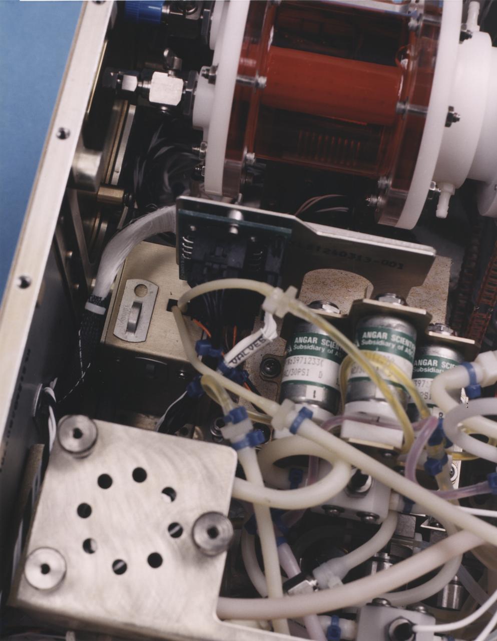
Close-up view of the interior of a NASA Bioreactor shows the plastic plumbing and valves (cylinders at right center) to control fluid flow. The rotating wall vessel is at top center. The NASA Bioreactor provides a low turbulence culture environment which promotes the formation of large, three-dimensional cell clusters. The Bioreactor is rotated to provide gentle mixing of fresh and spent nutrient without inducing shear forces that would damage the cells. Due to their high level of cellular organization and specialization, samples constructed in the bioreactor more closely resemble the original tumor or tissue found in the body. The work is sponsored by NASA's Office of Biological and Physical Research. The bioreactor is managed by the Biotechnology Cell Science Program at NASA's Johnson Space Center (JSC). NASA-sponsored bioreactor research has been instrumental in helping scientists to better understand normal and cancerous tissue development. In cooperation with the medical community, the bioreactor design is being used to prepare better models of human colon, prostate, breast and ovarian tumors. Cartilage, bone marrow, heart muscle, skeletal muscle, pancreatic islet cells, liver and kidney are just a few of the normal tissues being cultured in rotating bioreactors by investigators.
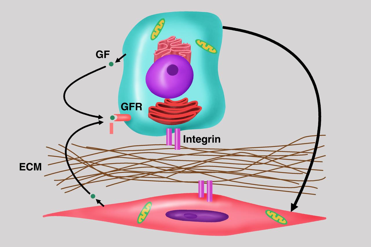
Diagram depicts the importance of cell-cell communication as central to the understanding of cancer growth and progression, the focus of the NASA bioreactor demonstration system (BDS-05) investigation. Microgravity studies will allow us to unravel the signaling and communication between these cells with the host and potential development of therapies for the treatment of cancer metastasis. The NASA Bioreactor provides a low turbulence culture environment which promotes the formation of large, three-dimensional cell clusters. Due to their high level of cellular organization and specialization, samples constructed in the bioreactor more closely resemble the original tumor or tissue found in the body. The Bioreactor is rotated to provide gentle mixing of fresh and spent nutrient without inducing shear forces that would damage the cells. The work is sponsored by NASA's Office of Biological and Physical Research. The bioreactor is managed by the Biotechnology Cell Science Program at NASA's Johnson Space Center (JSC). NASA-sponsored bioreactor research has been instrumental in helping scientists to better understand normal and cancerous tissue development. In cooperation with the medical community, the bioreactor design is being used to prepare better models of human colon, prostate, breast and ovarian tumors. Cartilage, bone marrow, heart muscle, skeletal muscle, pancreatic islet cells, liver and kidney are just a few of the normal tissues being cultured in rotating bioreactors by investigators. Credit: Emory University.
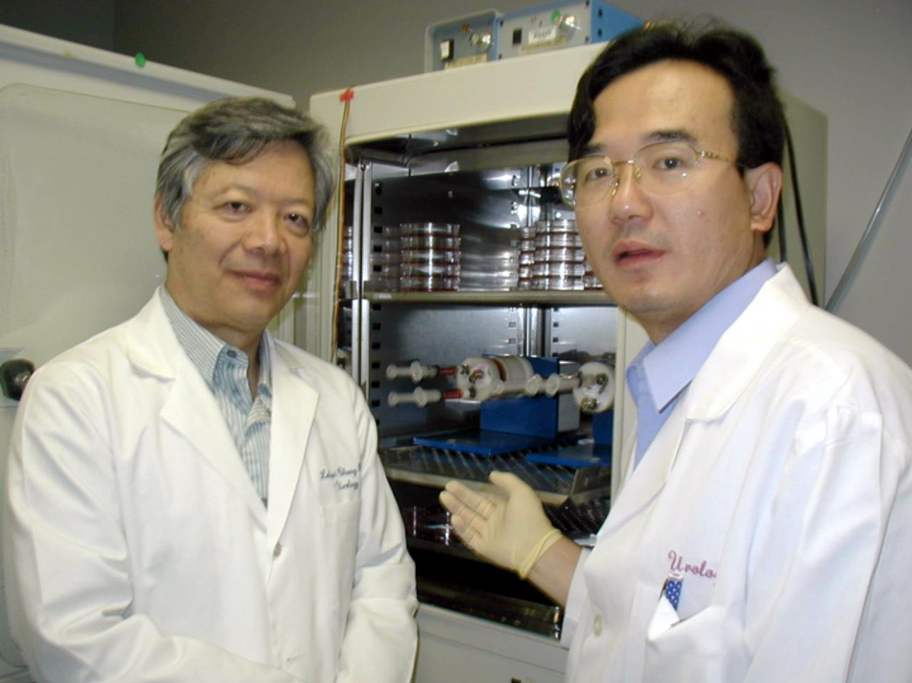
Leland W. K. Chung (left), Director, Molecular Urology Therapeutics Program at the Winship Cancer Institute at Emory University, is principal investigator for the NASA bioreactor demonstration system (BDS-05). With him is Dr. Jun Shu, an assistant professor of Orthopedics Surgery from Kuming Medical University China. The NASA Bioreactor provides a low turbulence culture environment which promotes the formation of large, three-dimensional cell clusters. Due to their high level of cellular organization and specialization, samples constructed in the bioreactor more closely resemble the original tumor or tissue found in the body. The Bioreactor is rotated to provide gentle mixing of fresh and spent nutrient without inducing shear forces that would damage the cells. The work is sponsored by NASA's Office of Biological and Physical Research. The bioreactor is managed by the Biotechnology Cell Science Program at NASA's Johnson Space Center (JSC). NASA-sponsored bioreactor research has been instrumental in helping scientists to better understand normal and cancerous tissue development. In cooperation with the medical community, the bioreactor design is being used to prepare better models of human colon, prostate, breast and ovarian tumors. Cartilage, bone marrow, heart muscle, skeletal muscle, pancreatic islet cells, liver and kidney are just a few of the normal tissues being cultured in rotating bioreactors by investigators. Credit: Emory University.
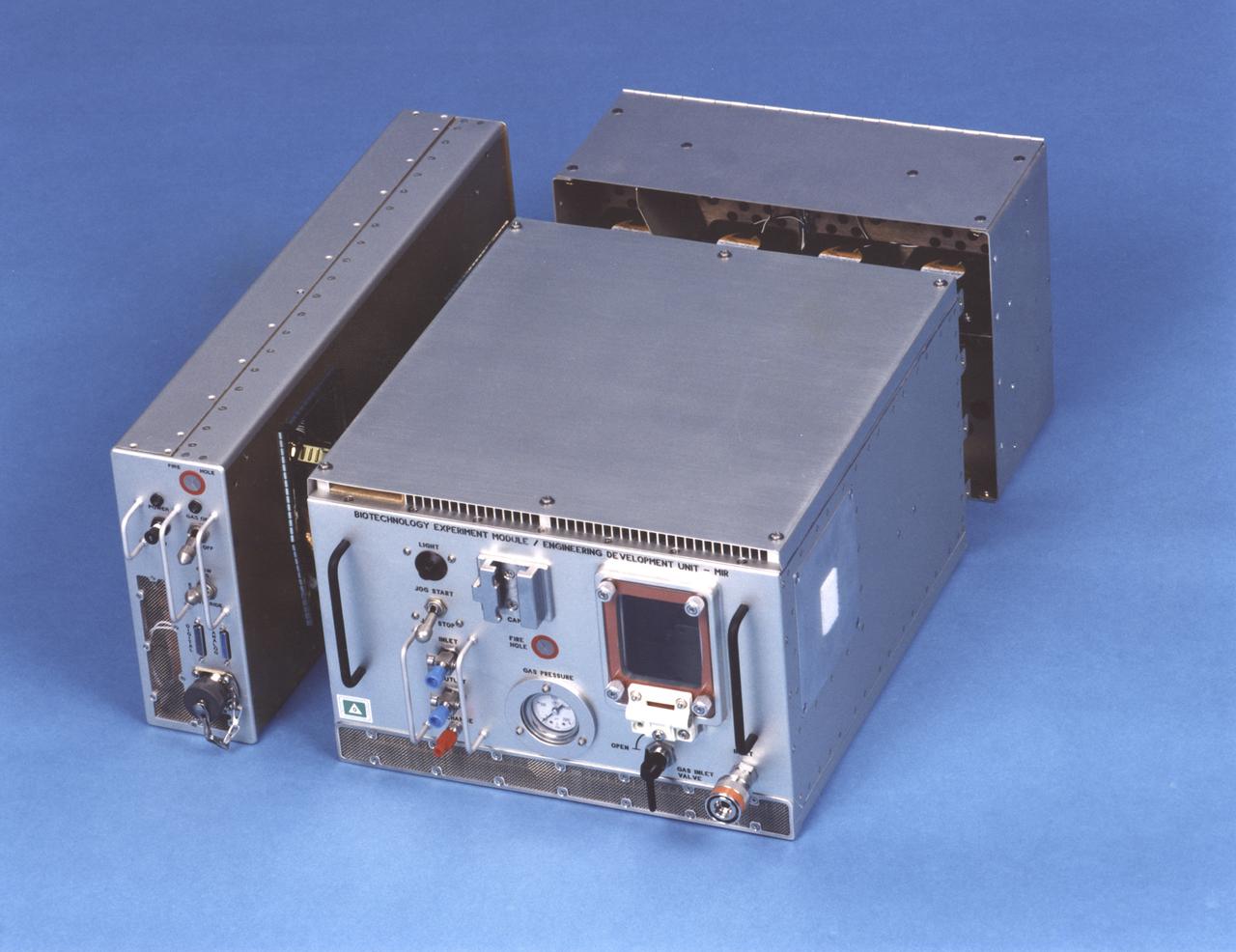
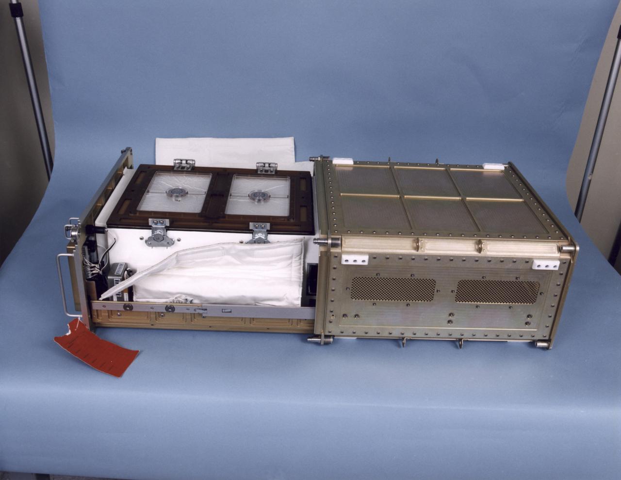
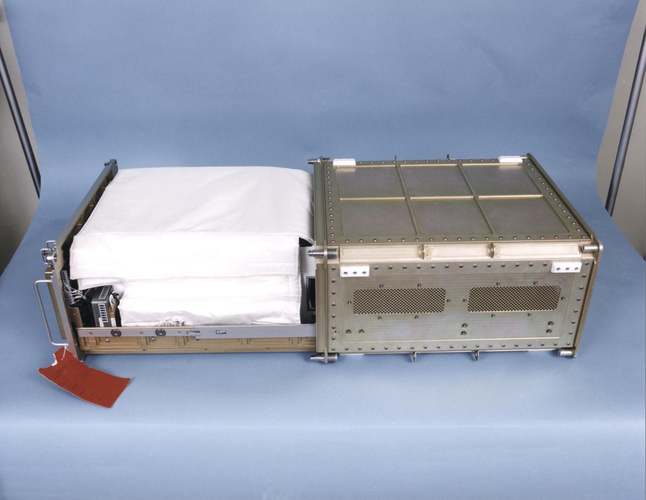
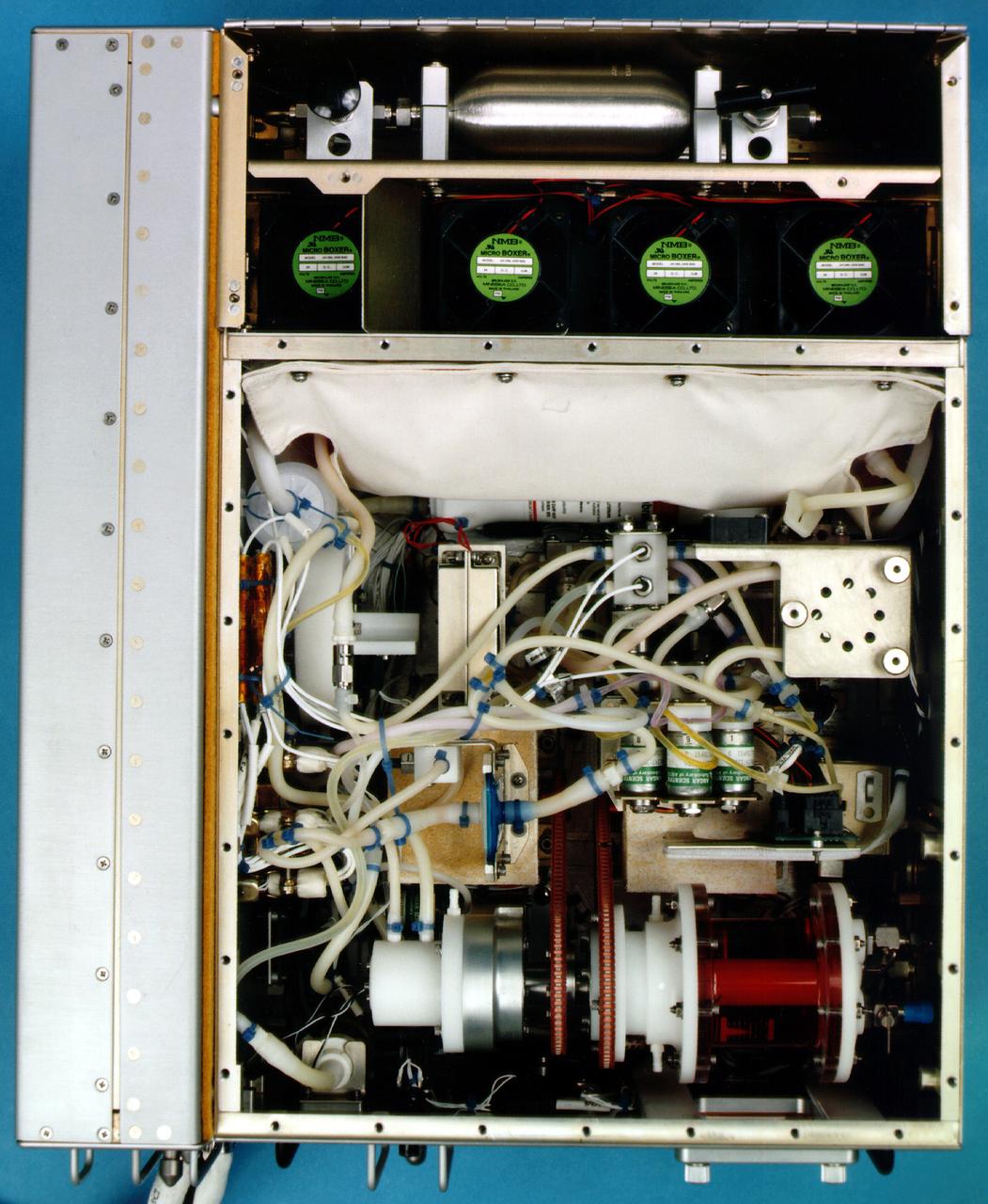
Exterior view of the NASA Bioreactor Engineering Development Unit flown on Mir. The rotating wall vessel is behind the window on the face of the large module. Control electronics are in the module at left; gas supply and cooling fans are in the module at back. The NASA Bioreactor provides a low turbulence culture environment which promotes the formation of large, three-dimensional cell clusters. The Bioreactor is rotated to provide gentle mixing of fresh and spent nutrient without inducing shear forces that would damage the cells. Due to their high level of cellular organization and specialization, samples constructed in the bioreactor more closely resemble the original tumor or tissue found in the body. The work is sponsored by NASA's Office of Biological and Physical Research. The bioreactor is managed by the Biotechnology Cell Science Program at NASA's Johnson Space Center (JSC). NASA-sponsored bioreactor research has been instrumental in helping scientists to better understand normal and cancerous tissue development. In cooperation with the medical community, the bioreactor design is being used to prepare better models of human colon, prostate, breast and ovarian tumors. Cartilage, bone marrow, heart muscle, skeletal muscle, pancreatic islet cells, liver and kidney are just a few of the normal tissues being cultured in rotating bioreactors by investigators.
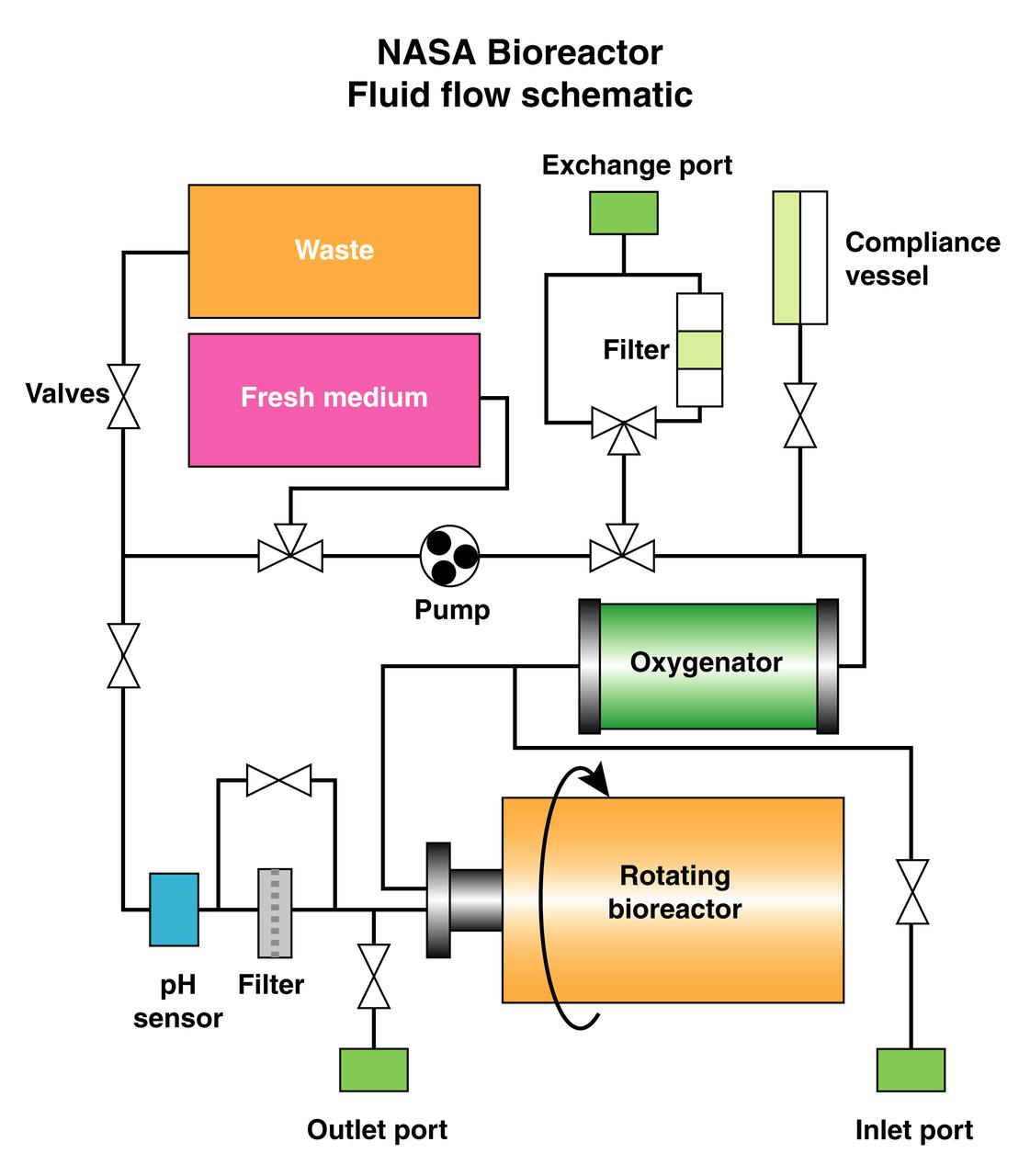
The schematic depicts the major elements and flow patterns inside the NASA Bioreactor system. Waste and fresh medium are contained in plastic bags placed side-by-side so the waste bag fills as the fresh medium bag is depleted. The compliance vessel contains a bladder to accommodate pressure transients that might damage the system. A peristolic pump moves fluid by squeezing the plastic tubing, thus avoiding potential contamination. The work is sponsored by NASA's Office of Biological and Physical Research. The bioreactor is managed by the Biotechnology Cell Science Program at NASA's Johnson Space Center (JSC). NASA-sponsored bioreactor research has been instrumental in helping scientists to better understand normal and cancerous tissue development. In cooperation with the medical community, the bioreactor design is being used to prepare better models of human colon, prostate, breast and ovarian tumors. Cartilage, bone marrow, heart muscle, skeletal muscle, pancreatic islet cells, liver and kidney are just a few of the normal tissues being cultured in rotating bioreactors by investigators.
Interior of a Biotechnology Refrigerator that preserves samples for use in (or after culturing in) the NASA Bioreactor. The unit is shown extracted from a middeck locker shell. The NASA Bioreactor provides a low turbulence culture environment which promotes the formation of large, three-dimensional cell clusters. The Bioreactor is rotated to provide gentle mixing of fresh and spent nutrient without inducing shear forces that would damage the cells. Due to their high level of cellular organization and specialization, samples constructed in the bioreactor more closely resemble the original tumor or tissue found in the body. The work is sponsored by NASA's Office of Biological and Physical Research. The bioreactor is managed by the Biotechnology Cell Science Program at NASA's Johnson Space Center (JSC). NASA-sponsored bioreactor research has been instrumental in helping scientists to better understand normal and cancerous tissue development. In cooperation with the medical community, the bioreactor design is being used to prepare better models of human colon, prostate, breast and ovarian tumors. Cartilage, bone marrow, heart muscle, skeletal muscle, pancreatic islet cells, liver and kidney are just a few of the normal tissues being cultured in rotating bioreactors by investigators.
Biotechnology Refrigerator that preserves samples for use in (or after culturing in) the NASA Bioreactor. The unit is shown extracted from a middeck locker shell and with thermal blankets partially removed. The NASA Bioreactor provides a low turbulence culture environment which promotes the formation of large, three-dimensional cell clusters. The Bioreactor is rotated to provide gentle mixing of fresh and spent nutrient without inducing shear forces that would damage the cells. Due to their high level of cellular organization and specialization, samples constructed in the bioreactor more closely resemble the original tumor or tissue found in the body. The work is sponsored by NASA's Office of Biological and Physical Research. The bioreactor is managed by the Biotechnology Cell Science Program at NASA's Johnson Space Center (JSC). NASA-sponsored bioreactor research has been instrumental in helping scientists to better understand normal and cancerous tissue development. In cooperation with the medical community, the bioreactor design is being used to prepare better models of human colon, prostate, breast and ovarian tumors. Cartilage, bone marrow, heart muscle, skeletal muscle, pancreatic islet cells, liver and kidney are just a few of the normal tissues being cultured in rotating bioreactors by investigators.
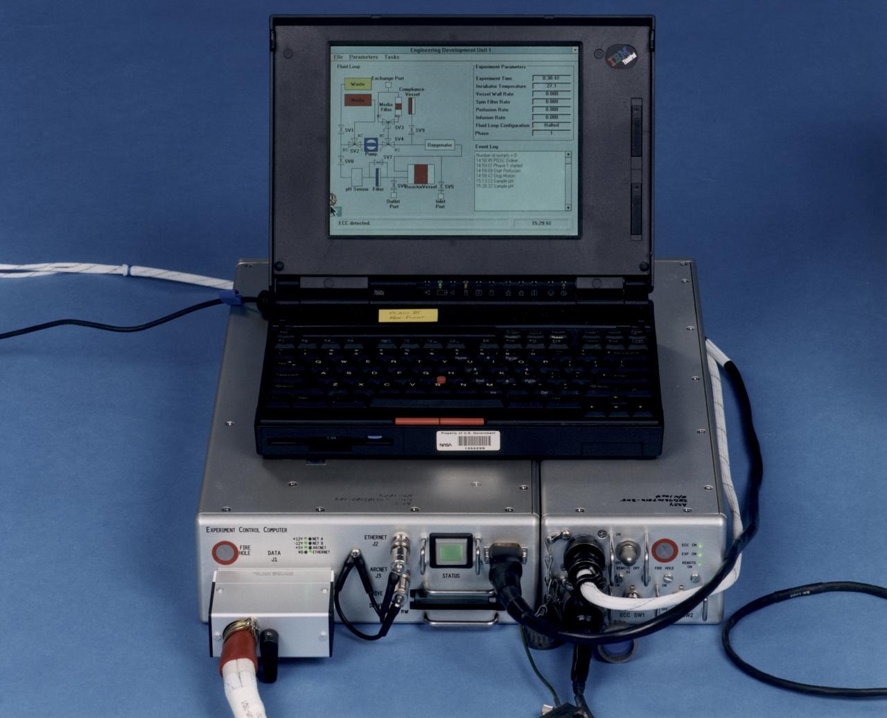
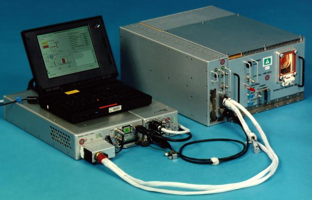
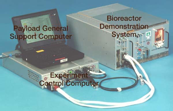
Laptop computer sits atop the Experiment Control Computer for a NASA Bioreactor. The flight crew can change operating conditions in the Bioreactor by using the graphical interface on the laptop. The NASA Bioreactor provides a low turbulence culture environment which promotes the formation of large, three-dimensional cell clusters. The Bioreactor is rotated to provide gentle mixing of fresh and spent nutrient without inducing shear forces that would damage the cells. Due to their high level of cellular organization and specialization, samples constructed in the bioreactor more closely resemble the original tumor or tissue found in the body. The work is sponsored by NASA's Office of Biological and Physical Research. The bioreactor is managed by the Biotechnology Cell Science Program at NASA's Johnson Space Center (JSC). NASA-sponsored bioreactor research has been instrumental in helping scientists to better understand normal and cancerous tissue development. In cooperation with the medical community, the bioreactor design is being used to prepare better models of human colon, prostate, breast and ovarian tumors. Cartilage, bone marrow, heart muscle, skeletal muscle, pancreatic islet cells, liver and kidney are just a few of the normal tissues being cultured in rotating bioreactors by investigators.
Close-up view of the interior of a NASA Bioreactor shows the plastic plumbing and valves (cylinders at center) to control fluid flow. A fresh nutrient bag is installed at top; a flattened waste bag behind it will fill as the nutrients are consumed during the course of operation. The drive chain and gears for the rotating wall vessel are visible at bottom center center. The NASA Bioreactor provides a low turbulence culture environment which promotes the formation of large, three-dimensional cell clusters. The Bioreactor is rotated to provide gentle mixing of fresh and spent nutrient without inducing shear forces that would damage the cells. Due to their high level of cellular organization and specialization, samples constructed in the bioreactor more closely resemble the original tumor or tissue found in the body. The work is sponsored by NASA's Office of Biological and Physical Research. The bioreactor is managed by the Biotechnology Cell Science Program at NASA's Johnson Space Center (JSC). NASA-sponsored bioreactor research has been instrumental in helping scientists to better understand normal and cancerous tissue development. In cooperation with the medical community, the bioreactor design is being used to prepare better models of human colon, prostate, breast and ovarian tumors. Cartilage, bone marrow, heart muscle, skeletal muscle, pancreatic islet cells, liver and kidney are just a few of the normal tissues being cultured in rotating bioreactors by investigators.
Interior view of the gas supply for the NASA Bioreactor. The NASA Bioreactor provides a low turbulence culture environment which promotes the formation of large, three-dimensional cell clusters. The Bioreactor is rotated to provide gentle mixing of fresh and spent nutrient without inducing shear forces that would damage the cells. Due to their high level of cellular organization and specialization, samples constructed in the bioreactor more closely resemble the original tumor or tissue found in the body. The work is sponsored by NASA's Office of Biological and Physical Research. The bioreactor is managed by the Biotechnology Cell Science Program at NASA's Johnson Space Center (JSC). NASA-sponsored bioreactor research has been instrumental in helping scientists to better understand normal and cancerous tissue development. In cooperation with the medical community, the bioreactor design is being used to prepare better models of human colon, prostate, breast and ovarian tumors. Cartilage, bone marrow, heart muscle, skeletal muscle, pancreatic islet cells, liver and kidney are just a few of the normal tissues being cultured in rotating bioreactors by investigators.
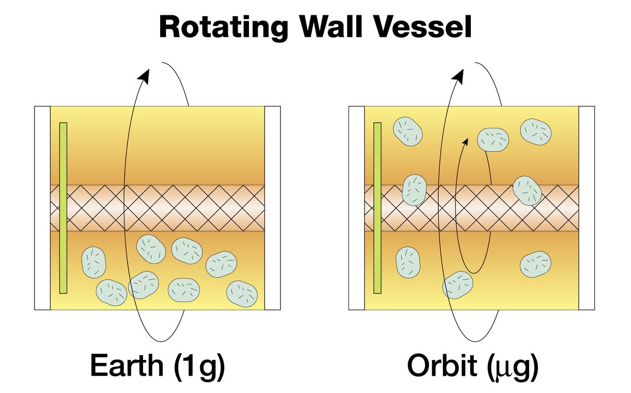
The NASA Bioreactor provides a low turbulence culture environment which promotes the formation of large, three-dimensional cell clusters. Due to their high level of cellular organization and specialization, samples constructed in the bioreactor more closely resemble the original tumor or tissue found in the body. The work is sponsored by NASA's Office of Biological and Physical Research. The bioreactor is managed by the Biotechnology Cell Science Program at NASA's Johnson Space Center (JSC). NASA-sponsored bioreactor research has been instrumental in helping scientists to better understand normal and cancerous tissue development. In cooperation with the medical community, the bioreactor design is being used to prepare better models of human colon, prostate, breast and ovarian tumors. Cartilage, bone marrow, heart muscle, skeletal muscle, pancreatic islet cells, liver and kidney are just a few of the normal tissues being cultured in rotating bioreactors by investigators. Cell constructs grown in a rotating bioreactor on Earth (left) eventually become too large to stay suspended in the nutrient media. In the microgravity of orbit, the cells stay suspended. Rotation then is needed for gentle stirring to replenish the media around the cells.
Paul Ducheyne, a principal investigator in the microgravity materials science program and head of the University of Pernsylvania's Center for Bioactive Materials and Tissue Engineering, is leading the trio as they use simulated microgravity to determine the optimal characteristics of tiny glass particles for growing bone tissue. The result could make possible a much broader range of synthetic bone-grafting applications. Even in normal gravity, bioactive glass particles enhance bone growth in laboratory tests with flat tissue cultures. Ducheyne and his team believe that using the bioactive microcarriers in a rotating bioreactor in microgravity will produce improved, three-dimensional tissue cultures. The work is sponsored by NASA's Office of Biological and Physical Research. The bioreactor is managed by the Biotechnology Cell Science Program at NASA's Johnson Space Center (JSC). NASA-sponsored bioreactor research has been instrumental in helping scientists to better understand normal and cancerous tissue development. In cooperation with the medical community, the bioreactor design is being used to prepare better models of human colon, prostate, breast and ovarian tumors. Cartilage, bone marrow, heart muscle, skeletal muscle, pancreatic islet cells, liver and kidney are just a few of the normal tissues being cultured in rotating bioreactors by investigators. Credit: NASA and University of Pennsylvania Center for Bioactive Materials and Tissue Engineering.
Biotechnology Refrigerator that preserves samples for use in (or after culturing in) the NASA Bioreactor. The unit is shown extracted from a middeck locker shell. The NASA Bioreactor provides a low turbulence culture environment which promotes the formation of large, three-dimensional cell clusters. The Bioreactor is rotated to provide gentle mixing of fresh and spent nutrient without inducing shear forces that would damage the cells. Due to their high level of cellular organization and specialization, samples constructed in the bioreactor more closely resemble the original tumor or tissue found in the body. The work is sponsored by NASA's Office of Biological and Physical Research. The bioreactor is managed by the Biotechnology Cell Science Program at NASA's Johnson Space Center (JSC). NASA-sponsored bioreactor research has been instrumental in helping scientists to better understand normal and cancerous tissue development. In cooperation with the medical community, the bioreactor design is being used to prepare better models of human colon, prostate, breast and ovarian tumors. Cartilage, bone marrow, heart muscle, skeletal muscle, pancreatic islet cells, liver and kidney are just a few of the normal tissues being cultured in rotating bioreactors by investigators.
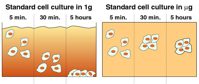
Cells cultured on Earth (left) typically settle quickly on the bottom of culture vessels due to gravity. In microgravity (right), cells remain suspended and aggregate to form three-dimensional tissue. The NASA Bioreactor provides a low turbulence culture environment which promotes the formation of large, three-dimensional cell clusters. The Bioreactor is rotated to provide gentle mixing of fresh and spent nutrient without inducing shear forces that would damage the cells. Due to their high level of cellular organization and specialization, samples constructed in the bioreactor more closely resemble the original tumor or tissue found in the body. The work is sponsored by NASA's Office of Biological and Physical Research. The bioreactor is managed by the Biotechnology Cell Science Program at NASA's Johnson Space Center (JSC). NASA-sponsored bioreactor research has been instrumental in helping scientists to better understand normal and cancerous tissue development. In cooperation with the medical community, the bioreactor design is being used to prepare better models of human colon, prostate, breast and ovarian tumors. Cartilage, bone marrow, heart muscle, skeletal muscle, pancreatic islet cells, liver and kidney are just a few of the normal tissues being cultured in rotating bioreactors by investigators.
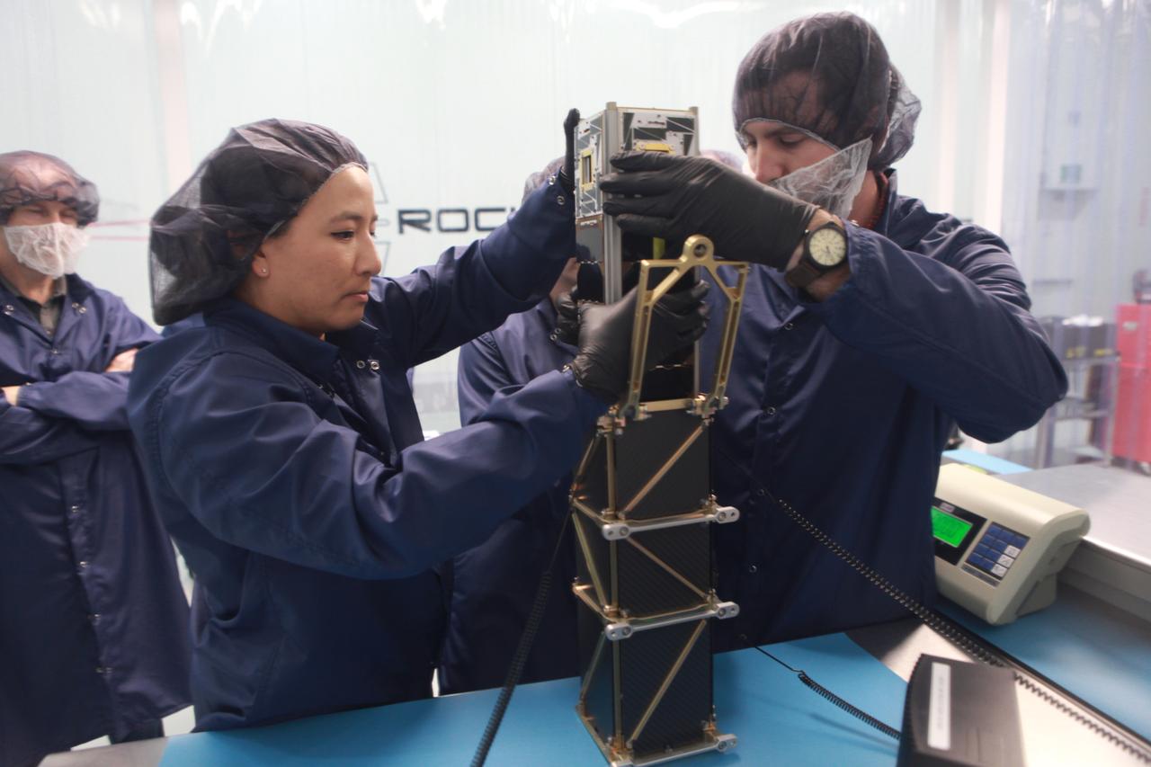
The goal of the CHOMPTT mission is to demonstrate new technologies that could be used for navigation and satellite networking in deep space. For future explorers and colonizers of the Moon or Mars, navigation systems like GPS here on Earth, will be essential. The key idea behind CHOMPTT is to use lasers to transfer time code data over long distances instead of radio waves. Because lasers can be more tightly beamed compared to radio waves, more of the transmitted energy reaches its intended target, making them more power-efficient. CHOMPTT takes advantage of this and of new miniature but very stable atomic clocks to produce a timing system with performance similar to that of GPS, but in a very compact and power efficient form factor. We will use a pulsed laser system, located at the Kennedy Space Center that will be synchronized with an atomic clock. Laser pulses will propagate from the ground to the orbiting CHOMPTT CubeSat and back. By precisely measuring the time of emission and detection of these pulses on the ground and in space we can calculate the time discrepancy between the ground atomic clock and the atomic clock on CHOMPTT. Our goal is to do this with an accuracy of 0.2 billionths of a second, or the time it takes light to travel just 6 centimeters. In the future, we envision using this technology on constellations or swarms of small satellites, for example orbiting the Moon, to equip them with precision navigation, networking, and ranging capabilities. CHOMPTT is a collaboration between the University of Florida and the NASA Ames Research Center. The CHOMPTT precision timing payload was designed and built by the Precision Space Systems Lab at the University of Florida, while the 3U CubeSat bus that has prior flight heritage, was provided by NASA Ames. The CHOMPTT mission has been funded by the Air Force Research Lab and by NASA.
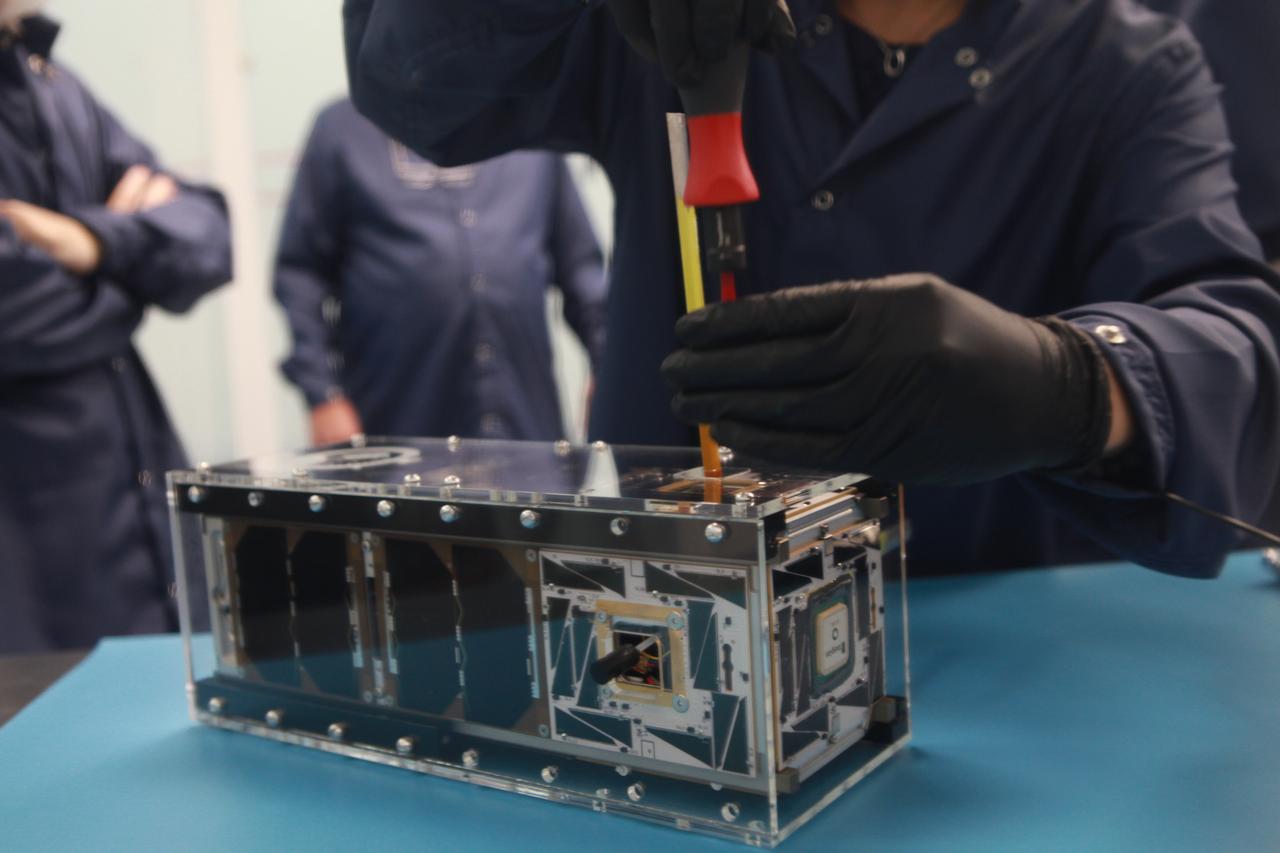
The goal of the CHOMPTT mission is to demonstrate new technologies that could be used for navigation and satellite networking in deep space. For future explorers and colonizers of the Moon or Mars, navigation systems like GPS here on Earth, will be essential. The key idea behind CHOMPTT is to use lasers to transfer time code data over long distances instead of radio waves. Because lasers can be more tightly beamed compared to radio waves, more of the transmitted energy reaches its intended target, making them more power-efficient. CHOMPTT takes advantage of this and of new miniature but very stable atomic clocks to produce a timing system with performance similar to that of GPS, but in a very compact and power efficient form factor. We will use a pulsed laser system, located at the Kennedy Space Center that will be synchronized with an atomic clock. Laser pulses will propagate from the ground to the orbiting CHOMPTT CubeSat and back. By precisely measuring the time of emission and detection of these pulses on the ground and in space we can calculate the time discrepancy between the ground atomic clock and the atomic clock on CHOMPTT. Our goal is to do this with an accuracy of 0.2 billionths of a second, or the time it takes light to travel just 6 centimeters. In the future, we envision using this technology on constellations or swarms of small satellites, for example orbiting the Moon, to equip them with precision navigation, networking, and ranging capabilities. CHOMPTT is a collaboration between the University of Florida and the NASA Ames Research Center. The CHOMPTT precision timing payload was designed and built by the Precision Space Systems Lab at the University of Florida, while the 3U CubeSat bus that has prior flight heritage, was provided by NASA Ames. The CHOMPTT mission has been funded by the Air Force Research Lab and by NASA.
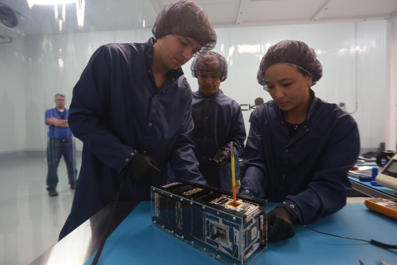
The goal of the CHOMPTT mission is to demonstrate new technologies that could be used for navigation and satellite networking in deep space. For future explorers and colonizers of the Moon or Mars, navigation systems like GPS here on Earth, will be essential. The key idea behind CHOMPTT is to use lasers to transfer time code data over long distances instead of radio waves. Because lasers can be more tightly beamed compared to radio waves, more of the transmitted energy reaches its intended target, making them more power-efficient. CHOMPTT takes advantage of this and of new miniature but very stable atomic clocks to produce a timing system with performance similar to that of GPS, but in a very compact and power efficient form factor. We will use a pulsed laser system, located at the Kennedy Space Center that will be synchronized with an atomic clock. Laser pulses will propagate from the ground to the orbiting CHOMPTT CubeSat and back. By precisely measuring the time of emission and detection of these pulses on the ground and in space we can calculate the time discrepancy between the ground atomic clock and the atomic clock on CHOMPTT. Our goal is to do this with an accuracy of 0.2 billionths of a second, or the time it takes light to travel just 6 centimeters. In the future, we envision using this technology on constellations or swarms of small satellites, for example orbiting the Moon, to equip them with precision navigation, networking, and ranging capabilities. CHOMPTT is a collaboration between the University of Florida and the NASA Ames Research Center. The CHOMPTT precision timing payload was designed and built by the Precision Space Systems Lab at the University of Florida, while the 3U CubeSat bus that has prior flight heritage, was provided by NASA Ames. The CHOMPTT mission has been funded by the Air Force Research Lab and by NASA.
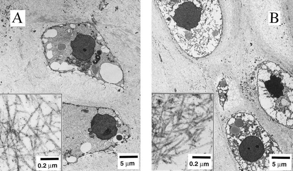
Dr. Lisa E. Freed of the Massachusetts Institute of Technology and her colleagues have reported that initially disc-like specimens tend to become spherical in space, demonstrating that tissues can grow and differentiate into distinct structures in microgravity. The Mir Increment 3 (Sept. 16, 1996 - Jan. 22, 1997) samples were smaller, more spherical, and mechanically weaker than Earth-grown control samples. These results demonstrate the feasibility of microgravity tissue engineering and may have implications for long human space voyages and for treating musculoskeletal disorders on earth. Final samples from Mir and Earth appeared histologically cartilaginous throughout their entire cross sections (5-8 mm thick), with the exception of fibrous outer capsules. Constructs grown on Earth (A) appeared to have a more organized extracellular matrix with more uniform collagen orientation as compared with constructs grown on Mir (B), but the average collagen fiber diameter was similar in the two groups (22 +- 2 nm) and comparable to that previously reported for developing articular cartilage. Randomly oriented collagen in Mir samples would be consistent with previous reports that microgravity disrupts fibrillogenesis. These are transmission electron micrographs of constructs from Mir (A) and Earth (B) groups at magnifications of x3,500 and x120,000 (Inset). The work is sponsored by NASA's Office of Biological and Physical Research. The bioreactor is managed by the Biotechnology Cell Science Program at NASA's Johnson Space Center (JSC). NASA-sponsored bioreactor research has been instrumental in helping scientists to better understand normal and cancerous tissue development. In cooperation with the medical community, the bioreactor design is being used to prepare better models of human colon, prostate, breast and ovarian tumors. Credit: Proceedings of the National Academy of Sciences.
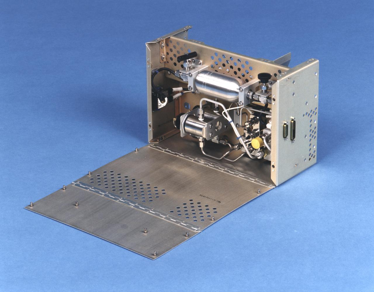
Bioreactor Demonstration System (BDS) comprises an electronics module, a gas supply module, and the incubator module housing the rotating wall vessel and its support systems. Nutrient media are pumped through an oxygenator and the culture vessel. The shell rotates at 0.5 rpm while the irner filter typically rotates at 11.5 rpm to produce a gentle flow that ensures removal of waste products as fresh media are infused. Periodically, some spent media are pumped into a waste bag and replaced by fresh media. When the waste bag is filled, an astronaut drains the waste bag and refills the supply bag through ports on the face of the incubator. Pinch valves and a perfusion pump ensure that no media are exposed to moving parts. An Experiment Control Computer controls the Bioreactor, records conditions, and alerts the crew when problems occur. The crew operates the system through a laptop computer displaying graphics designed for easy crew training and operation. The work is sponsored by NASA's Office of Biological and Physical Research. The bioreactor is managed by the Biotechnology Cell Science Program at NASA's Johnson Space Center (JSC). NASA-sponsored bioreactor research has been instrumental in helping scientists to better understand normal and cancerous tissue development. In cooperation with the medical community, the bioreactor design is being used to prepare better models of human colon, prostate, breast and ovarian tumors. Cartilage, bone marrow, heart muscle, skeletal muscle, pancreatic islet cells, liver and kidney are just a few of the normal tissues being cultured in rotating bioreactors by investigators. See No. 0101825 for a version with major elements labeled, and No. 0103180 for an operational schematic. 0101816
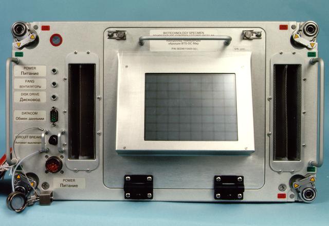
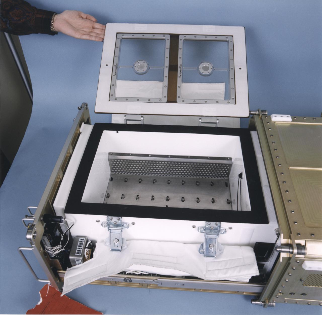
Biotechnology Specimen Temperature Controller (BSTC) will cultivate cells until their turn in the bioreactor; it can also be used in culturing experiments that do not require the bioreactor. The BSTC comprises four incubation/refrigeration chambers individually set at 4 to 50 deg. C (near-freezing to above body temperature). Each chamber holds three rugged tissue chamber modules (12 total), clear Teflon bags holding 30 ml of growth media, all positioned by a metal frame. Every 7 to 21 days (depending on growth rates), an astronaut uses a shrouded syringe and the bags' needleless injection ports to transfer a few cells to a fresh media bag, and to introduce a fixative so that the cells may be studied after flight. The design also lets the crew sample the media to measure glucose, gas, and pH levels, and to inspect cells with a microscope. The controller is monitored by the flight crew through a 23-cm (9-inch) color computer display on the face of the BSTC. This view shows the BTSC with the front panel open. The work is sponsored by NASA's Office of Biological and Physical Research. The bioreactor is managed by the Biotechnology Cell Science Program at NASA's Johnson Space Center (JSC). NASA-sponsored bioreactor research has been instrumental in helping scientists to better understand normal and cancerous tissue development. In cooperation with the medical community, the bioreactor design is being used to prepare better models of human colon, prostate, breast and ovarian tumors. Cartilage, bone marrow, heart muscle, skeletal muscle, pancreatic islet cells, liver and kidney are just a few of the normal tissues being cultured in rotating bioreactors by investigators.
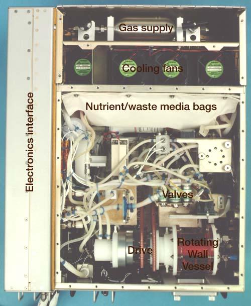
Bioreactor Demonstration System (BDS) comprises an electronics module, a gas supply module, and the incubator module housing the rotating wall vessel and its support systems. Nutrient media are pumped through an oxygenator and the culture vessel. The shell rotates at 0.5 rpm while the irner filter typically rotates at 11.5 rpm to produce a gentle flow that ensures removal of waste products as fresh media are infused. Periodically, some spent media are pumped into a waste bag and replaced by fresh media. When the waste bag is filled, an astronaut drains the waste bag and refills the supply bag through ports on the face of the incubator. Pinch valves and a perfusion pump ensure that no media are exposed to moving parts. An Experiment Control Computer controls the Bioreactor, records conditions, and alerts the crew when problems occur. The crew operates the system through a laptop computer displaying graphics designed for easy crew training and operation. The work is sponsored by NASA's Office of Biological and Physical Research. The bioreactor is managed by the Biotechnology Cell Science Program at NASA's Johnson Space Center (JSC). NASA-sponsored bioreactor research has been instrumental in helping scientists to better understand normal and cancerous tissue development. In cooperation with the medical community, the bioreactor design is being used to prepare better models of human colon, prostate, breast and ovarian tumors. Cartilage, bone marrow, heart muscle, skeletal muscle, pancreatic islet cells, liver and kidney are just a few of the normal tissues being cultured in rotating bioreactors by investigators. See No. 0101816 for a version without labels, and No. 0103180 for an operational schematic.
Bioreactor Demonstration System (BDS) comprises an electronics module, a gas supply module, and the incubator module housing the rotating wall vessel and its support systems. Nutrient media are pumped through an oxygenator and the culture vessel. The shell rotates at 0.5 rpm while the irner filter typically rotates at 11.5 rpm to produce a gentle flow that ensures removal of waste products as fresh media are infused. Periodically, some spent media are pumped into a waste bag and replaced by fresh media. When the waste bag is filled, an astronaut drains the waste bag and refills the supply bag through ports on the face of the incubator. Pinch valves and a perfusion pump ensure that no media are exposed to moving parts. An Experiment Control Computer controls the Bioreactor, records conditions, and alerts the crew when problems occur. The crew operates the system through a laptop computer displaying graphics designed for easy crew training and operation. The work is sponsored by NASA's Office of Biological and Physical Research. The bioreactor is managed by the Biotechnology Cell Science Program at NASA's Johnson Space Center (JSC). NASA-sponsored bioreactor research has been instrumental in helping scientists to better understand normal and cancerous tissue development. In cooperation with the medical community, the bioreactor design is being used to prepare better models of human colon, prostate, breast and ovarian tumors. Cartilage, bone marrow, heart muscle, skeletal muscle, pancreatic islet cells, liver and kidney are just a few of the normal tissues being cultured in rotating bioreactors by investigators. See No. 0101824 for a version with labels, and No. 0103180 for an operational schematic.

Lisa Freed and Gordana Vunjak-Novakovic, both of the Massachusetts Institute of Technology (MIT), have taken the first steps toward engineering heart muscle tissue that could one day be used to patch damaged human hearts. Cells isolated from very young animals are attached to a three-dimensional polymer scaffold, then placed in a NASA bioreactor. The cells do not divide, but after about a week start to cornect to form a functional piece of tissue. Here, a transmission electron micrograph of engineered tissue shows a number of important landmarks present in functional heart tissue: (A) well-organized myofilaments (Mfl), z-lines (Z), and abundant glycogen granules (Gly); and (D) intercalcated disc (ID) and desmosomes (DES). The NASA Bioreactor provides a low turbulence culture environment which promotes the formation of large, three-dimensional cell clusters. The Bioreactor is rotated to provide gentle mixing of fresh and spent nutrient without inducing shear forces that would damage the cells. Due to their high level of cellular organization and specialization, samples constructed in the bioreactor more closely resemble the original tumor or tissue found in the body. NASA-sponsored bioreactor research has been instrumental in helping scientists to better understand normal and cancerous tissue development. In cooperation with the medical community, the bioreactor design is being used to prepare better models of human colon, prostate, breast and ovarian tumors. Cartilage, bone marrow, heart muscle, skeletal muscle, pancreatic islet cells, liver and kidney are just a few of the normal tissues being cultured in rotating bioreactors by investigators. The work is sponsored by NASA's Office of Biological and Physical Research. The bioreactor is managed by the Biotechnology Cell Science Program at NASA's Johnson Space Center (JSC). Credit: MIT
Bioreactor Demonstration System (BDS) comprises an electronics module, a gas supply module, and the incubator module housing the rotating wall vessel and its support systems. Nutrient media are pumped through an oxygenator and the culture vessel. The shell rotates at 0.5 rpm while the irner filter typically rotates at 11.5 rpm to produce a gentle flow that ensures removal of waste products as fresh media are infused. Periodically, some spent media are pumped into a waste bag and replaced by fresh media. When the waste bag is filled, an astronaut drains the waste bag and refills the supply bag through ports on the face of the incubator. Pinch valves and a perfusion pump ensure that no media are exposed to moving parts. An Experiment Control Computer controls the Bioreactor, records conditions, and alerts the crew when problems occur. The crew operates the system through a laptop computer displaying graphics designed for easy crew training and operation. The work is sponsored by NASA's Office of Biological and Physical Research. The bioreactor is managed by the Biotechnology Cell Science Program at NASA's Johnson Space Center (JSC). NASA-sponsored bioreactor research has been instrumental in helping scientists to better understand normal and cancerous tissue development. In cooperation with the medical community, the bioreactor design is being used to prepare better models of human colon, prostate, breast and ovarian tumors. Cartilage, bone marrow, heart muscle, skeletal muscle, pancreatic islet cells, liver and kidney are just a few of the normal tissues being cultured in rotating bioreactors by investigators. See No. 0101823 for a version without labels, and No. 0103180 for an operational schematic.
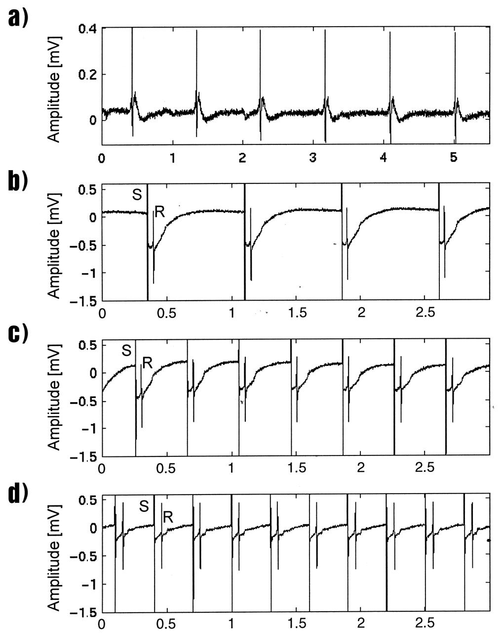
Lisa Freed and Gordana Vunjak-Novakovic, both of the Massachusetts Institute of Technology (MIT), have taken the first steps toward engineering heart muscle tissue that could one day be used to patch damaged human hearts. Cells isolated from very young animals are attached to a three-dimensional polymer scaffold, then placed in a NASA bioreactor. The cells do not divide, but after about a week start to cornect to form a functional piece of tissue. Functionally connected heart cells that are capable of transmitting electrical signals are the goal for Freed and Vunjak-Novakovic. Electrophysiological recordings of engineered tissue show spontaneous contractions at a rate of 70 beats per minute (a), and paced contractions at rates of 80, 150, and 200 beats per minute respectively (b, c, and d). The NASA Bioreactor provides a low turbulence culture environment which promotes the formation of large, three-dimensional cell clusters. The Bioreactor is rotated to provide gentle mixing of fresh and spent nutrient without inducing shear forces that would damage the cells. Due to their high level of cellular organization and specialization, samples constructed in the bioreactor more closely resemble the original tumor or tissue found in the body. NASA-sponsored bioreactor research has been instrumental in helping scientists to better understand normal and cancerous tissue development. In cooperation with the medical community, the bioreactor design is being used to prepare better models of human colon, prostate, breast and ovarian tumors. Cartilage, bone marrow, heart muscle, skeletal muscle, pancreatic islet cells, liver and kidney are just a few of the normal tissues being cultured in rotating bioreactors by investigators. The work is sponsored by NASA's Office of Biological and Physical Research. The bioreactor is managed by the Biotechnology Cell Science Program at NASA's Johnson Space Center (JSC). Credit: NASA and MIT.

STS099-753-032 (11-22 February 2000) ---This 70mm photograph, photographed from the Space Shuttle Endeavour, centers on the two westernmost Galapagos Islands--seahorse-shaped Isla Isabela and the smaller round Isla Fernandina to its west. All of the 19 islands in the chain are volcanic in origin, and the craters of several of the shield volcanoes are visible as circular features on each of the islands. The two islands shown in this picture contain the most active volcanoes of the Galapagos. Fernandina last erupted in January-February 1995, with red-hot lava pouring into the sea. After 20 years of inactivity, Cerro Azul on Isla Isabela, last erupted in September-October 1998. Cerro Azul is the southwesternmost volcano on Isla Isabela. At 82 miles long, Isla Isabela is the largest of the islands, and comprises half of the land area of the archipelago. The islands are famous for their unique flora and fauna. Charles Darwin's observations of these species in 1835 contributed to the formation of his ideas on natural selection. Some of the most unique species include flightless cormorants, Galapagos penguins, giant land tortoises, and Galapagos finches. The range of Galapagos penguins is restricted to these western islands where upwelling enriches the ocean productivity, and the adaptation of a typically Antarctic bird family to the equator is an ecological marvel. Giant land tortoises are thought to have the oldest lifespans of any animal on Earth, but, scientists say, they have been driven near to extinction. During the most recent eruption of Cerro Azul, one tortoise was killed and many had to be relocated. The 13 species of Galapagos finches on the islands, although varied in form and lifestyle, are the descendants of an ancestor that happened to colonize this isolated archipelago. The human population of the entire archipelago is about 10,000.
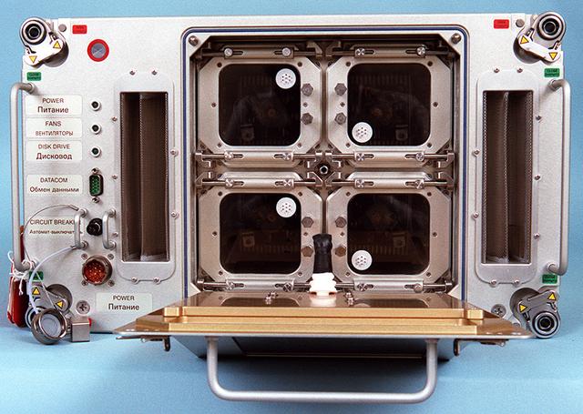
Biotechnology Specimen Temperature Controller (BSTC) will cultivate cells until their turn in the bioreactor; it can also be used in culturing experiments that do not require the bioreactor. The BSTC comprises four incubation/refrigeration chambers individually set at 4 to 50 degreesC (near-freezing to above body temperature). Each chamber holds three rugged tissue chamber modules (12 total), clear Teflon bags holding 30 ml of growth media, all positioned by a metal frame. Every 7 to 21 days (depending on growth rates), an astronaut uses a shrouded syringe and the bags' needleless injection ports to transfer a few cells to a fresh media bag, and to introduce a fixative so that the cells may be studied after flight. The design also lets the crew sample the media to measure glucose, gas, and pH levels, and to inspect cells with a microscope. The controller is monitored by the flight crew through a 23-cm (9-inch) color computer display on the face of the BSTC. This view shows the BTSC with the front panel open. The work is sponsored by NASA's Office of Biological and Physical Research. The bioreactor is managed by the Biotechnology Cell Science Program at NASA's Johnson Space Center (JSC). NASA-sponsored bioreactor research has been instrumental in helping scientists to better understand normal and cancerous tissue development. In cooperation with the medical community, the bioreactor design is being used to prepare better models of human colon, prostate, breast and ovarian tumors. Cartilage, bone marrow, heart muscle, skeletal muscle, pancreatic islet cells, liver and kidney are just a few of the normal tissues being cultured in rotating bioreactors by investigators.