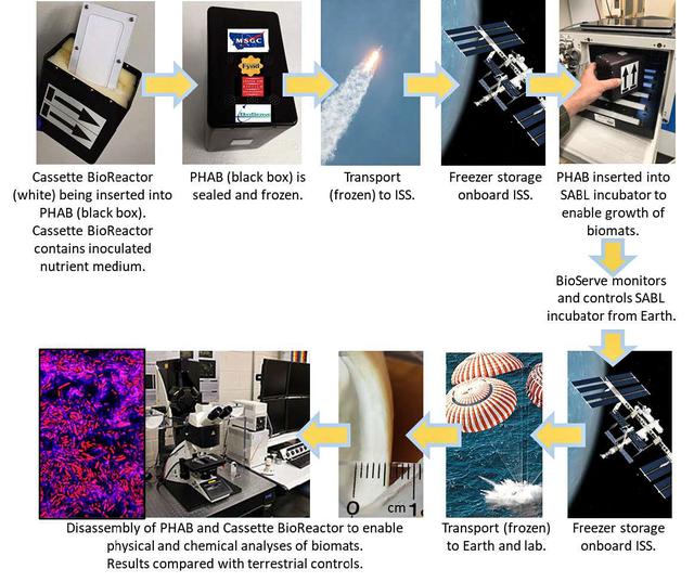
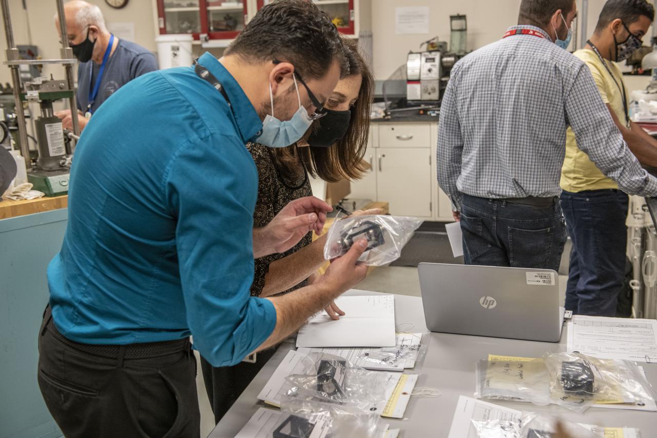
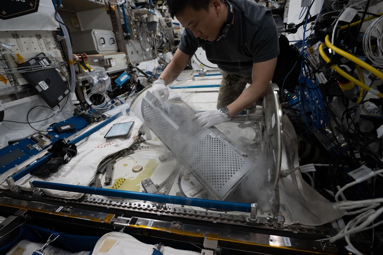
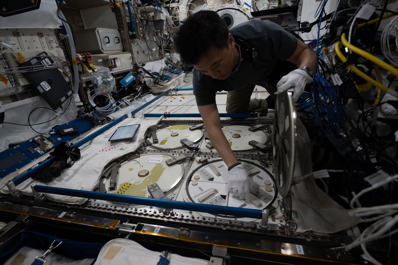
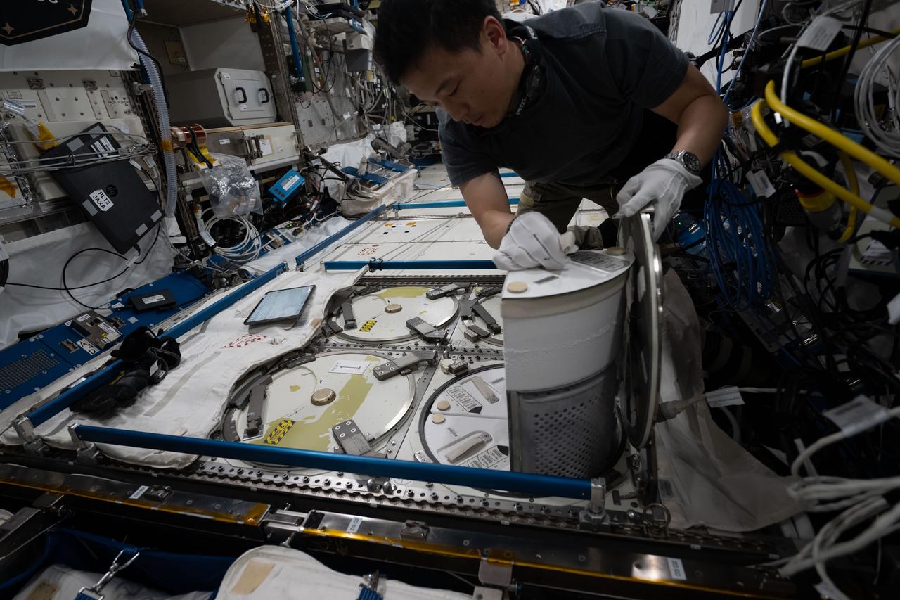
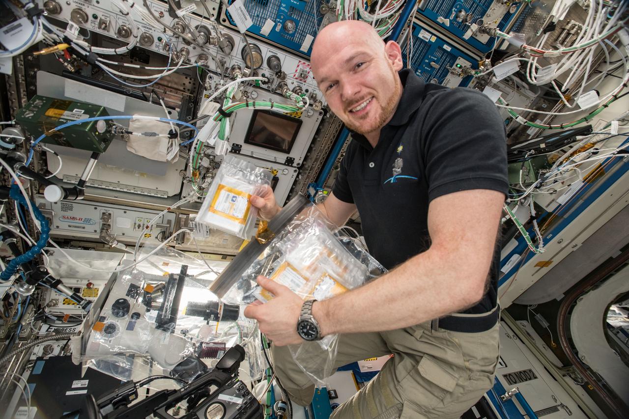
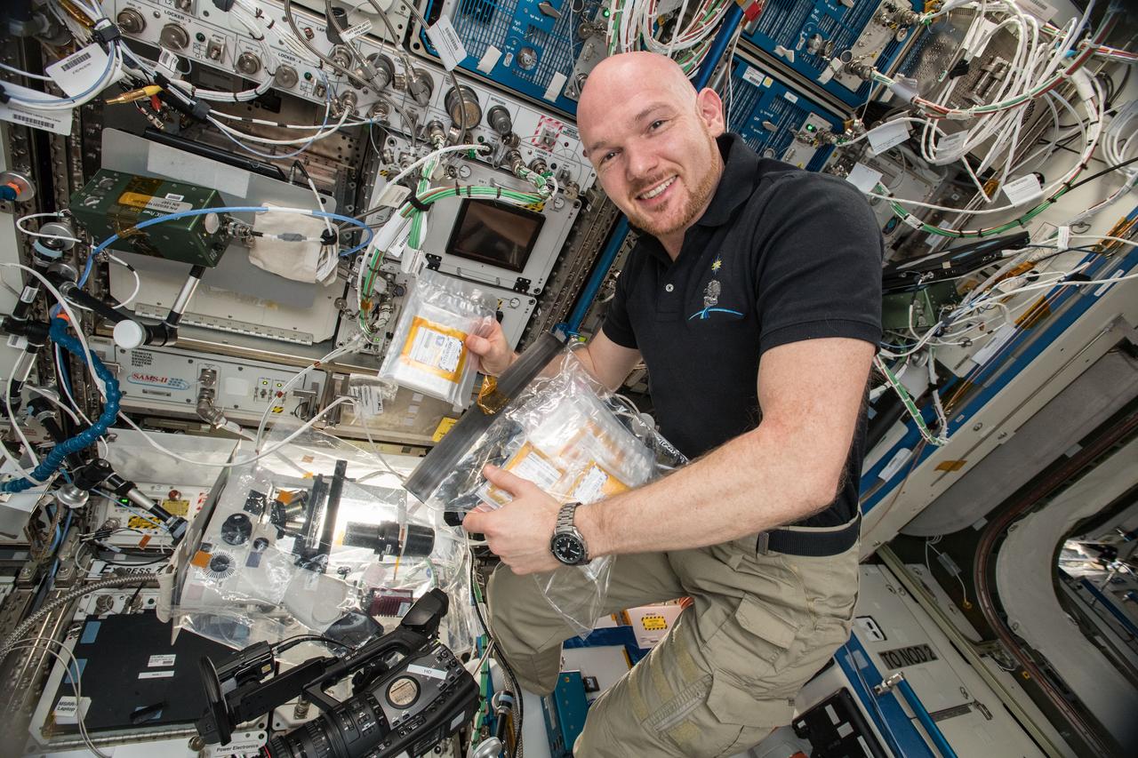
jsc2022e031226 (4/26/2022) --- A mission overview of the Protein Manufacturing investigation shows hardware, operations, and scientific details. The Protein Manufacturing project demonstrates and tests the operation of a novel bioreactor technology to support robust fungal growth for the production of high-protein food in a low-Earth orbit, space environment. Image courtesy of BioServe.
The major storage protein of leguminous plants and a major source of dietary protein for humans and domestic animals. It is studied in efforts to enhance nutritional value of proteins through protein engineerings. It is isolated from Jack Bean because of it's potential as a nutritional substance. Principal Investigator was Alexander McPherson.
(PCG) Protein Crystal Growth Canavalin. The major storage protein of leguminous plants and a major source of dietary protein for humans and domestic animals. It is studied in efforts to enhance nutritional value of proteins through protein engineerings. It is isolated from Jack Bean because of it's potential as a nutritional substance. Principal Investigator on STS-26 was Alex McPherson.
(PCG) Protein Crystal Growth C-reactive Protein. Plays a major role in human immune system response. Principal Investigator on STS-26 was Charles Bugg.
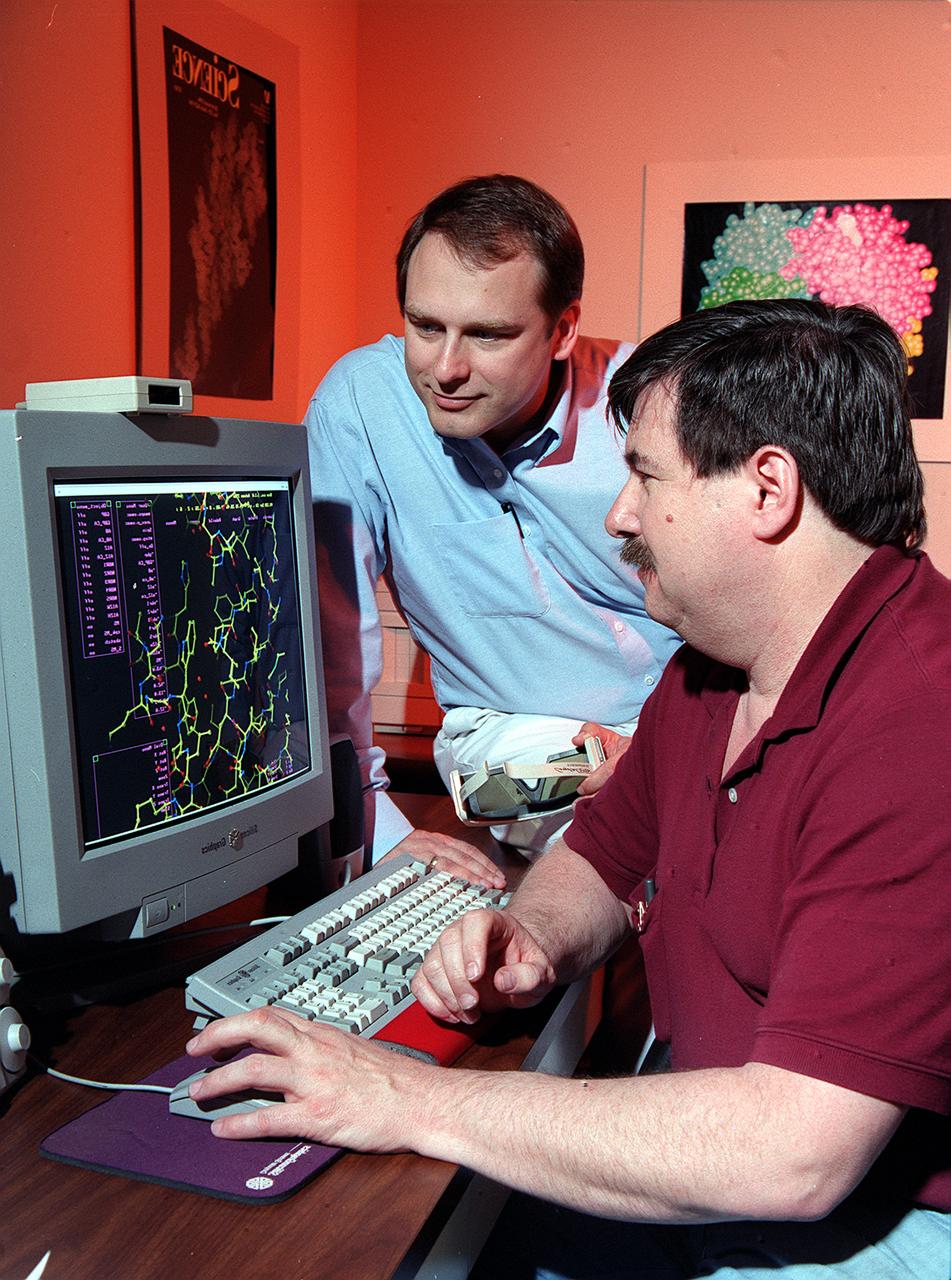
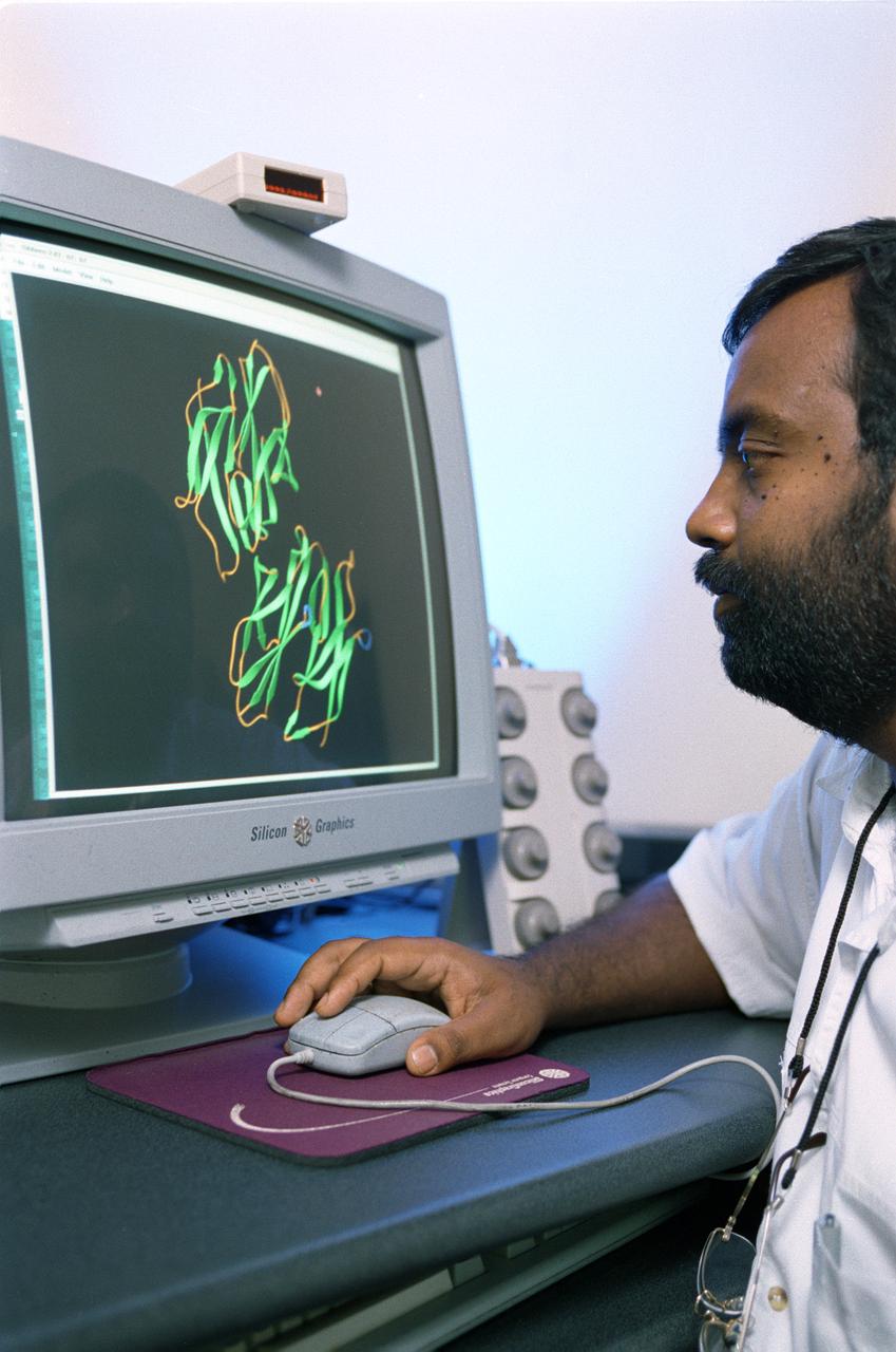
Dr. Marc Pusey (seated) and Dr. Craig Kundrot use computers to analyze x-ray maps and generate three-dimensional models of protein structures. With this information, scientists at Marshall Space Flight Center can learn how proteins are made and how they work. The computer screen depicts a proten structure as a ball-and-stick model. Other models depict the actual volume occupied by the atoms, or the ribbon-like structures that are crucial to a protein's function.
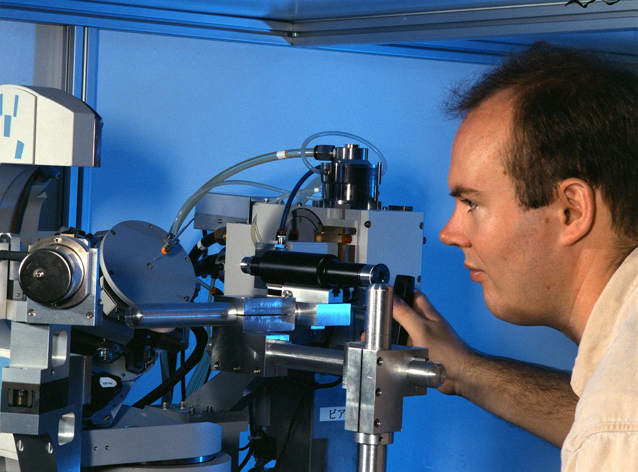
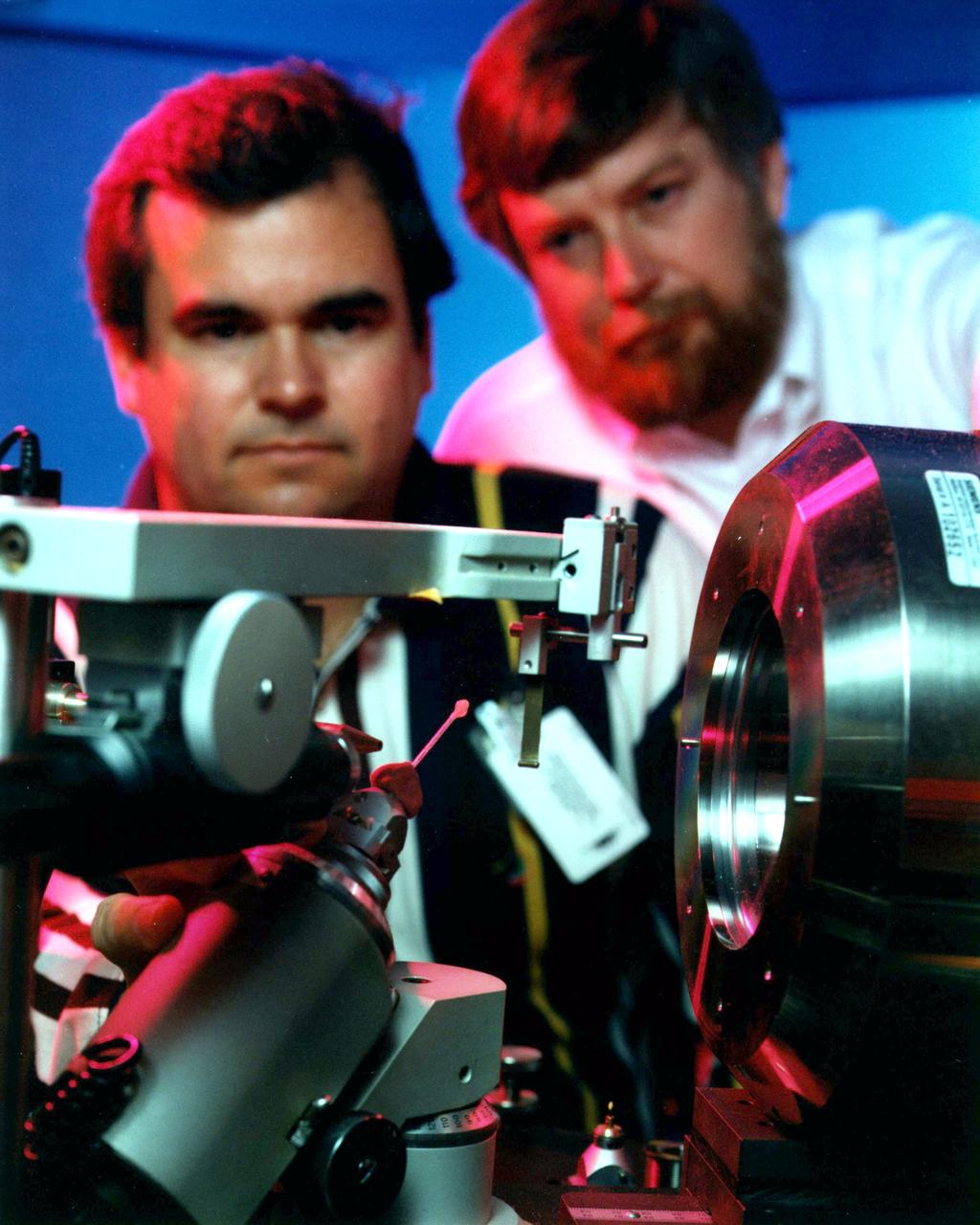
Eddie Snell, Post-Doctoral Fellow the National Research Council (NRC) uses a reciprocal space mapping diffractometer for macromolecular crystal quality studies. The diffractometer is used in mapping the structure of macromolecules such as proteins to determine their structure and thus understand how they function with other proteins in the body. This is one of several analytical tools used on proteins crystallized on Earth and in space experiments. Photo credit: NASA/Marshall Space Flight Center (MSFC)
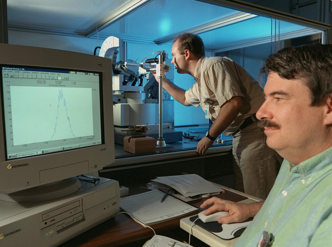
Eddie Snell (standing), Post-Doctoral Fellow the National Research Council (NRC),and Marc Pusey of Marshall Space Flight Center (MSFC) use a reciprocal space mapping diffractometer for marcromolecular crystal quality studies. The diffractometer is used in mapping the structure of marcromolecules such as proteins to determine their structure and thus understand how they function with other proteins in the body. This is one of several analytical tools used on proteins crystalized on Earth and in space experiments. Photo credit: NASA/Marshall Space Flight Center (MSFC)
iss047e055613 (4/11/2016) --- A view of the JAXA Protein Crystal Growth (PCG) Demo Sample, in the Japanese Experiment Module (JEM) Pressurized Module (JPM) aboard the International space Station (ISS). The objective of JAXA High Quality Protein Crystal Growth Demonstration Experiment (JAXA PCG-Demo) is to grow high quality protein crystals in microgravity.

iss047e055611 (4/11/2016) --- A view of the JAXA Protein Crystal Growth (PCG) Demo Sample, in the Japanese Experiment Module (JEM) Pressurized Module (JPM) aboard the International space Station (ISS). The objective of JAXA High Quality Protein Crystal Growth Demonstration Experiment (JAXA PCG-Demo) is to grow high quality protein crystals in microgravity.
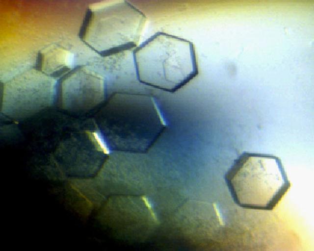
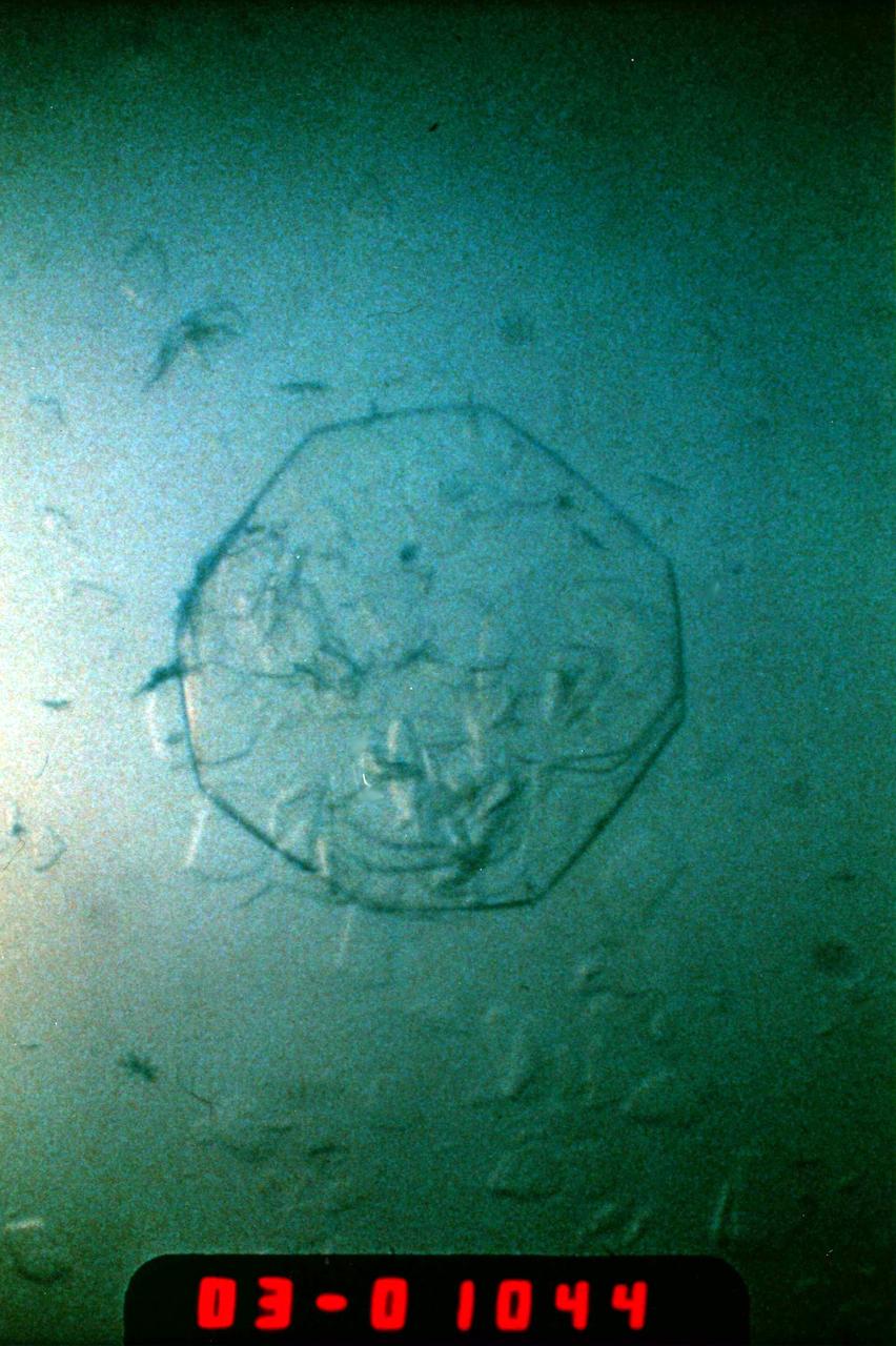
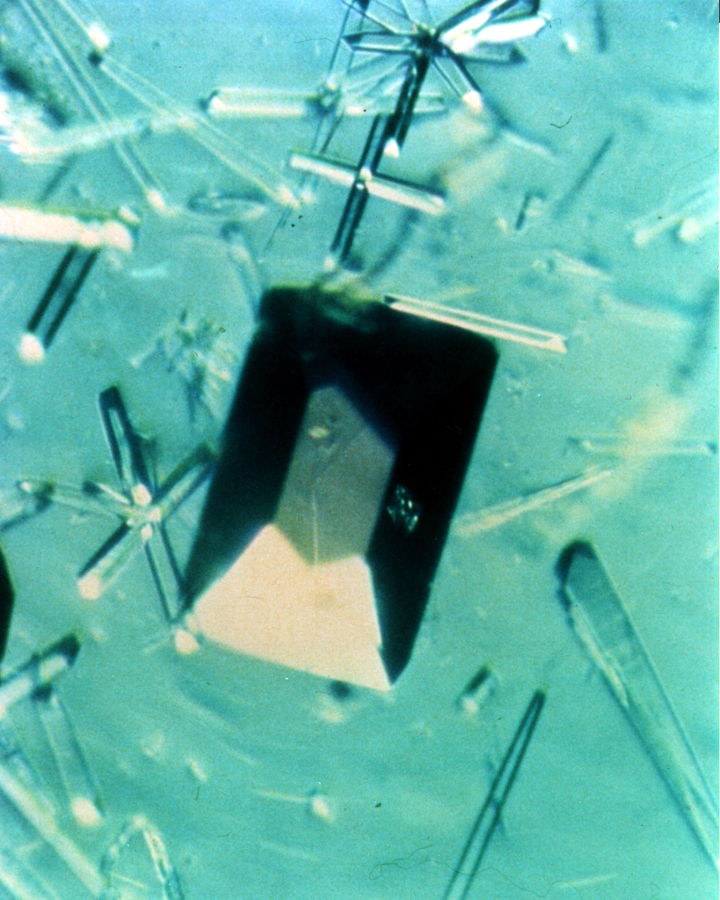
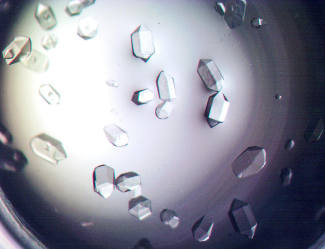
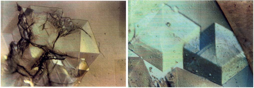
![Atomic force microscopy uses laser technology to reveal a defect, a double-screw dislocation, on the surface of this crystal of canavalin, a major source of dietary protein for humans and domestic animals. When a crystal grows, attachment kinetics and transport kinetics are competing for control of the molecules. As a molecule gets close to the crystal surface, it has to attach properly for the crystal to be usable. NASA has funded investigators to look at those attachment kinetics from a theoretical standpoint and an experimental standpoint. Dr. Alex McPherson of the University of California, Irvine, is one of those investigators. He uses X-ray diffraction and atomic force microscopy in his laboratory to answer some of the many questions about how protein crystals grow. Atomic force microscopy provides a means of looking at how individual molecules are added to the surface of growing protein crystals. This helps McPherson understand the kinetics of protein crystal growth. McPherson asks, How fast do crystals grow? What are the forces involved? Investigators funded by NASA have clearly shown that such factors as the level of supersaturation and the rate of growth all affect the habit [characteristic arrangement of facets] of the crystal and the defects that occur in the crystal.](https://images-assets.nasa.gov/image/0101744/0101744~small.jpg)
Atomic force microscopy uses laser technology to reveal a defect, a double-screw dislocation, on the surface of this crystal of canavalin, a major source of dietary protein for humans and domestic animals. When a crystal grows, attachment kinetics and transport kinetics are competing for control of the molecules. As a molecule gets close to the crystal surface, it has to attach properly for the crystal to be usable. NASA has funded investigators to look at those attachment kinetics from a theoretical standpoint and an experimental standpoint. Dr. Alex McPherson of the University of California, Irvine, is one of those investigators. He uses X-ray diffraction and atomic force microscopy in his laboratory to answer some of the many questions about how protein crystals grow. Atomic force microscopy provides a means of looking at how individual molecules are added to the surface of growing protein crystals. This helps McPherson understand the kinetics of protein crystal growth. McPherson asks, How fast do crystals grow? What are the forces involved? Investigators funded by NASA have clearly shown that such factors as the level of supersaturation and the rate of growth all affect the habit [characteristic arrangement of facets] of the crystal and the defects that occur in the crystal.
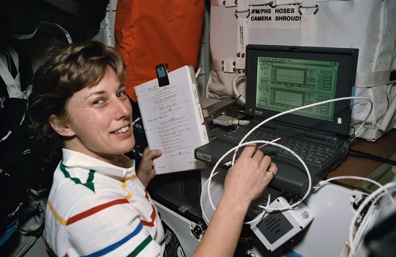
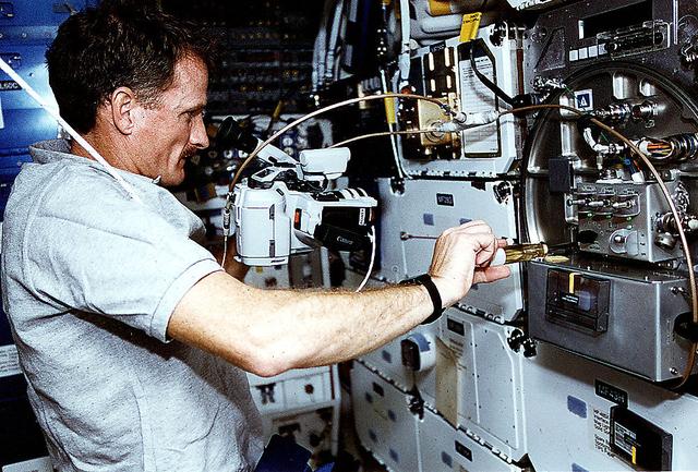
STS060-21-031 (3-11 Feb 1994) --- Using a lap top computer, astronaut N. Jan Davis monitors systems for the Commercial Protein Crystal Growth (CPCG) experiment onboard the Space Shuttle Discovery. Davis joined four other NASA astronauts and a Russian cosmonaut for eight days in space aboard Discovery.
As the most abundant protein in the circulatory system albumin contributes 80% to colloid osmotic blood pressure. Albumin is also chiefly responsible for the maintenance of blood pH. It is located in every tissue and bodily secretion, with extracellular protein comprising 60% of total albumin. Perhaps the most outstanding property of albumin is its ability to bind reversibly to an incredible variety of ligands. It is widely accepted in the pharmaceutical industry that the overall distribution, metabolism, and efficiency of many drugs are rendered ineffective because of their unusually high affinity for this abundant protein. An understanding of the chemistry of the various classes of pharmaceutical interactions with albumin can suggest new approaches to drug therapy and design. Principal Investigator: Dan Carter/New Century Pharmaceuticals
Dan Carter and Charles Sisk center a Lysozyme Protein crystal grown aboard the USML-2 shuttle mission. Protein isolated from hen egg-white and functions as a bacteriostatic enzyme by degrading bacterial cell walls. First enzyme ever characterized by protein crystallography. It is used as an excellent model system for better understanding parameters involved in microgravity crystal growth experiments. The goal is to compare kinetic data from microgravity experiments with data from laboratory experiments to study the equilibrium.
Malic Enzyme is a target protein for drug design because it is a key protein in the life cycle of intestinal parasites. After 2 years of effort on Earth, investigators were unable to produce any crystals that were of high enough quality and for this reason the structure of this important protein could not be determined. Crystals obtained from one STS-50 were of superior quality allowing the structure to be determined. This is just one example why access to space is so vital for these studies. Principal Investigator is Larry DeLucas.
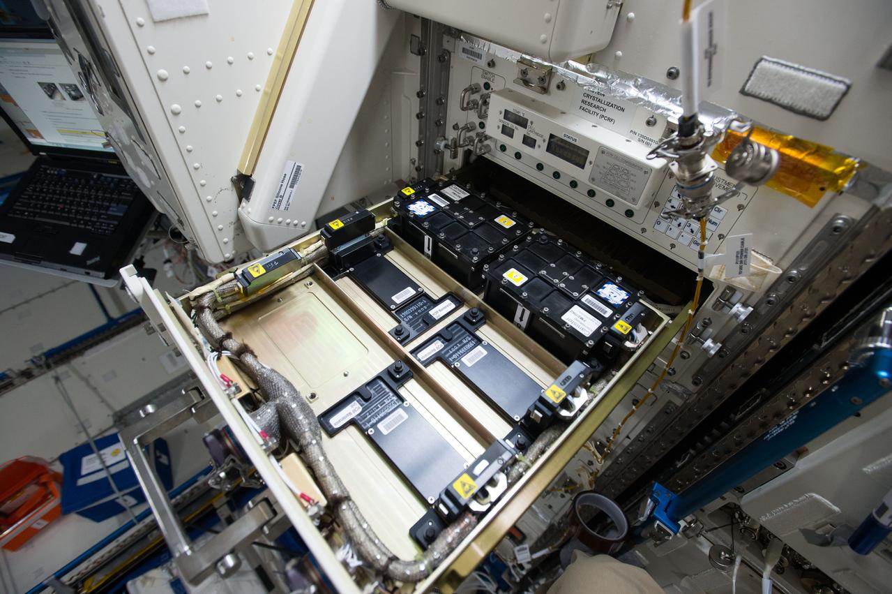
iss062e087808 (3/11/2020) --- A view of Protein Crystal Growth-10 experiment hardware inside JAXA's (Japan Aerospace Exploration Agency) Kibo laboratory module aboard the International Space Station (ISS). Microgravity Crystallization of Glycogen Synthase-Glycogenin Protein Complex (CASIS PCG 10) crystallizes human glycogen synthase proteins on the space station. Determining the structure of the human glycogen synthase and full-length glycogenin protein complex could facilitate the development of treatments on Earth for metabolic disorders such as Type 2 diabetes, obesity, rare genetic disorders, and some forms of cancer.
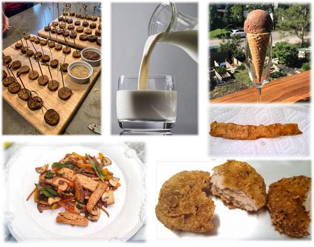
jsc2022e031225 (4/26/2022) --- For the Protein Manufacturing investigations, examples are shown of alternative meat and dairy products prepared from fungal strain MK7 biomats by Nature’s Fynd. Delicious foods developed by world renown chefs, including Sebastian Canonne, founder of the French Pastry School. Image courtesy of BioServe.
Astronaut Michael Clifford places a liquid nitrogen Dewar containing frozen protein solutions aboard Russia's space station Mir during a visit by the Space Shuttle (STS-76). The protein samples were flash-frozen on Earth and will be allowed to thaw and crystallize in the microgravity environment on Mir Space Station. A later crew will return the Dewar to Earth for sample analysis. Dr. Alexander McPherson of the University of California at Riverside is the principal investigator. Photo credit: NASA/Johnson Space Center.
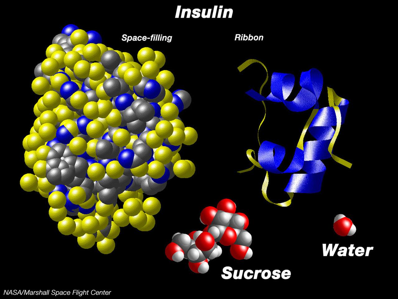
This computer graphic depicts the relative complexity of crystallizing large proteins in order to study their structures through x-ray crystallography. Insulin is a vital protein whose structure has several subtle points that scientists are still trying to determine. Large molecules such as insuline are complex with structures that are comparatively difficult to understand. For comparison, a sugar molecule (which many people have grown as hard crystals in science glass) and a water molecule are shown. These images were produced with the Macmolecule program. Photo credit: NASA/Marshall Space Flight Center (MSFC)
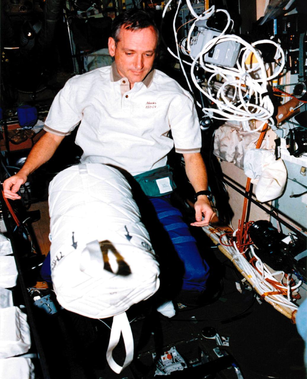
Astronaut Tom Akers places a liquid nitrogen Dewar containing frozen protein solutions aboard Russia's space Station Mir during a visit by the Space Shuttle (STS-79). The protein samples were flash-frozen on Earth and will be allowed to thaw and crystallize in the microgravity environment on Mir Space Station. A later crew will return the Dewar to Earth for sample analysis. Dr. Alexander McPherson of the University of California at Riverside is the principal investigator. Photo credit: NASA/Johnson Space Center.
Type II restriction enzymes, such as Eco R1 endonulease, present a unique advantage for the study of sequence-specific recognition because they leave a record of where they have been in the form of the cleaved ends of the DNA sites where they were bound. The differential behavior of a sequence -specific protein at sites of differing base sequence is the essence of the sequence-specificity; the core question is how do these proteins discriminate between different DNA sequences especially when the two sequences are very similar. Principal Investigator: Dan Carter/New Century Pharmaceuticals
High school students screen crystals of various proteins that are part of the ground-based work that supports Alexander McPherson's protein crystal growth experiment. The students also prepared and stored samples in the Enhanced Gaseous Nitrogen Dewar, which was launched on the STS-98 mission for delivery to the ISS. The crystals grown on the ground will be compared with crystals grown in orbit. Participants include Joseph Negron (shown), of Terry Parker High School, Jacksonville, Florida; Megan Miskowski, of Ridgeview High School, Orange Park, Florida; and Sam Swank, of Fletcher High School, Neptune Beach, Florida. The proteins are placed in plastic tubing that is heat-sealed at the ends, then flash-frozen and preserved in a liquid nitrogen Dewar. Aboard the ISS, the nitrogen will be allowed to evaporated so the samples thaw and then slowly crystallize. They will be analyzed after return to Earth. Photo credit: NASA/Marshall Space Flight Center.
High school students screen crystals of various proteins that are part of the ground-based work that supports Alexander McPherson's protein crystal growth experiment. The students also prepared and stored samples in the Enhanced Gaseous Nitrogen Dewar, which was launched on the STS-98 mission for delivery to the ISS. The crystals grown on the ground will be compared with crystals grown in orbit. Participants include Joseph Negron, of Terry Parker High School, Jacksonville, Florida; Megan Miskowski (shown), of Ridgeview High School, Orange Park, Florida; and Sam Swank, of Fletcher High School, Neptune Beach, Florida. The proteins are placed in plastic tubing that is heat-sealed at the ends, then flash-frozen and preserved in a liquid nitrogen Dewar. Aboard the ISS, the nitrogen will be allowed to evaporated so the samples thaw and then slowly crystallize. They will be analyzed after return to Earth. Photo credit: NASA/Marshall Space Flight Center.
High school students screen crystals of various proteins that are part of the ground-based work that supports Alexander McPherson's protein crystal growth experiment. The students also prepared and stored samples in the Enhanced Gaseous Nitrogen Dewar, which was launched on the STS-98 mission for delivery to the ISS. The crystals grown on the ground will be compared with crystals grown in orbit. Participants include Joseph Negron, of Terry Parker High School, Jacksonville, Florida; Megan Miskowski, of Ridgeview High School, Orange Park, Florida; and Sam Swank (shown), of Fletcher High School, Neptune Beach, Florida. The proteins are placed in plastic tubing that is heat-sealed at the ends, then flash-frozen and preserved in a liquid nitrogen Dewar. Aboard the ISS, the nitrogen will be allowed to evaporated so the samples thaw and then slowly crystallize. They will be analyzed after return to Earth. Photo credit: NASA/Marshall Space Flight Center.
61B-02-014 (26 Nov-3 Dec 1985) --- Payload Specialist Charles D. Walker works with the handheld protein growth experiment -- one of a series of tests being flown to study the possibility of crystallizing biological materials. Walker rests the experiment against the larger continuous flow electrophoresis systems experiment.
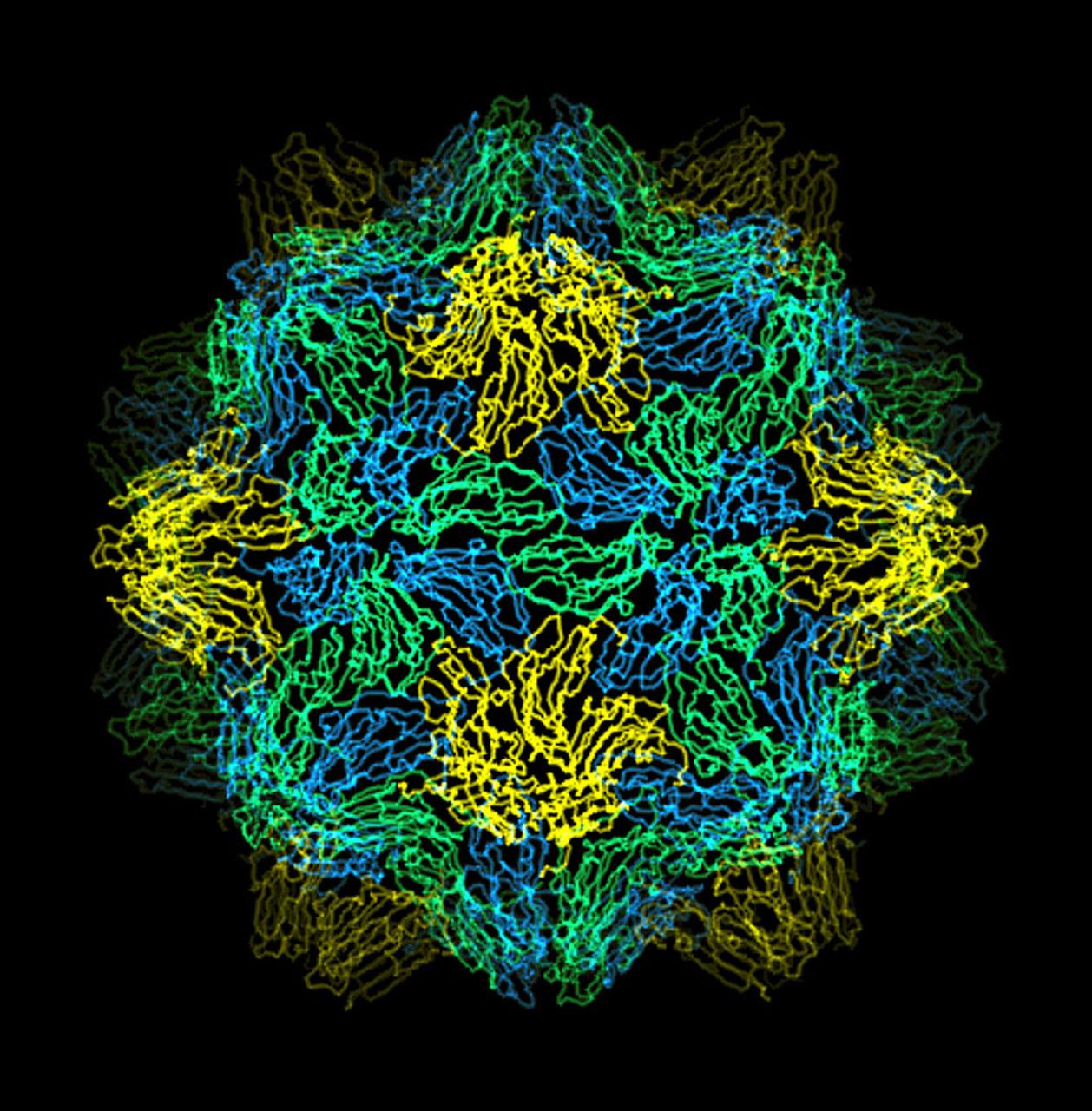
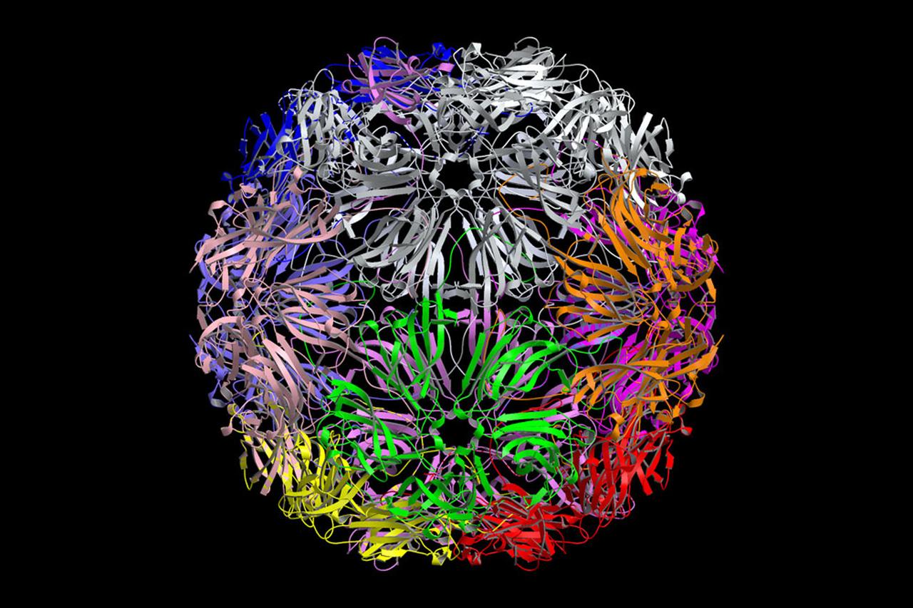
The bumpy exterior of the turnip yellow mosaic virus (TYMV) protein coat, or capsid, was defined in detail by Dr. Alexander McPherson of the University of California, Irvin using protein crystallized in space for analysis on Earth. TYMV is an icosahedral virus constructed from 180 copies of the same protein arranged into 12 clusters of five proteins (pentamers), and 20 clusters of six proteins (hexamers). The final TYMV structure led to the enexpected hypothesis that the virus release its RNA by essentially chemical-mechanical means. Most viruses have farly flat coats, but in TYMV, the fold in each protein, called the jellyroll, is clustered at the points where the protein pentamers and hexamers join. The jellyrolls are almost standing on end, producing a bumpy surface with knobs at all of the pentamers and hexamers. At the inside surface of the pentamers is a void that is not present at the hexamers. The coating had been seen in early studies of TYMV, but McPhereson's atomic structure shows much more detail. The inside surface is strikingly, and unexpectedly, different than the outside. While the pentamers contain a central viod on the inside, the hexameric units contain peptides liked to each other, forming a ring or, more accurately, rings to fill the voild. Credit: Dr. Alexander McPherson, University of California, Irvine.
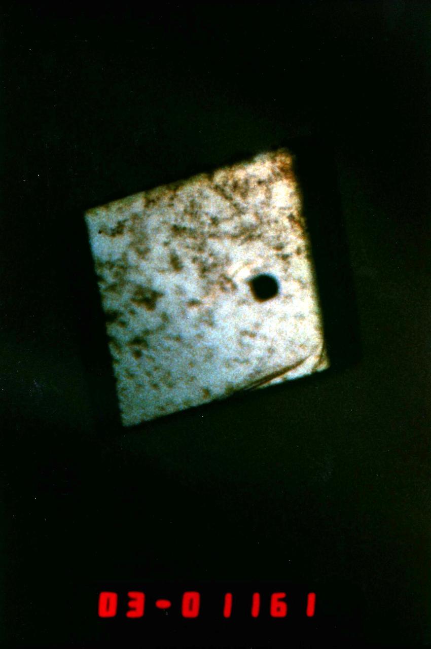
Orbital Documentation of Porcine Elastase grown in (PCG) Protein Crystal Growth (RIM) Refrigerator Incubator Module
Scientist photographs STS- 26 Post-flight (VDA) Vapor Diffusion Apparatus Tray with (PCG) Protein Crystal Growth Samples.
iss049e045287 (10/21/2016) --- Photographic documentation taken during JAXA Protein Crystal Growth (PCG) Installation into the Protein Crystallization Research Facility (PCRF) of the Ryutai Rack.
(PCG) Protein Crystal Growth Human Serum Albumin. Contributes to many transport and regulatory processes and has multifunctional binding properties which range from various metals, to fatty acids, hormones, and a wide spectrum of therapeutic drugs. The most abundant protein of the circulatory system. It binds and transports an incredible variety of biological and pharmaceutical ligands throughout the blood stream. Principal Investigator on STS-26 was Larry DeLucas.
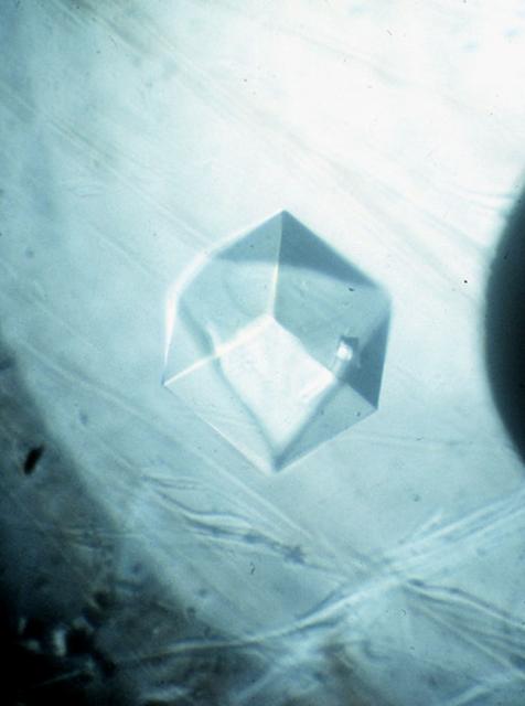
Horse Serum Albumin crystals grown during the USML-1 (STS-50) mission's Protein Crystal Growth Glovebox Experiment. These crystals were grown using a vapor diffusion technique at 22 degrees C. The crystals were allowed to grow for nine days while in orbit. Crystals of 1.0 mm in length were produced. The most abundant blood serum protein, regulates blood pressure and transports ions, metabolites, and therapeutic drugs. Principal Investigator was Edward Meehan.
(PCG) Protein Crystal Growth Porcine Elastase. This enzyme is associated with the degradation of lung tissue in people suffering from emphysema. It is useful in studying causes of this disease. Principal Investigator on STS-26 was Charles Bugg.
(PCG) Protein Crystal Growth Renin. Enzyme produced by the kidneys, plays a major role in the chemical reaction that controls blood pressure. Principal Investigator on STS-26 was Charles Bugg.

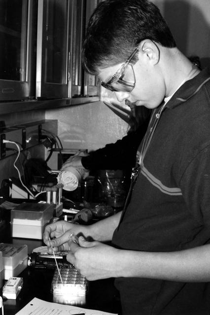
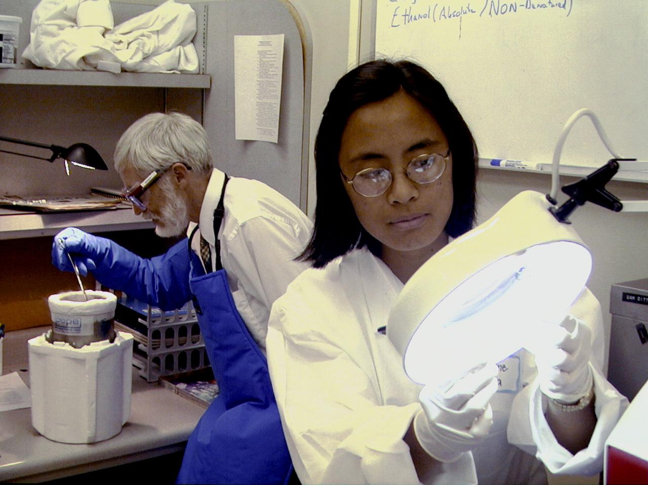
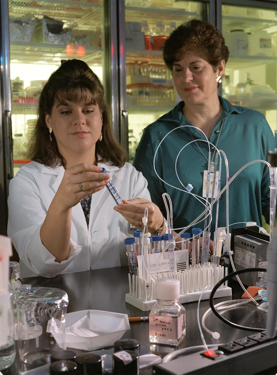
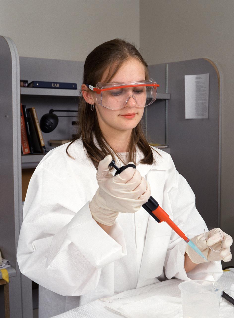
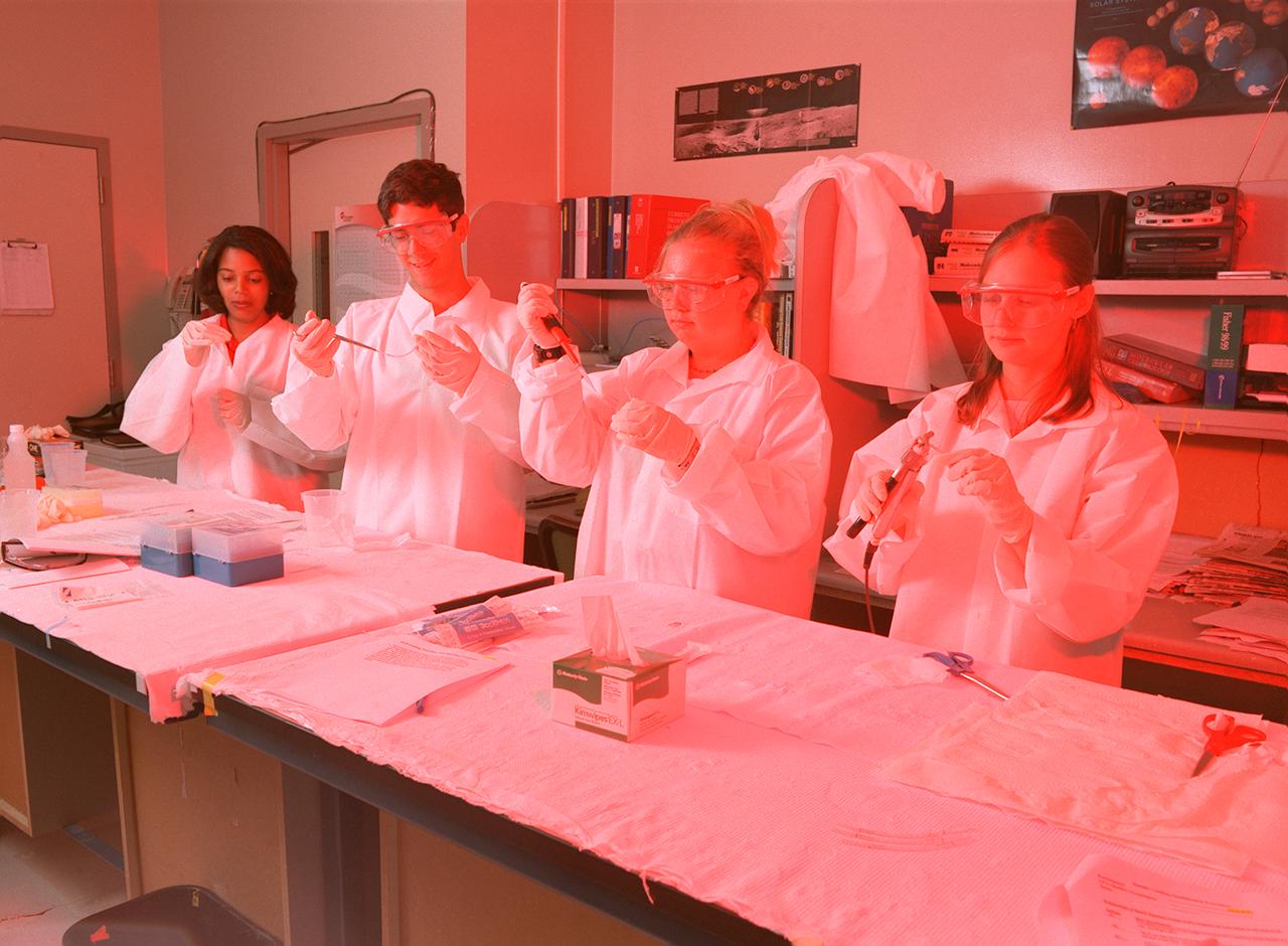
Cindy Barnes of University Space Research Association (USRA) at NASA's Marshall Space Flight Center pipettes a protein solution in preparation to grow crystals as part of NASA's structural biology program. Research on Earth helps scientists define conditions and specimens they will use in space experiments.
61C-05-036 (12-18 Jan. 1986) --- U.S. Representative Bill Nelson (Democrat - Florida), STS-61C payload specialist, prepares to photograph individual samples in the Handheld Protein Crystal Growth Experiment (HPCG) on Columbia's middeck. The operations involve the use of four pieces of equipment to attempt the growth of 60 different types of crystals -- 12 by means of dialysis and 48 via the vapor diffusion method. The photo was used by members of the STS-61C crew at their Jan. 23, 1986, Post-Flight Press Conference.
The bumpy exterior of the turnip yellow mosaic virus (TYMV) protein coat, or capsid, was defined in detail by Dr. Alexander McPherson of the University of California, Irvin using proteins crystallized in space for analysis on Earth. TYMV is an icosahedral virus constructed from 180 copies of the same protein arranged into 12 clusters of five proteins (pentamers), and 20 clusters of six proteins (hexamers). The final TYMV structure led to the unexpected hypothesis that the virus releases its RNA by essentially chemical-mechanical means. Most viruses have fairly flat coats, but in TYNV, the fold in each protein, called the jellyroll, is clustered at the points where the protein pentamers and hexamers join. The jellyrolls are almost standing on end, producing a bumpy surface with knobs at all of the pentamers and hexamers. At the inside surface of the pentamers is a void that is not present at the hexamers. The coating had been seen in early stuties of TYMV, but McPherson's atomic structure shows much more detail. The inside surface is strikingly, and unexpectedly, different than the outside. While the pentamers contain a central void on the inside, the hexameric units contain peptides linked to each other, forming a ring or, more accurately, rings to fill the void. Credit: Dr. Alexander McPherson, University of California, Irvine
Christiane Gumera, right, a student at Stanton College Preparatory High School in Jacksonville, AL, examines a protein sample while preparing an experiment for flight on the International Space Station (ISS). Merle Myers, left, a University of California, Irvine, researcher, prepares to quick-freeze protein samples in nitrogen. The proteins are in a liquid nitrogen Dewar. Aboard the ISS, the nitrogen will be allowed to evaporated so the samples thaw and then slowly crystallize. They will be anlyzed after return to Earth. Photo credit: NASA/Marshall Space Flight Center (MSFC)
On the Space Shuttle Orbiter Atlantis' middeck, Astronaut Joseph R. Tarner, mission specialist, works at an area amidst several lockers which support the Protein Crystal Growth (PCG) experiment during the STS-66 mission. This particular section is called the Crystal Observation System, housed in the Thermal Enclosure System (COS/TES). Together with the Vapor Diffusion Apparatus (VDA), housed in Single Locker Thermal Enclosure (SLTES), the COS/TES represents the continuing research into the structure of proteins and other macromolecules such as viruses.
Lisa Crawford, a graduate research assistant from the University of Toledo, works with Laurel Karr of Marshall Space Flight Center (MSFC) in the molecular biology laboratory. They are donducting genetic manipulation of bacteria and yeast for the production of large amount of desired protein. Photo credit: NASA/Marshall Space Flight Center (MSFC)
(PCG) Protein Crystal Growth Isocitrate Lysase. Target enzyme for fungicides. A better understanding of this enzyme should lead to the discovery of more potent fungicides to treat serious crop diseases such as rice blast. It regulates the flow of metabolic intermediates required for cell growth. Principal Investigator on STS-26 was Charles Bugg.
(PCG) Protein Crystal Growth Isocitrate Lyase. Target enzyme for fungicides. A better understanding of this enzyme should lead to the discovery of more potent fungicides to treat serious crop diseases such as rice blast. It regulates the flow of metabolic intermediates required for cell growth. Principal Investigator for STS-26 was Charles Bugg.
iss056e075928 (7/3/2018) --- Astronaut Alexander Gerst of ESA (European Space Agency), during the JAXA Protein Crystal Growth (PCG) sample retrieval from the Freezer-Refrigerator Of Stirling Cycle 2 (FROST2) and initiation of the crystallization of the samples before inserting them back into the FROST2, where crystallization will continue.
A Memphis student working at the University of Alabama in Huntsville prepares samples for the first protein crystal growth experiments plarned to be performed aboard the International Space Station (ISS). The proteins are placed in plastic tubing that is heat-sealed at the ends, then flash-frozen and preserved in a liquid nitrogen Dewar. Aboard the ISS, the nitrogen will be allowed to evaporated so the samples thaw and then slowly crystallize. They will be analyzed after return to Earth. Photo credit: NASA/Marshall Space Flight Center (MSFC)
A Memphis student working at the University of Alabama in Huntsville prepares samples for the first protein crystal growth experiments plarned to be performed aboard the International Space Station (ISS). The proteins are placed in plastic tubing that is heat-sealed at the ends, then flash-frozen and preserved in a liquid nitrogen Dewar. Aboard the ISS, the nitrogen will be allowed to evaporated so the samples thaw and then slowly crystallize. They will be analyzed after return to Earth. Photo credit: NASA/Marshall Space Flight Center (MSFC)
Memphis students working at the University of Alabama in Huntsville prepare samples for the first protein crystal growth experiments plarned to be performed aboard the International Space Station (ISS). The proteins are placed in plastic tubing that is heat-sealed at the ends, then flash-frozen and preserved in a liquid nitrogen Dewar. Aboard the ISS, the nitrogen will be allowed to evaporated so the samples thaw and then slowly crystallize. They will be analyzed after return to Earth. Photo credit: NASA/Marshall Space Flight Center (MSFC)
Onboard Space Shuttle Columbia (STS-73) Mission Specialists Catherine Cady Coleman works at the glovebox facility in support of the Protein Crystal Growth Glovebox (PCG-GBX) experiment in the United States Microgravity Laboratory 2 (USML-2) Spacelab science module.
Diabetic patients may someday reduce their insulin injections and lead more normal lives because of new insights gained through irnovative space research in which insulin crystals were grown on the Space Shuttle. Results from a 1994 insulin crystal growth experiment in space are leading to a new understanding of protein insulin. Lack of insulin is the cause of diabetes, a desease that accounts for one-seventh of the nation's health care costs. Dr. Marianna Long, associate director of the Center of Macromolecular Crystallography at the University of Alabama at Birmingham, is a co-investigator on the research. Photo credit: NASA/Marshall Space Flight Center (MSFC)
Diabetic patients may someday reduce their insulin injections and lead more normal lives because of new insights gained through innovative space research in which insulin crystals were grown on the Space Shuttle. Results from a 1994 insulin crystals growth experiment in space are leading to a new understanding of protein insulin. Lack of insulin is the cause of diabetes, a disease that accounts for one-seventh of the nation's health care costs. Champion Deivanaygam, a researcher at the Center for Macromolecular Crystallography at the University of Alabama in Birmingham, assists in this work. Photo credit: NASA/Marshall Space Flight Center (MSFC)
(PCG) Protein Crystal Growth Gamma-Interferon. Stimulates the body's immune system and is used clinically in the treatment of cancer. Potential as an anti-tumor agent against solid tumors as well as leukemia's and lymphomas. It has additional utility as an anti-ineffective agent, including antiviral, anti-bacterial, and anti-parasitic activities. Principal Investigator on STS-26 was Charles Bugg.
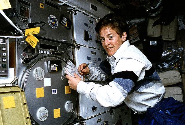
Astronaut Wendy B. Lawrence, flight engineer and mission specialist for STS-67, scribbles notes on the margin of a checklist while monitoring an experiment on the Space Shuttle Endeavour's mid-deck. The experiment is the Protein Crystal Growth (PCG), which takes up locker space near the Commercial Materials Dispersion Apparatus Instruments Technology Associates Experiment (CMIX).
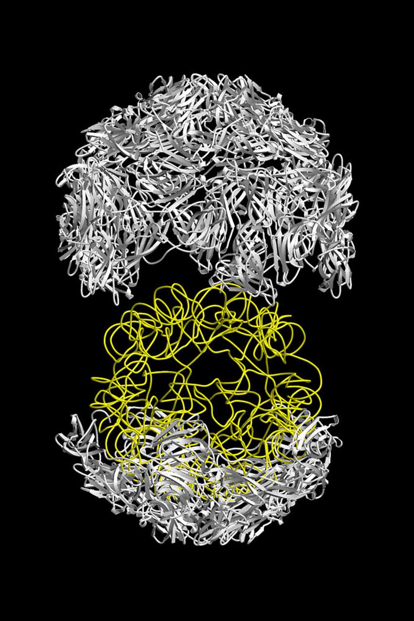
The structure of the Satellite Tobacco Mosaic Viurus (STMV)--one of the smallest viruses known--has been successfully reduced using STMV crystals grown aboard the Space Shuttle in 1992 and 1994. The STMV crystals were up to 30 times the volume of any seen in the laboratory. At the time they gave the best resolution data ever obtained on any virus crystal. STMV is a small icosahedral plant virus, consisting of a protein shell made up of 60 identical protein subunits of molecular weight 17,500. Particularly noteworthy is the fact that, in contrast to the crystals grown on Earth, the crystals grown under microgravity conditions were visually perfect, with no striations or clumping of crystals. Furthermore, the x-ray diffraction data obtained from the space-grown crystals was of a much higher quality than the best data available at that time from ground-based crystals. This stylized ribbon model shows the protein coat in white and the nucleic acid in yellow. STMV is used because it is a simple protein to work with; studies are unrelated to tobacco. Credit: Dr. Alex McPherson, University of California at Irvin.
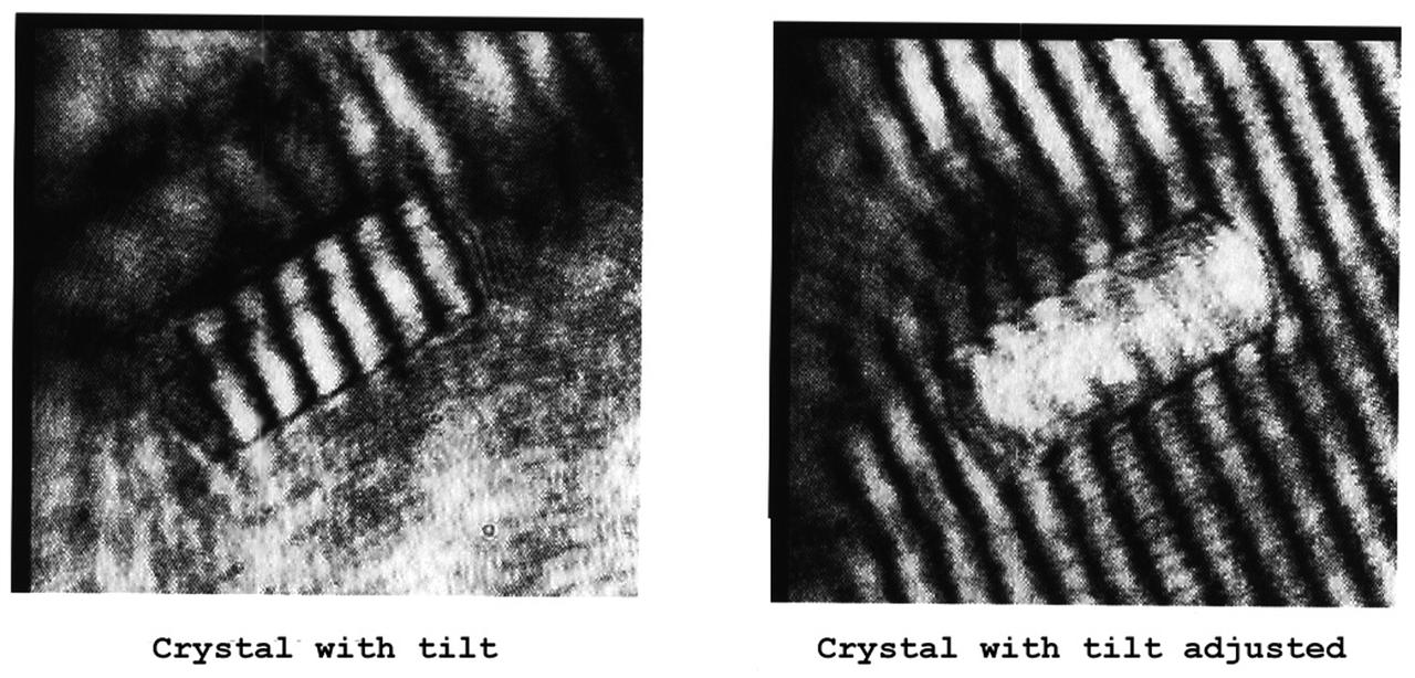
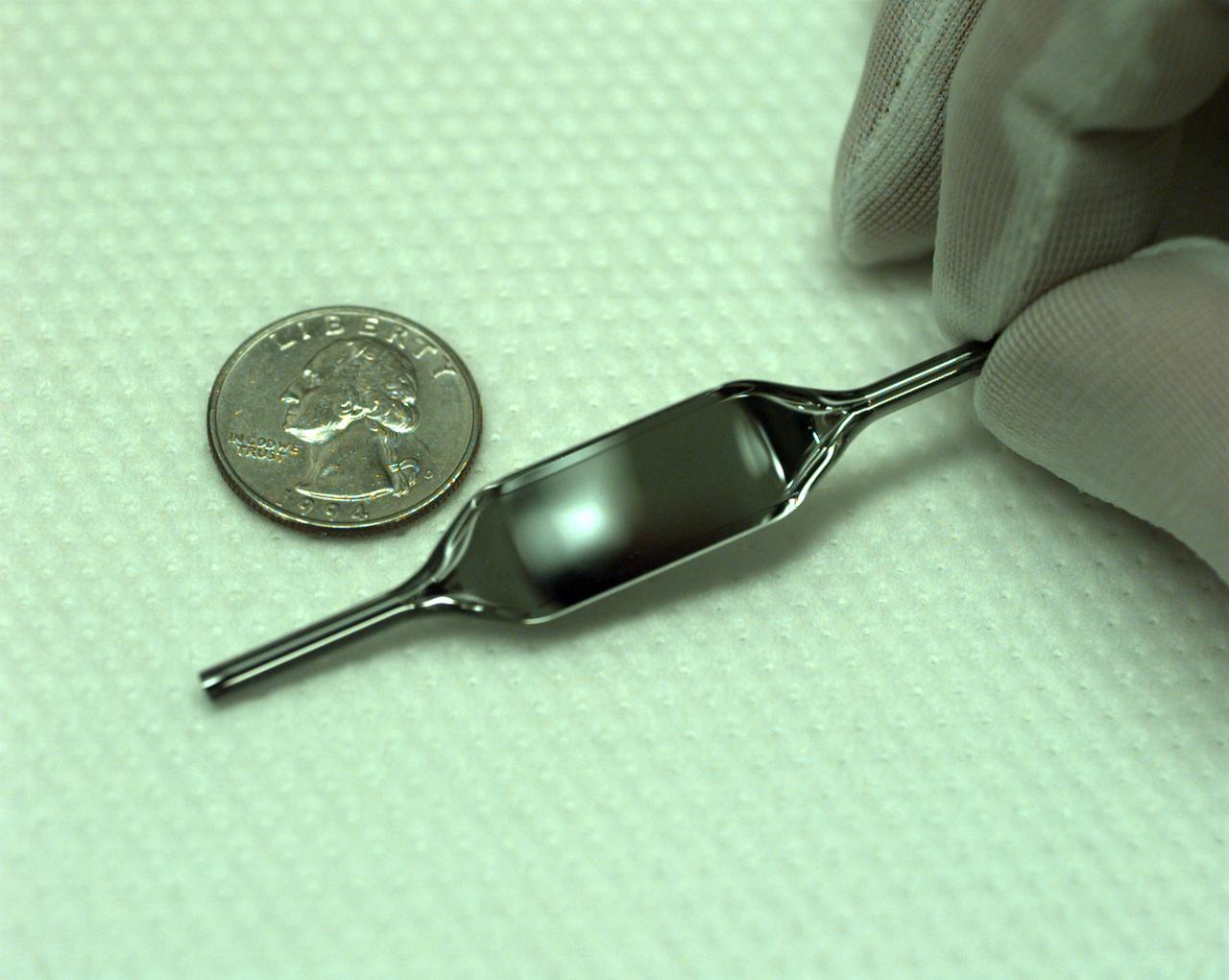
The Interferometer Protein Crystal Growth (IPCG) experiment was designed to measure details of how protein molecules move through a fluid. It was flown on the STS-86 mission for use aboard Russian Space Station Mir in 1998. It studied aspects of how crystals grow - and what conditions lead to the best crystals, details that remain a mystery. IPCG produces interference patterns by spilitting then recombining laser light. This let scientists see how fluid densities - and molecular diffusion - change around a crystal as it grows in microgravity. The heart of the IPCG apparatus is the interferometer cell comprising the optical bench, microscope, other optics, and video camera. IPCG experiment cells are made of optical glass and silvered on one side to serve as a mirror in the interferometer system that visuzlizes crystals and conditions around them as they grow inside the cell. This view shows interferograms produced in ground tests. The principal investigator was Dr. Alexander McPherson of University of California, Irvine. Co-investigators are William Witherow and Dr. Marc Pusey of NASA's Marshall Space Flight Center (MSFC).
The Interferometer Protein Crystal Growth (IPCG) experiment was designed to measure details of how protein molecules move through a fluid. It was flown on the STS-86 mission for use aboard Russian Space Station Mir in 1998. It studied aspects of how crystals grow - and what conditions lead to the best crystals, details that remain a mystery. IPCG produces interference patterns by spilitting then recombining laser light. This let scientists see how fluid densities - and molecular diffusion - change around a crystal as it grows in microgravity. The heart of the IPCG apparatus is the interferometer cell comprising the optical bench, microscope, other optics, and video camera. IPCG experiment cells are made of optical glass and silvered on one side to serve as a mirror in the interferometer system that visuzlizes crystals and conditions around them as they grow inside the cell. This view shows a large growth cell. The principal investigator was Dr. Alexander McPherson of University of California, Irvine. Co-investigators are William Witherow and Dr. Marc Pusey of NASA's Marshall Space Flight Center (MSFC).
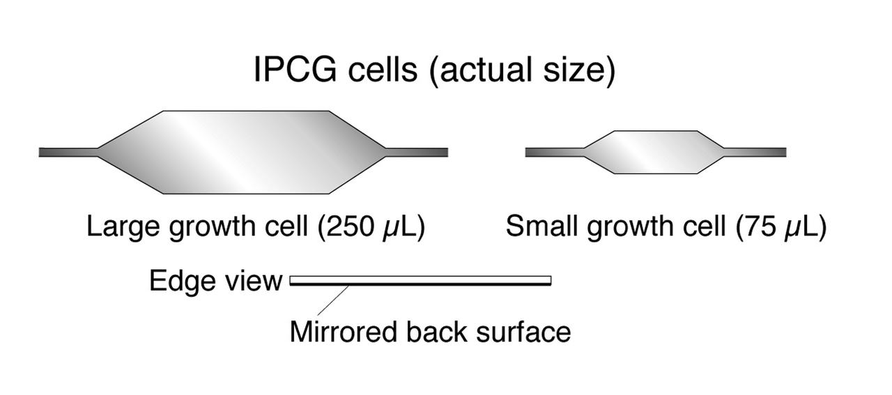
The Interferometer Protein Crystal Growth (IPCG) experiment was designed to measure details of how protein molecules move through a fluid. It was flown on the STS-86 mission for use aboard Russian Space Station Mir in 1998. It studied aspects of how crystals grow - and what conditions lead to the best crystals, details that remain a mystery. IPCG produces interference patterns by spilitting then recombining laser light. This let scientists see how fluid densities - and molecular diffusion - change around a crystal as it grows in microgravity. The heart of the IPCG apparatus is the interferometer cell comprising the optical bench, microscope, other optics, and video camera. IPCG experiment cells are made of optical glass and silvered on one side to serve as a mirror in the interferometer system that visuzlizes crystals and conditions around them as they grow inside the cell. This diagram shows the growth cells. The principal investigator was Dr. Alexander McPherson of University of California, Irvine. Co-investigators are William Witherow and Dr. Marc Pusey of NASA's Marshall Space Flight Center (MSFC).
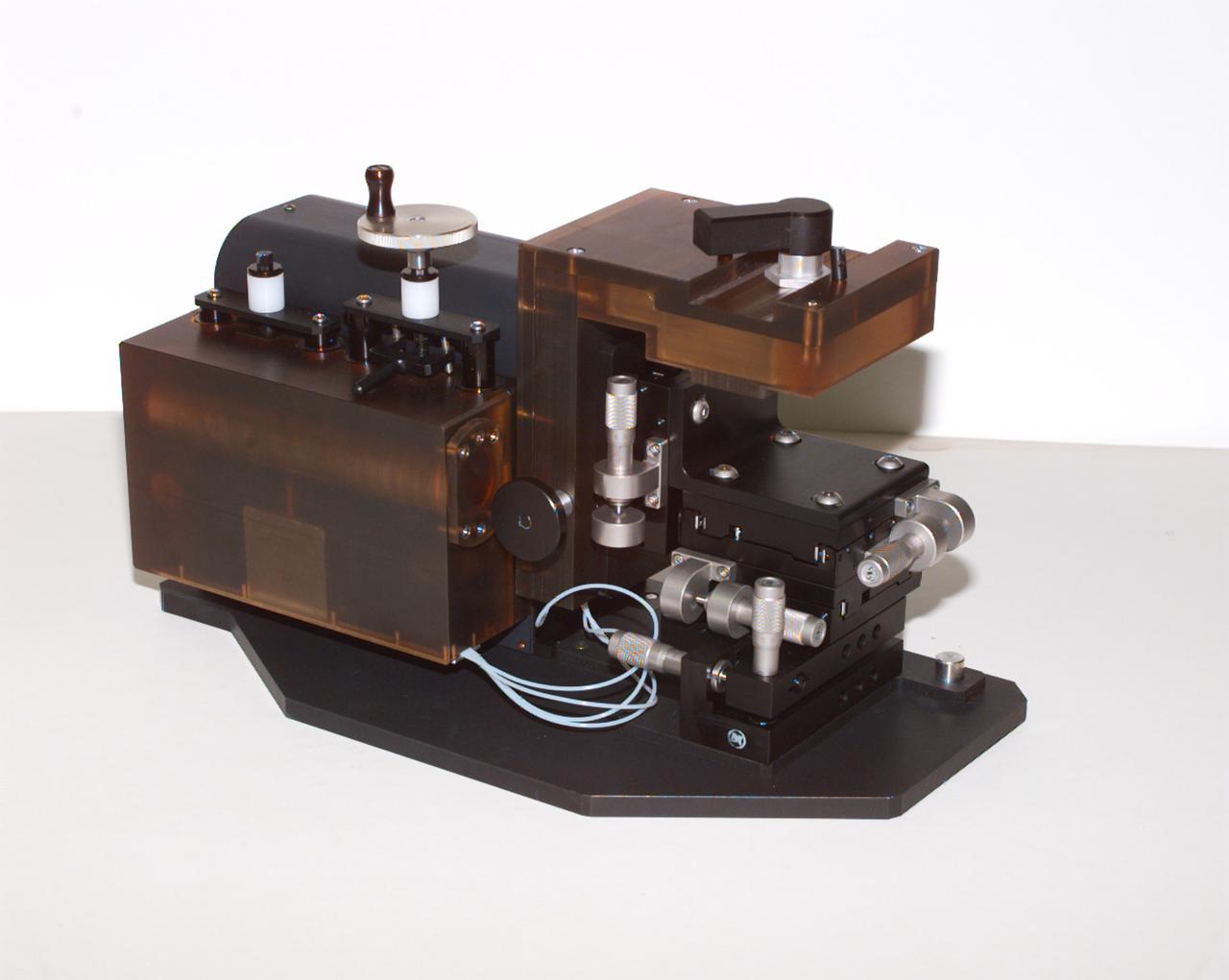
The Interferometer Protein Crstal Growth (IPCG) experiment was designed to measure details of how protein molecules move through a fluid. It was flown on the STS-86 mission for use aboard Russin Space Station Mir in 1998. It studied aspects of how crystals grow - and what conditions lead to the best crystals, details that remain a mystery. IPCG produces interference patterns by splitting then recombining laser light. This let scientists see how fluid densities - and molecular diffusion - change around a crystal as it grows in microgravity. The heart of the IPCG apparatus is the interferometer cell comprising the optical bench, microscope, other optics, and video camera. IPCG experiment cells are made of optical glass and silvered on one side to serve as a mirror in the interferometer system that visualizes crystals and conditions around them as they grow inside the cell. This view shows the complete apparatus. The principal investigator was Dr. Alexander McPherson of the University of California, Irvin. Co-investigators are William Witherow and Dr. Marc Pusey of NASA's Marshall Space Flight Center
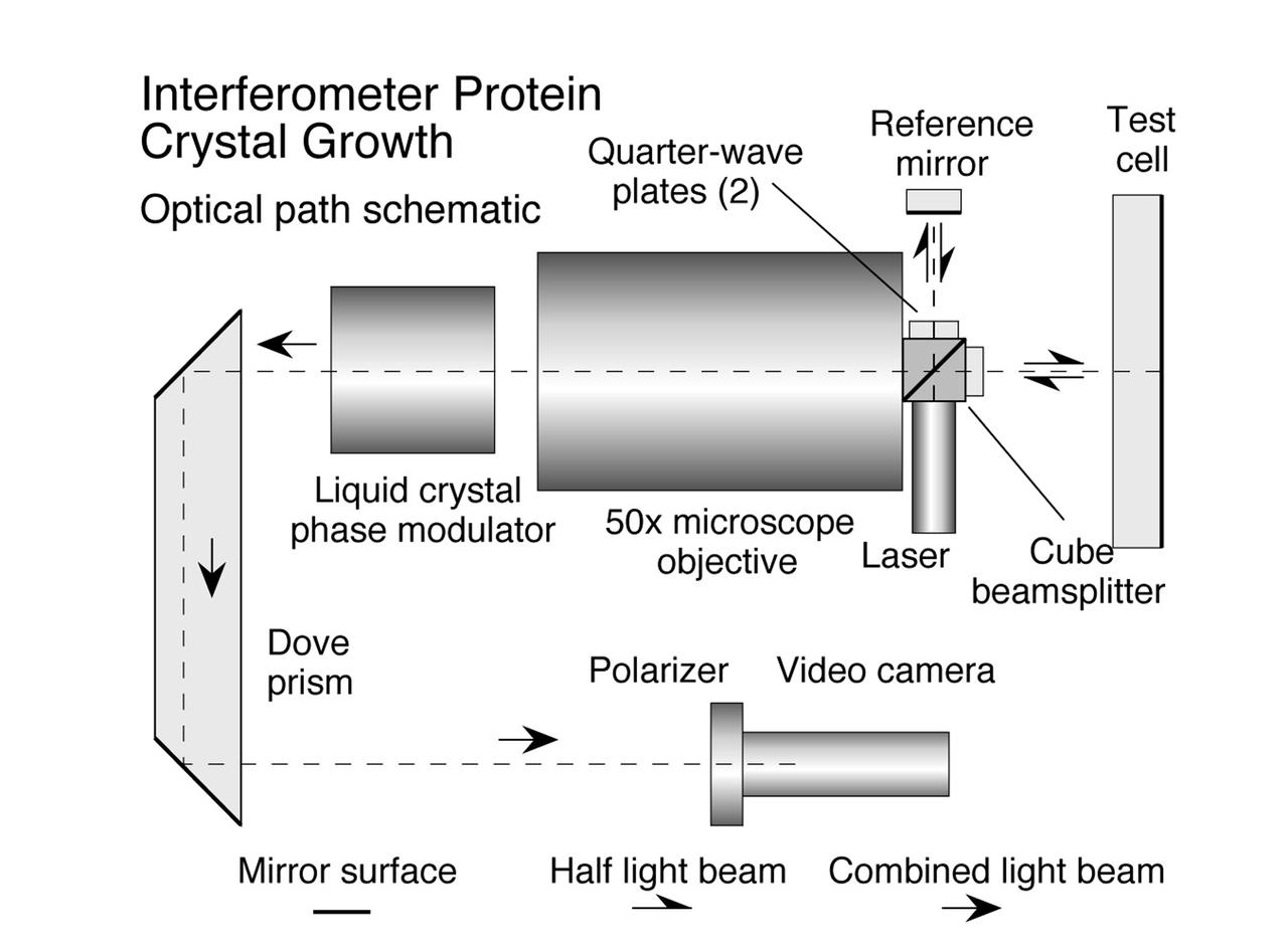
The Interferometer Protein Crystal Growth (IPCG) experiment was designed to measure details of how protein molecules move through a fluid. It was flown on the STS-86 mission for use aboard Russian Space Station Mir in 1998. It studied aspects of how crystals grow - and what conditions lead to the best crystals, details that remain a mystery. IPCG produces interference patterns by spilitting then recombining laser light. This let scientists see how fluid densities - and molecular diffusion - change around a crystal as it grows in microgravity. The heart of the IPCG apparatus is the interferometer cell comprising the optical bench, microscope, other optics, and video camera. IPCG experiment cells are made of optical glass and silvered on one side to serve as a mirror in the interferometer system that visuzlizes crystals and conditions around them as they grow inside the cell. This diagram shows the optical layout. The principal investigator was Dr. Alexander McPherson of University of California, Irvine. Co-investigators are William Witherow and Dr. Marc Pusey of NASA's Marshall Space Flight Center (MSFC).
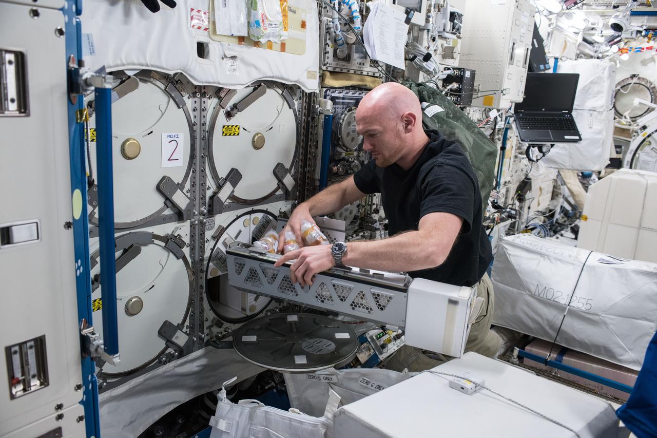
iss056e075950 (July 3, 2018) --- Astronaut Alexander Gerst of ESA (European Space Agency) works inside the Japanese Kibo laboratory module retrieving Protein Crystal Growth samples from a science freezer, also known as the Minus Eighty-Degree Laboratory Freezer for ISS (MELFI).
Kim Nelson, left, of Sandalwood High School in Jacksonville, FL, helps Steven Nepowada, right, of Terry Parker High School in Jacksonville, practice loading a protein sample into a thermos-like container, known as Dewar. Students from Jacksonville worked with researchers from NASA/Marshall Space Flight Center (MSFC), as well as universities, in Huntsville, AL, on an experiment for the International Space Station (ISS). The proteins are placed in plastic tubing that is heat-sealed at the ends, then flash-frozen and preserved in a liquid nitrogen Dewar. Aboard the ISS, the nitrogen will be allowed to evaporated so the samples thaw and then slowly crystallize. They will be analyzed after return to Earth. Photo credit: NASA/Marshall Space Flight Center (MSFC)
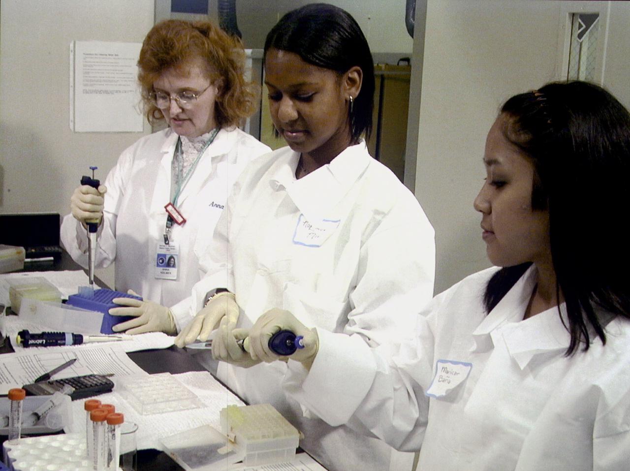
Chemist Arna Holmes, left, from the University of Alabama in Huntsville, teaches NaLonda Moorer, center, and Maricar Bana, right, both from Terry Parker High School in Jacksonville, Fl, procedures for preparing protein crystal growth samples for flight aboard the International Space Station (ISS). NASA/Marshall Space Flight Center in Huntsville, AL, is a sponsor for this educational activity. The proteins are placed in plastic tubing that is heat-sealed at the ends, then flash-frozen and preserved in a liquid nitrogen Dewar. Aborad the ISS, the nitrogen will be allowed to evaporated so the samples thaw and then slowly crystallize. They will be analyzed after return to Earth. Photo credit: NASA/Marshall Space Flight Center (MSFC)
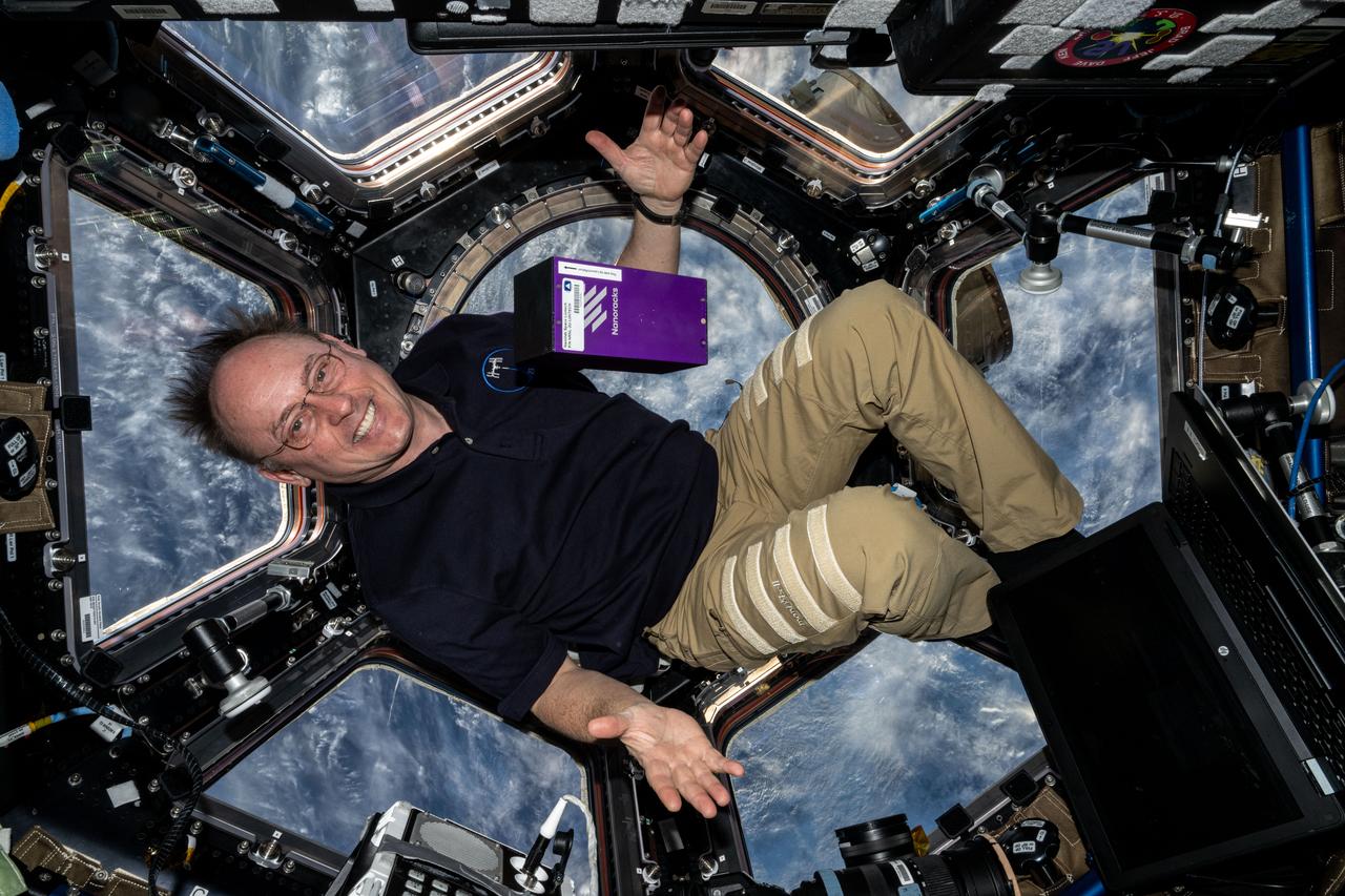
iss073e0548503 (Aug. 28, 2025) --- NASA astronaut and Expedtion 73 Flight Engineer Mike Fincke displays the Nanoracks' Nanolab Space Liintech research hardware inside the International Space Station's cupola while orbiting 262 miles above a cloudy United States. Nanolab Space LiinTech tests a platform that uses optical technology to monitor the process of producing protein crystals in microgravity. This investigation could lead to the development of technology for crystallizing proteins in microgravity to produce pharmaceuticals for use in space and Earth.

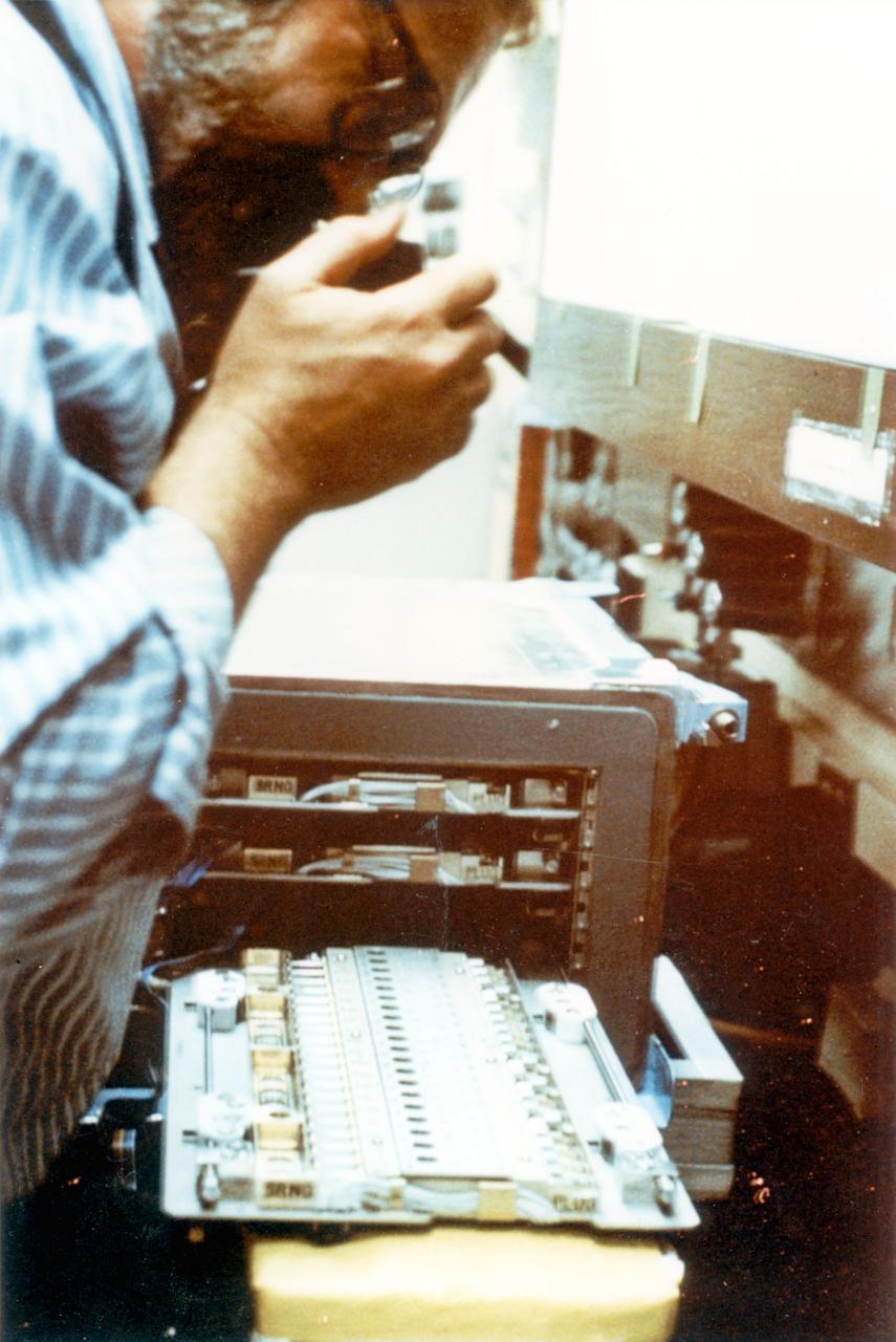
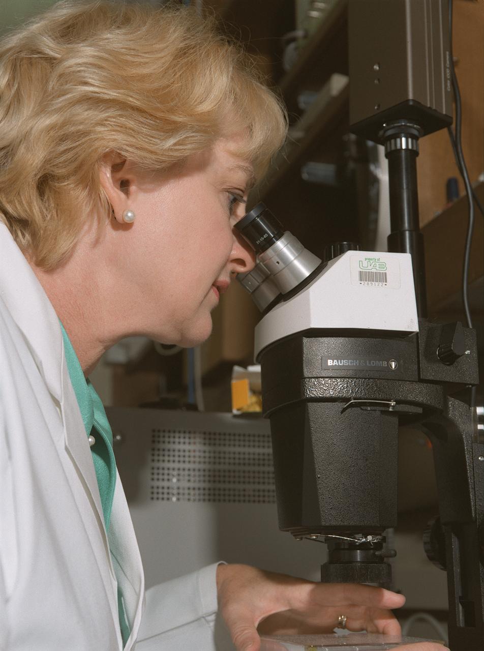
Edward Snell, a National Research Council research fellow at NASA's Marshall Space Flight Center (MSFC), prepares a protein crystal for analysis by x-ray crystallography as part of NASA's structural biology program. The small, individual crystals are bombarded with x-rays to produce diffraction patterns, a map of the intensity of the x-rays as they reflect through the crystal.
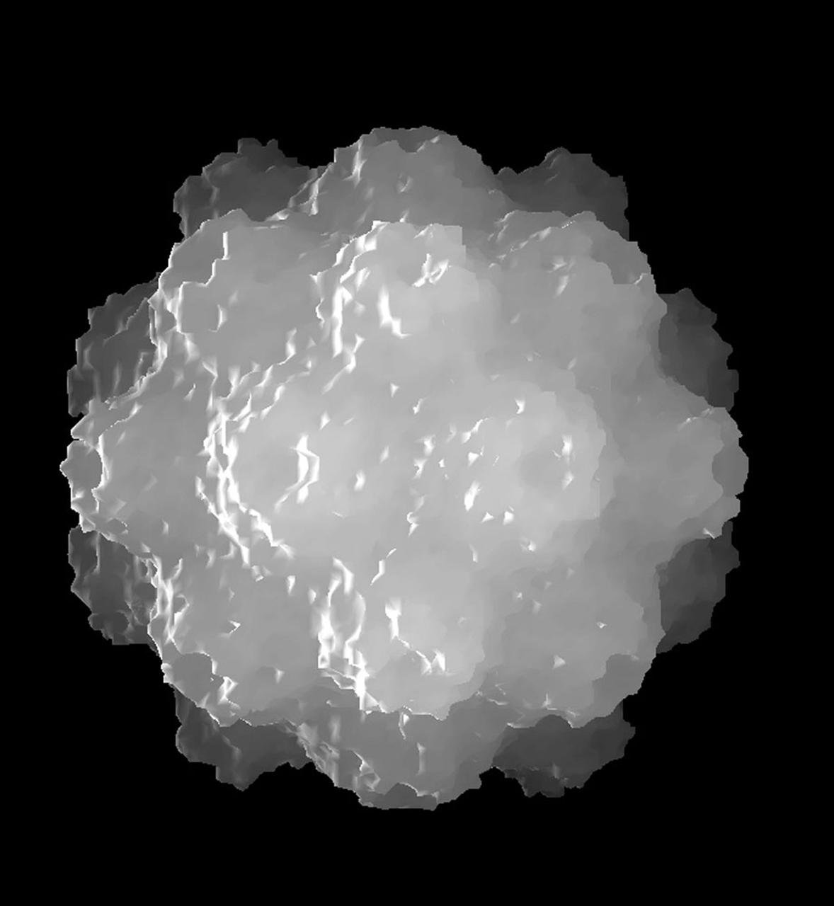
The structure of the Satellite Tobacco Mosaic Virus (STMV)--one of the smallest viruses known--has been successfully deduced using STMV crystals grown aboard the Space Shuttle in 1992 and 1994. The STMV crystals were up to 30 times the volume of any seen in the laboratory. At the same time they gave the best resolution data ever obtained on any virus crystal. STMV is a small icosahedral plant virus, consisting of a protein shell made up of 60 identical protein subunits of molecular weight 17,500. Particularly noteworthy is the fact that, in contrast to the crystal grown on Earth, the crystals grown under microgravity conditions were viusally perfect, with no striations or clumping of crystals. Furthermore, the X-ray diffraction data obtained from the space-grown crystals was of a much higher quality than the best data available at that time from ground-based crystals. This computer model shows the external coating or capsid. STMV is used because it is a simple protein to work with; studies are unrelated to tobacco. Credit: Dr. Alex McPherson, Univeristy of California at Irvin.
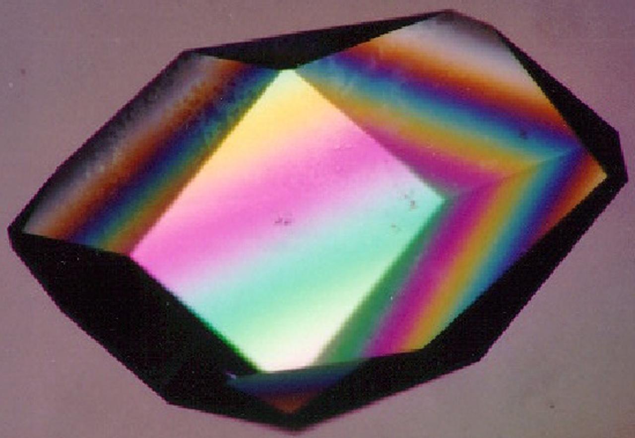
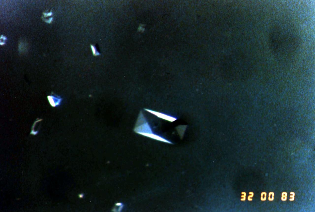
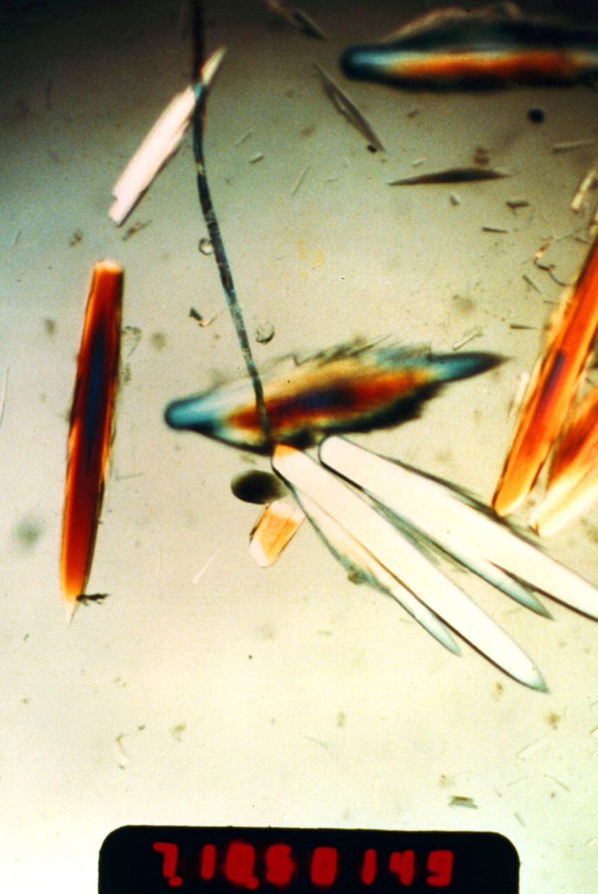
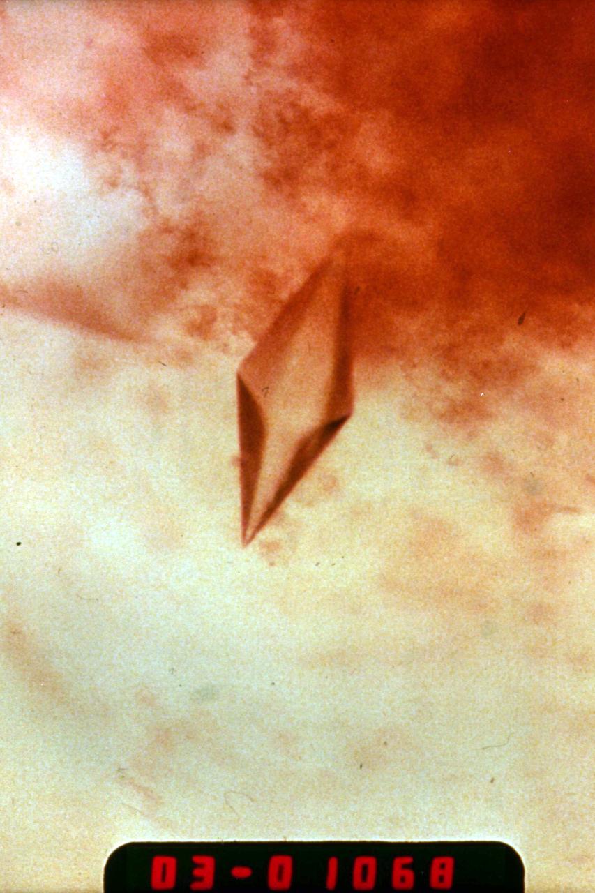
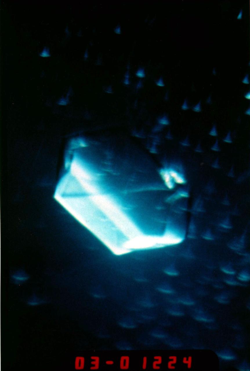
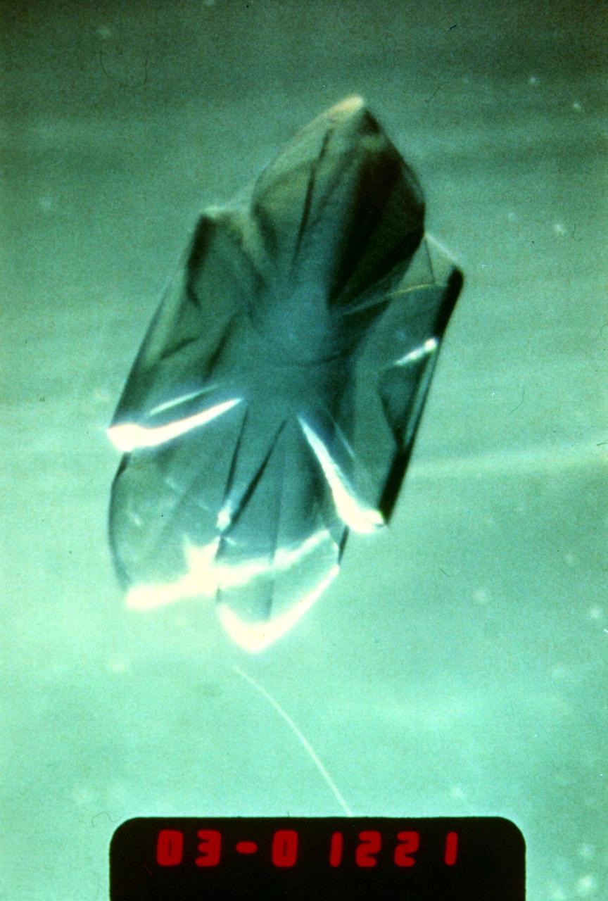
To the crystallographer, this may not be a diamond but it is just as priceless. A Lysozyme crystal grown in orbit looks great under a microscope, but the real test is X-ray crystallography. The colors are caused by polarizing filters. Proteins can form crystals generated by rows and columns of molecules that form up like soldiers on a parade ground. Shining X-rays through a crystal will produce a pattern of dots that can be decoded to reveal the arrangement of the atoms in the molecules making up the crystal. Like the troops in formation, uniformity and order are everything in X-ray crystallography. X-rays have much shorter wavelengths than visible light, so the best looking crystals under the microscope won't necessarily pass muster under the X-rays. In order to have crystals to use for X-ray diffraction studies, crystals need to be fairly large and well ordered. Scientists also need lots of crystals since exposure to air, the process of X-raying them, and other factors destroy them. Growing protein crystals in space has yielded striking results. Lysozyme's structure is well known and it has become a standard in many crystallization studies on Earth and in space.
iss056e075951 (July 3, 2018) --- Astronaut Alexander Gerst of ESA (European Space Agency) works inside the Japanese Kibo laboratory module retrieving Protein Crystal Growth samples from a science freezer, also known as the Minus Eighty-Degree Laboratory Freezer for ISS (MELFI).
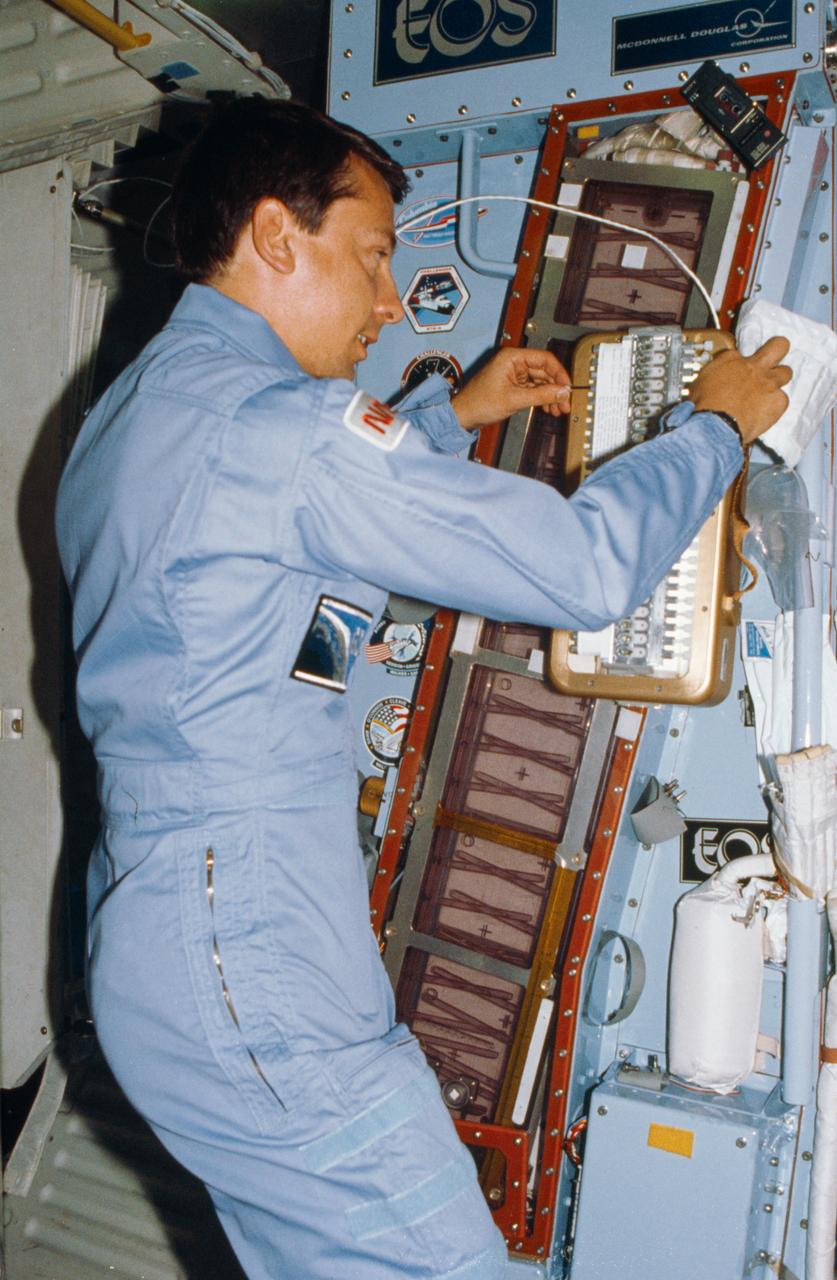
STS072-310-007 (11-20 Jan. 1996) --- Astronauts Brent W. Jett Jr. (left) and Koichi Wakata work with the Protein Crystal Growth (PCG) experiment at the Single Locker Thermal Enclosure System (STES) on the Space Shuttle Endeavour’s mid-deck. Jett, making his first flight in space, served as the crew’s pilot, while Wakata served as a mission specialist. Wakata, also a first time Shuttle crew member, represents Japan’s National Space Development Agency (NASDA).
STS073-351-009 (20 October - 5 November 1995) --- Astronaut Kent V. Rominger, STS-73 pilot, retrieves a protein sample on the middeck of the Earth-orbiting Space Shuttle Columbia. Rominger, along with four other NASA astronauts and two guest researchers, spent 16 full days in space in support of the United States Microgravity Laboratory (USML-2) mission.
During the STS-90 shuttle flight in April 1998, cultured renal cortical cells revealed new information about genes. Timothy Hammond, an investigator in NASA's microgravity biotechnology program was interested in culturing kidney tissue to study the expression of proteins useful in the treatment of kidney diseases. Protein expression is linked to the level of differentiation of the kidney cells, and Hammond had difficulty maintaining differentiated cells in vitro. Intrigued by the improvement in cell differentiation that he observed in rat renal cells cultured in NASA's rotating wall vessel (a bioreactor that simulates some aspects of microgravity) and during an experiment performed on the Russian Space Station Mir, Hammond decided to sleuth out which genes were responsible for controlling differentiation of kidney cells. To do this, he compared the gene activity of human renal cells in a variety of gravitational environments, including the microgravity of the space shuttle and the high-gravity environment of a centrifuge. Hammond found that 1,632 genes out of 10,000 analyzed changed their activity level in microgravity, more than in any of the other environments. These results have important implications for kidney research as well as for understanding the basic mechanism for controlling cell differentiation.
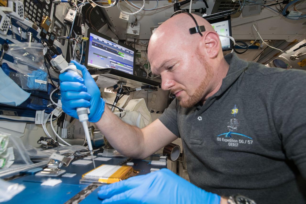
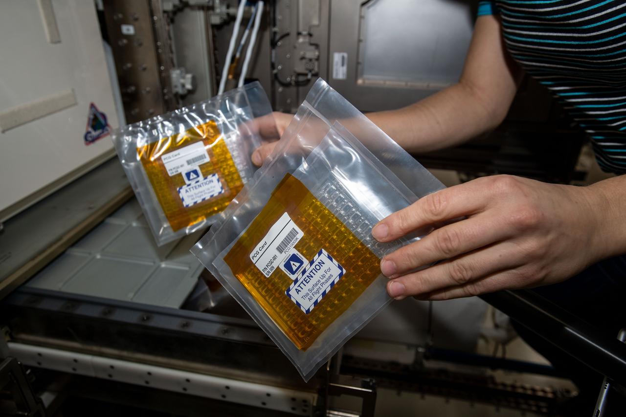
iss057e106232 (Nov. 26, 2018) --- Commander Alexander Gerst uses a uses a pipette to transfer a protein solution into the Protein Crystal Growth Card for an experiment observing protein crystals associated with Parkinson’s disease to potentially improve treatments on Earth.
Scientists at NASA's Marshall Space Flight Center in Huntsville, Alabama, clean equipment and prepare for shipment of the ring sheared drop payload currently set for launch on Northrop Grumman 16 the first week in August, 2021. The payload studies the formation of potentially destructive amyloid fibrils, or protein clusters, like those found in the brain tissue of patients battling neurodegenerative diseases. Such illnesses may damage neurons, the drivers of the human nervous system. Experimentation in microgravity provides the opportunity to study amyloid fibril formation in conditions more analogous to those found in the human body than can be studied in a ground-based laboratory environment.
Scientists at NASA's Marshall Space Flight Center in Huntsville, Alabama, clean equipment and prepare for shipment of the ring sheared drop payload currently set for launch on Northrop Grumman 16 the first week in August, 2021. The payload studies the formation of potentially destructive amyloid fibrils, or protein clusters, like those found in the brain tissue of patients battling neurodegenerative diseases. Such illnesses may damage neurons, the drivers of the human nervous system. Experimentation in microgravity provides the opportunity to study amyloid fibril formation in conditions more analogous to those found in the human body than can be studied in a ground-based laboratory environment.

Scientists at NASA's Marshall Space Flight Center in Huntsville, Alabama, clean equipment and prepare for shipment of the ring sheared drop payload currently set for launch on Northrop Grumman 16 the first week in August, 2021. The payload studies the formation of potentially destructive amyloid fibrils, or protein clusters, like those found in the brain tissue of patients battling neurodegenerative diseases. Such illnesses may damage neurons, the drivers of the human nervous system. Experimentation in microgravity provides the opportunity to study amyloid fibril formation in conditions more analogous to those found in the human body than can be studied in a ground-based laboratory environment.
Crystals grown in the hand-held Protein Crystallization Apparatus for Microgravity (PCAM) onboard STS-61C. The PCAM has a pedestal in the center of a circular chamber, the surrounding chamber holds an absorbent reservoir that contains a solution of the precipitant. Vapor pressure differences between the protein solution and the reservoir solution force water to move from the protein solution to the reservoir. As protein concentrations increase, protein crystals begin to nucleate and grow.
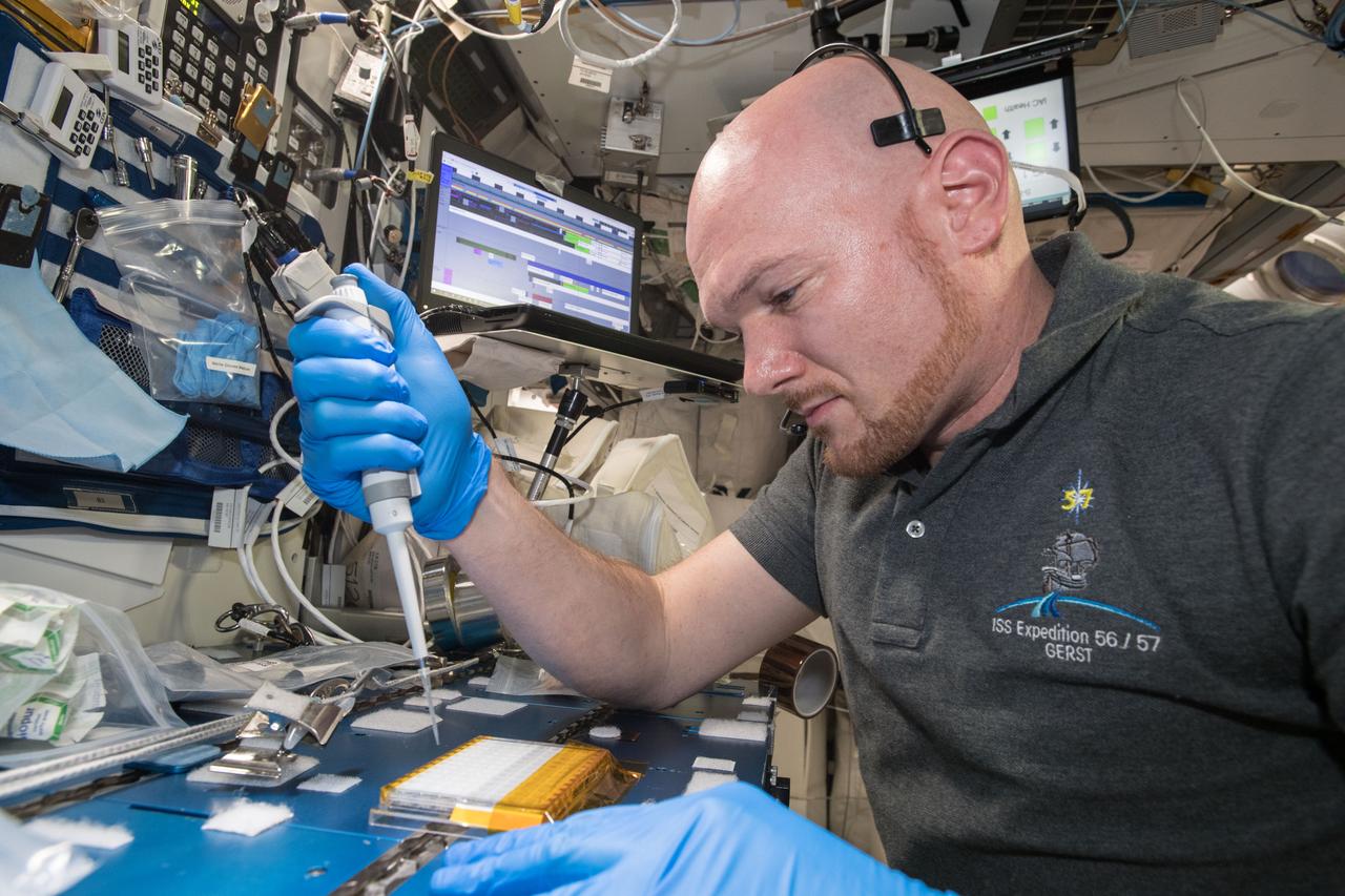
iss057e106231 (Nov. 26, 2018) --- European Space Agency (ESA) asrtonaut Alexander Gerst uses a uses a pipette to transfer a protein solution into the Protein Crystal Growth Card for an experiment observing protein crystals associated with Parkinson’s disease to potentially improve treatments on Earth. Crystallization of LRRK2 Under Microgravity Conditions-2 (CASIS PCG 16) evaluates growth of Leucine-rich repeat kinase 2 (LRRK2) protein crystals in microgravity. LRRK2 is implicated in Parkinson’s disease, but crystals of the protein grown on Earth are too small and compact to study. Detailed analysis of larger, space-grown crystals can define the protein’s exact shape and morphology and help scientists better understand the disease’s pathology.
DCAM, developed by MSFC, grows crystals by the dialysis and liquid-liquid diffusion methods. In both methods, protein crystal growth is induced by changing conditions in the protein. In dialysis, a semipermeable membrane retains the protein solution in one compartment, while allowing molecules of precipitant to pass freely through the membrane from an adjacent compartment. As the precipitant concentration increases within the protein compartment, crystallization begins. In liquid-liquid diffusion, a protein solution and a precipitant solution are layered in a container and allowed to diffuse into each other. This leads to conditions which may induce crystallization of the protein. Liquid-liquid diffusion is difficult on Earth because density and temperature differences cause the solutions to mix rapidly.
iss073e0071617 (May 16, 2025) --- NASA astronaut and Expedition 73 Flight Engineer Jonny Kim removes a cryogenic storage unit, called a dewar, containing frozen protein crystal samples from a science freezer located inside International Space Station's Kibo laboratory module. The research activities were part of a technology demonstration potentially enabling the synthesis of medications during deep space missions and improving the pharmaceutical industry on Earth.
iss073e0071610 (May 16, 2025) --- NASA astronaut and Expedition 73 Flight Engineer Jonny Kim removes a cryogenic storage unit, called a dewar, containing frozen protein crystal samples from a science freezer located inside International Space Station's Kibo laboratory module. The research activities were part of a technology demonstration potentially enabling the synthesis of medications during deep space missions and improving the pharmaceutical industry on Earth.
iss073e0071611 (May 16, 2025) --- NASA astronaut and Expedition 73 Flight Engineer Jonny Kim removes a cryogenic storage unit, called a dewar, containing frozen protein crystal samples from a science freezer located inside International Space Station's Kibo laboratory module. The research activities were part of a technology demonstration potentially enabling the synthesis of medications during deep space missions and improving the pharmaceutical industry on Earth.
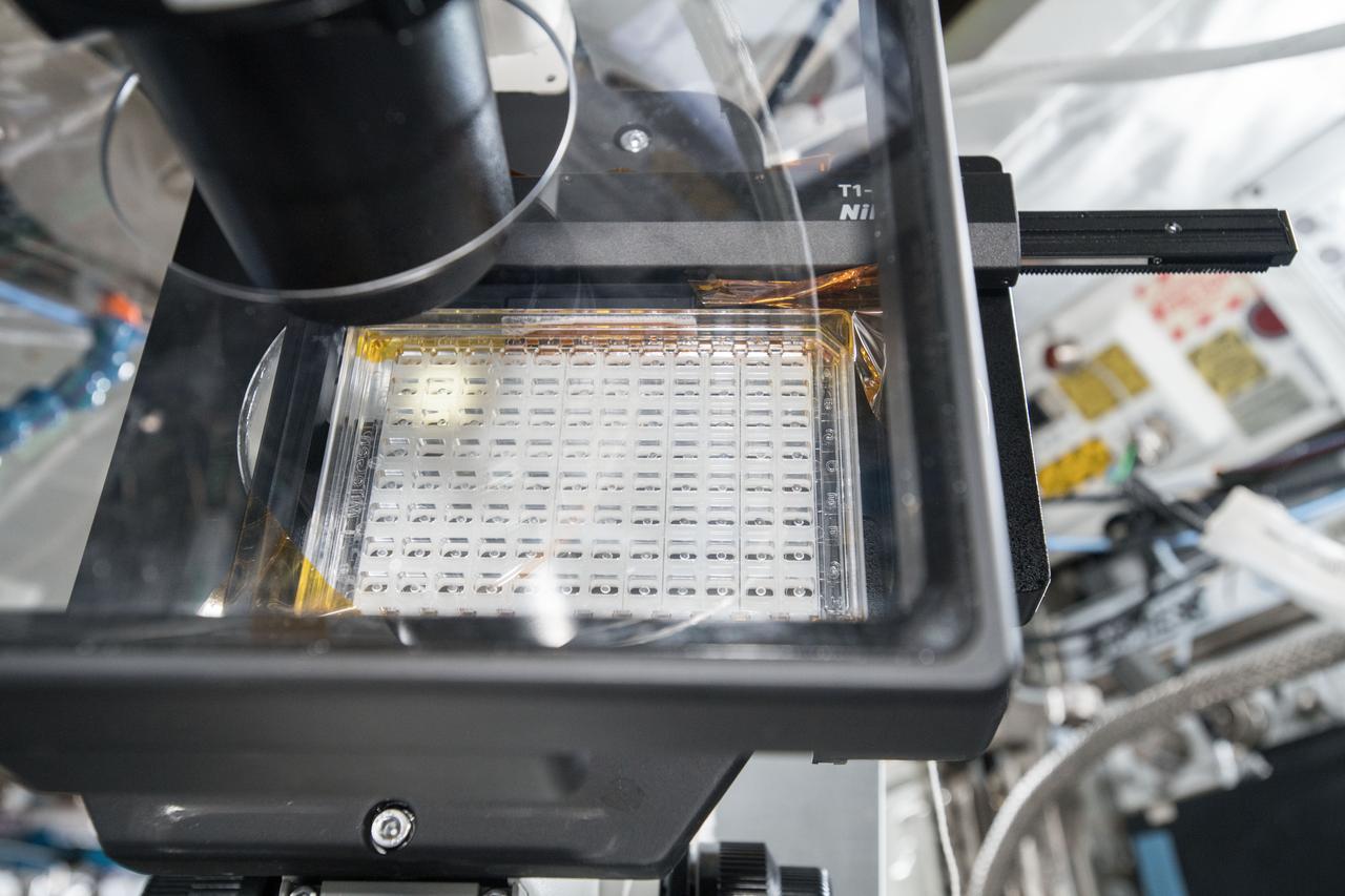
iss064e039017 (March 2, 2021) --- NASA astronaut Michael Hopkins loads protein crystallography plates with protein solutions for the Phase II Real-time Protein Crystal Growth experiment, a space commercialization study, that could benefit the pharmaceutical and biotechnology industries.
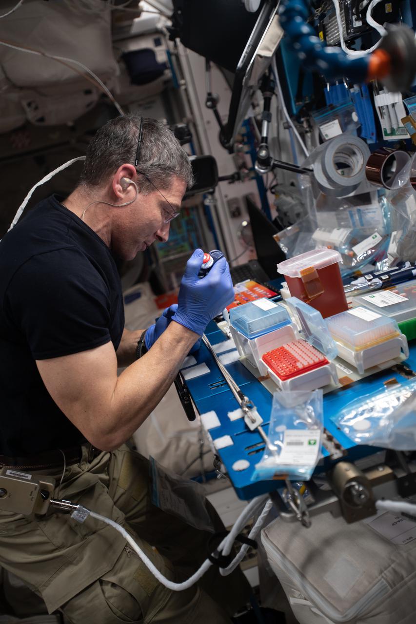
iss064e039273 (March 2, 2021) --- NASA astronaut and Expedition 64 Flight Engineer Michael Hopkins loads protein crystallography plates with protein solutions for the Phase II Real-time Protein Crystal Growth experiment, a space commercialization study, that could benefit the pharmaceutical and biotechnology industries.

iss064e038995 (March 2, 2021) --- NASA astronaut and Expedition 64 Flight Engineer Michael Hopkins loads protein crystallography plates with protein solutions for the Phase II Real-time Protein Crystal Growth experiment, a space commercialization study, that could benefit the pharmaceutical and biotechnology industries.
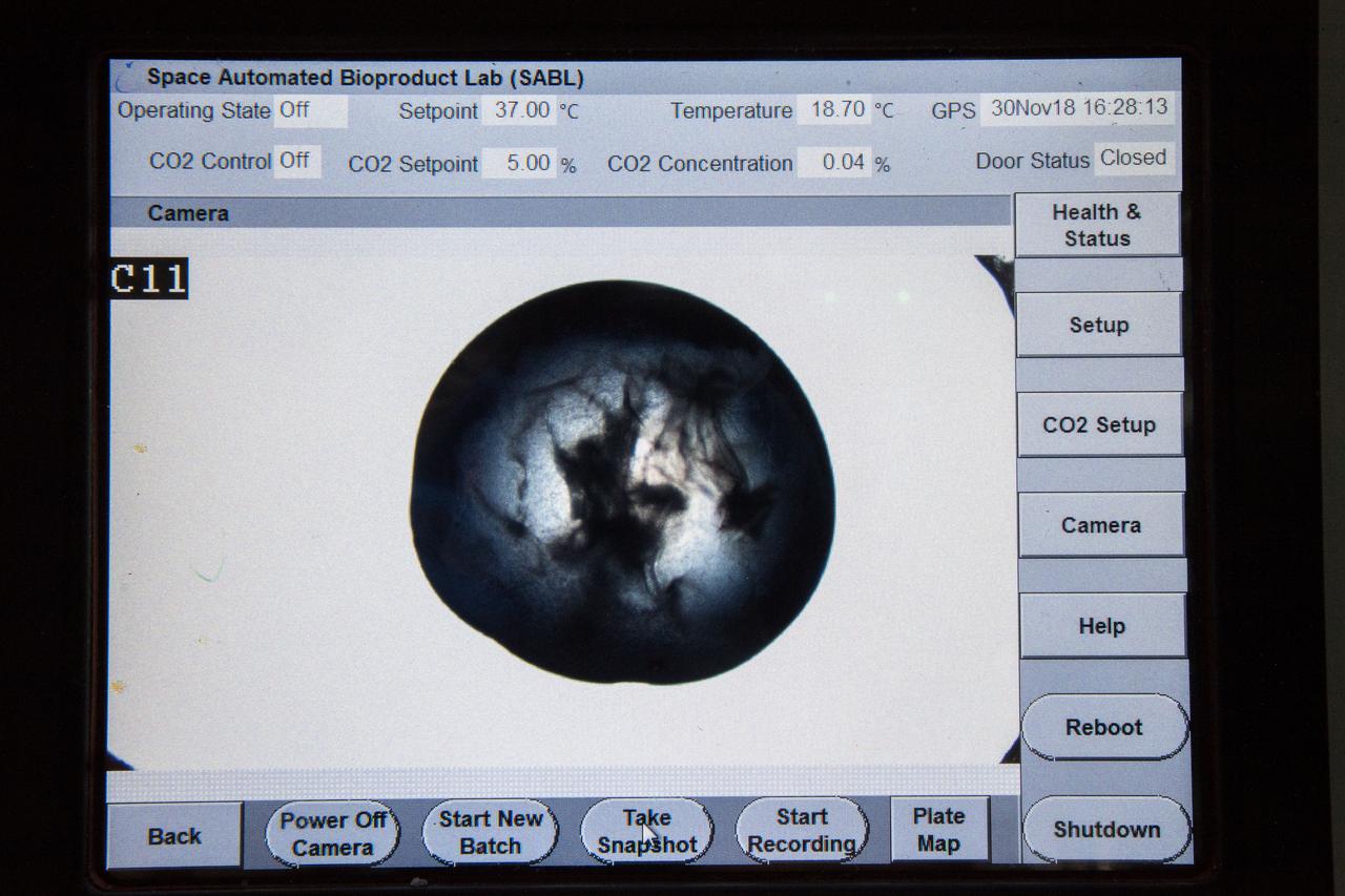
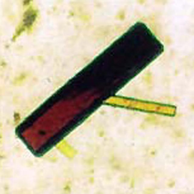
iss057e106417 (Nov. 30, 2018) --- Samples from the Protein Crystal Growth Card are examined using a microscope for an experiment observing protein crystals associated with Parkinson’s disease to potentially improve treatments on Earth. Crystallization of LRRK2 Under Microgravity Conditions-2 (CASIS PCG 16) evaluates growth of Leucine-rich repeat kinase 2 (LRRK2) protein crystals in microgravity. LRRK2 is implicated in Parkinson’s disease, but crystals of the protein grown on Earth are too small and compact to study. Detailed analysis of larger, space-grown crystals can define the protein’s exact shape and morphology and help scientists better understand the disease’s pathology.
iss057e106419 (Nov. 30, 2018) --- Samples from the Protein Crystal Growth Card are examined using a microscope for an experiment observing protein crystals associated with Parkinson’s disease to potentially improve treatments on Earth. Crystallization of LRRK2 Under Microgravity Conditions-2 (CASIS PCG 16) evaluates growth of Leucine-rich repeat kinase 2 (LRRK2) protein crystals in microgravity. LRRK2 is implicated in Parkinson’s disease, but crystals of the protein grown on Earth are too small and compact to study. Detailed analysis of larger, space-grown crystals can define the protein’s exact shape and morphology and help scientists better understand the disease’s pathology.
Along with hemoglobin the primary oxygen storage and transport proteins in all higher animals including humans. Important for medical reasons because they are primary blood proteins.
iss057e114766 (12/9/2018) --- European Space Agency (ESA) astronaut Alex Gerst is photographed with the Crystallization of RAS in Space (CASIS PCG 17) investigation. CASIS PCG 17grows crystals of KRAS proteins, which have a pivotal role in cell growth and death. Mutations in KRAS proteins are responsible for a third of all cancers and identifying the structure of these proteins is critical to developing therapeutics and treatments. Protein crystals grow larger and more perfectly in microgravity, allowing for detailed laboratory analysis of their structure back on Earth.
jsc2021e007777 - Aeropyrum pernix Flap Endonuclease-1 (FEN-1) protein crystals are shown grown under Earth gravity conditions. FEN-1 serves as the experimental protein for the Phase II Real-time Protein Crystal Growth on Board the International Space Station (Real-Time Protein Crystal Growth-2) investigation. Image courtesy of University of Toledo.
iss057e114765 (12/9/2018) --- European Space Agency (ESA) astronaut Alex Gerst is photographed with the Crystallization of RAS in Space (CASIS PCG 17) investigation. CASIS PCG 17grows crystals of KRAS proteins, which have a pivotal role in cell growth and death. Mutations in KRAS proteins are responsible for a third of all cancers and identifying the structure of these proteins is critical to developing therapeutics and treatments. Protein crystals grow larger and more perfectly in microgravity, allowing for detailed laboratory analysis of their structure back on Earth.
iss065e144296 (June 14, 2021) --- NASA astronaut and Expedition 65 Flight Engineer Megan McArthur loads protein crystallography plates with protein solutions, mixes them with custom salt solutions, then seals and transfers the plates for incubation for the Real-Time Protein Crystal Growth-2 experiment. The biotechnology study looks at new ways to produce high-quality protein crystals which could lead to new disease therapies on Earth.
iss065e085491 (June 3, 2021) --- NASA astronaut and Expedition 65 Flight Engineer Megan McArthur loads protein crystallography plates with protein solutions for the Real-time Protein Crystal Growth experiment. The biotechnology study demonstrates new methods for producing high-quality protein crystals in microgravity. Results may help identify new targets and develop better drugs to treat a variety of diseases on Earth and advance the commercialization of low-Earth orbit.
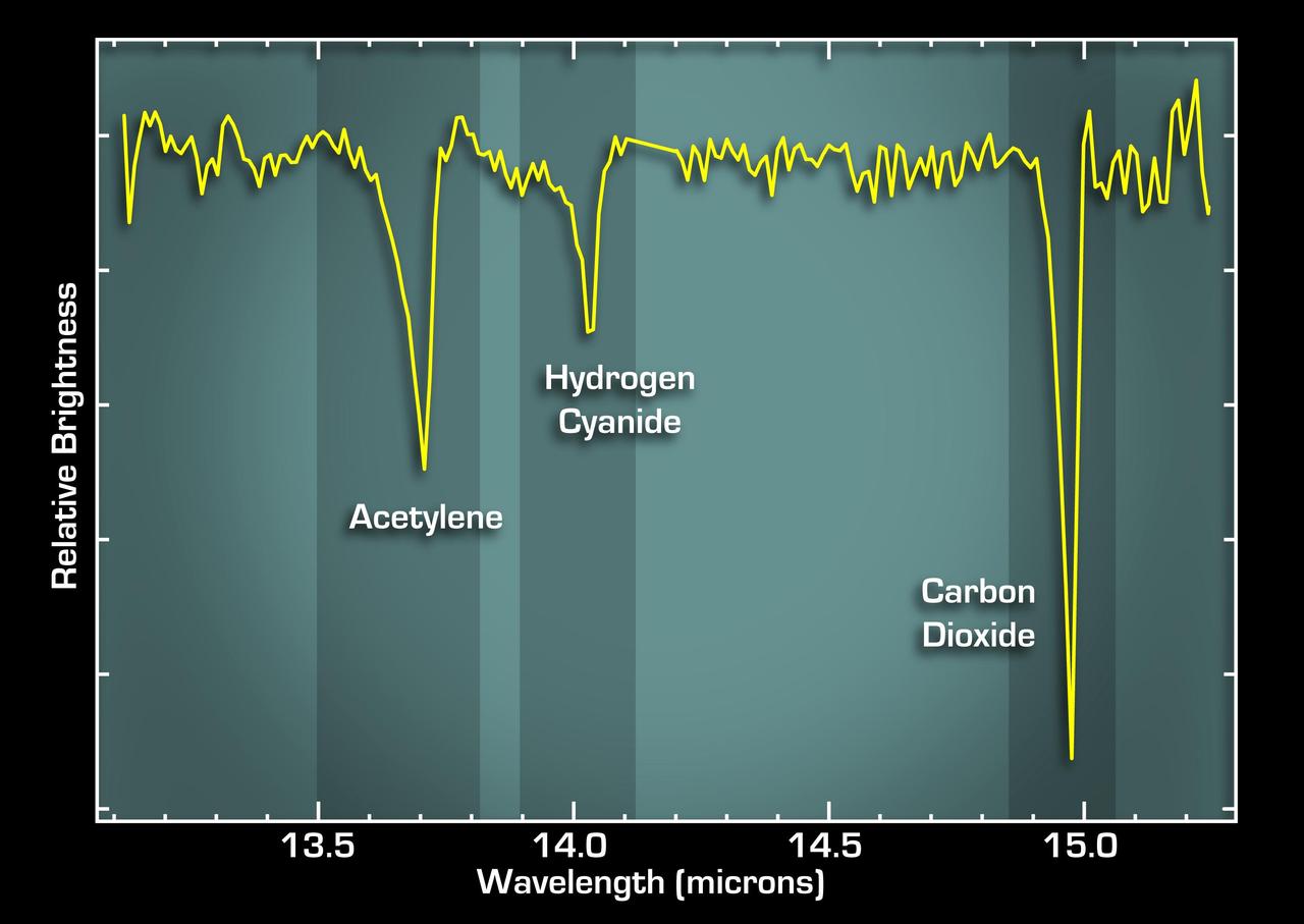
This graph, or spectrum, from NASA Spitzer Space Telescope tells astronomers that some of the most basic ingredients of DNA and protein are concentrated in a dusty planet-forming disk circling a young sun-like star called IRS 46.
iss058e001945 (Jan. 3, 2019) --- Expedition 58 Flight Engineer and astronaut Anne McClain of NASA peers into a microscope and takes photographs for the Protein Crystal Growth-16 experiment that is exploring therapies for Parkinson's disease.
iss058e001880 (Jan. 2, 2019) --- NASA astronaut and Expedition 58 Flight Engineer Anne McClain works inside the Unity module conducting research operations for the Protein Crystal Experiment-16 that is exploring therapies for Parkinson's disease.
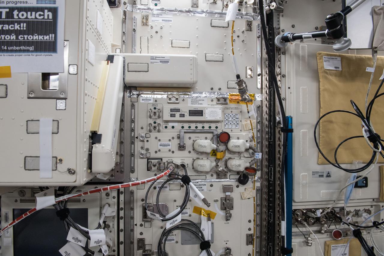
iss055e004890 (3/24/2018) --- Photographic documentation taken during JAXA Protein Crystal Growth (PCG) Installation into the Protein Crystallization Research Facility (PCRF) of the Ryutai Rack.

Rosetta Stone Protein Model

Rosetta Stone Protein Model
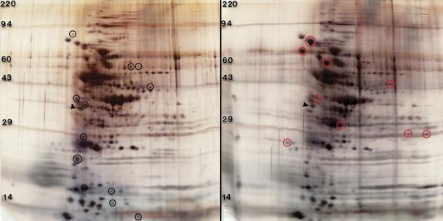
These gels were obtained by two-dimensional (2D) electrophoresis, in which proteins move different substances through a polyacrylamide gel matrix based on their molecular weight and total charge in an electric field. The gels illustrate principal investigator David Niesel’s findings that exposure to modeled microgravity results in some Streptoccoccus Pneumonia’s proteins being upregulated and others being downregulated. In 2D protein profiles of whole cell lysates of Streptoccoccus Pneumonia, 6,304 cultured under normal gravity (left), appear to be expressed at higher levels indicated with black circles. Red circles (right) indicate proteins that were grown under modeled microgravity in a high aspect ratio vessel HARV).
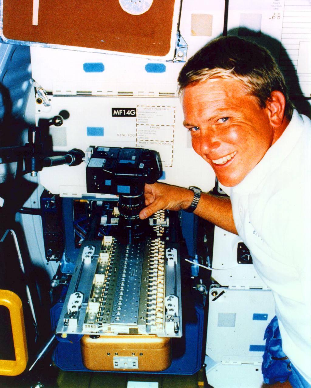
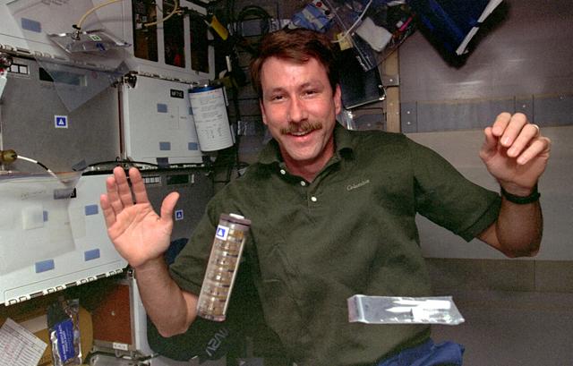
Mission Specialist George (Pinky) D. Nelson uses a 35 mm camera to photograph a protein crystal grown during the STS-26 Protein Crystal Growth (PCG-II-01) experiment. The protein crystal growth (PCG) carrier is shown deployed from the PCG Refrigerator/Incubator Mocule (R/IM) located in the middeck forward locker. The R/IM contained three Vapor Diffusion Apparatus (VDS) trays (one of which is shown). A total of sixty protein crystal samples were processed during the STS-26 mission.
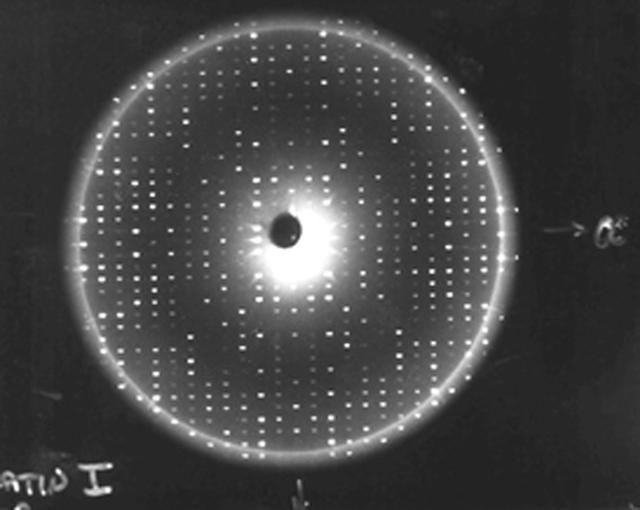
X-rays diffracted from a well-ordered protein crystal create sharp patterns of scattered light on film. A computer can use these patterns to generate a model of a protein molecule. To analyze the selected crystal, an X-ray crystallographer shines X-rays through the crystal. Unlike a single dental X-ray, which produces a shadow image of a tooth, these X-rays have to be taken many times from different angles to produce a pattern from the scattered light, a map of the intensity of the X-rays after they diffract through the crystal. The X-rays bounce off the electron clouds that form the outer structure of each atom. A flawed crystal will yield a blurry pattern; a well-ordered protein crystal yields a series of sharp diffraction patterns. From these patterns, researchers build an electron density map. With powerful computers and a lot of calculations, scientists can use the electron density patterns to determine the structure of the protein and make a computer-generated model of the structure. The models let researchers improve their understanding of how the protein functions. They also allow scientists to look for receptor sites and active areas that control a protein's function and role in the progress of diseases. From there, pharmaceutical researchers can design molecules that fit the active site, much like a key and lock, so that the protein is locked without affecting the rest of the body. This is called structure-based drug design.
Proteins are the building blocks of our bodies and the living world around us. Within our bodies proteins make it possible for red blood cells to carry oxygen throughout the body. Others help transmit nerve impulses so we can hear, smell and feel the world around us. While others play a crucial role in preventing or causing disease. If the structure of a protein is known, then companies can develop new or improved drugs to fight the disease of which the protein is a part. To determine protein structure, researchers must grow near-perfect crystals of the protein. On Earth convection currents, sedimentation and other gravity-induced phenomena hamper crystal growth efforts. In microgravity researchers can grow near-perfect crystals in an environment free of these effects. Because of the enormous potential for new pharmaceutical products the Center for Macromolecular Crystallography--the NASA Commercial Space Center responsible for commercial protein crystal growth efforts has more than fifty major industry and academic partners. Research on crystals of human insulin could lead to improved treatments for diabetes.
Johnathan Trent looking at Rosetta Stone Protein Model
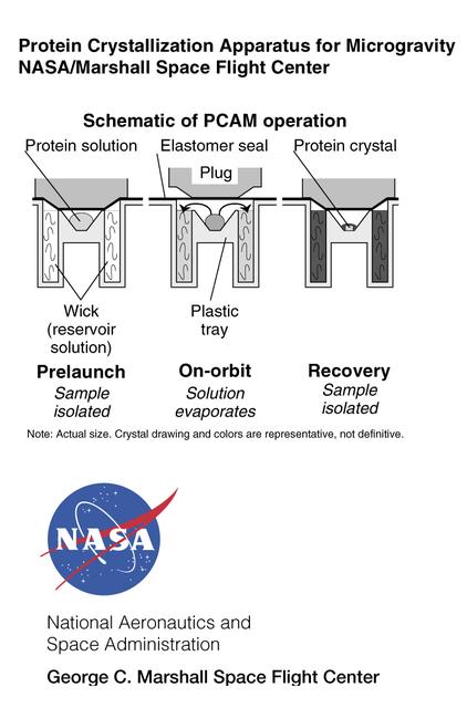
The Protein Crystallization for Microgravity (DCAM) was developed at NASA's Marshall Space Flight Center. A droplet of solution with protein molecules dissolved in it is isolated in the center of a small well. In orbit, an elastomer seal is lifted so the solution can evaporate and be absorbed by a wick material. This raises the concentration of the solution, thus prompting protein molecules in the solution to form crystals. The principal investigator is Dr. Dan Carter of New Century Pharmaceuticals in Huntsville, AL.
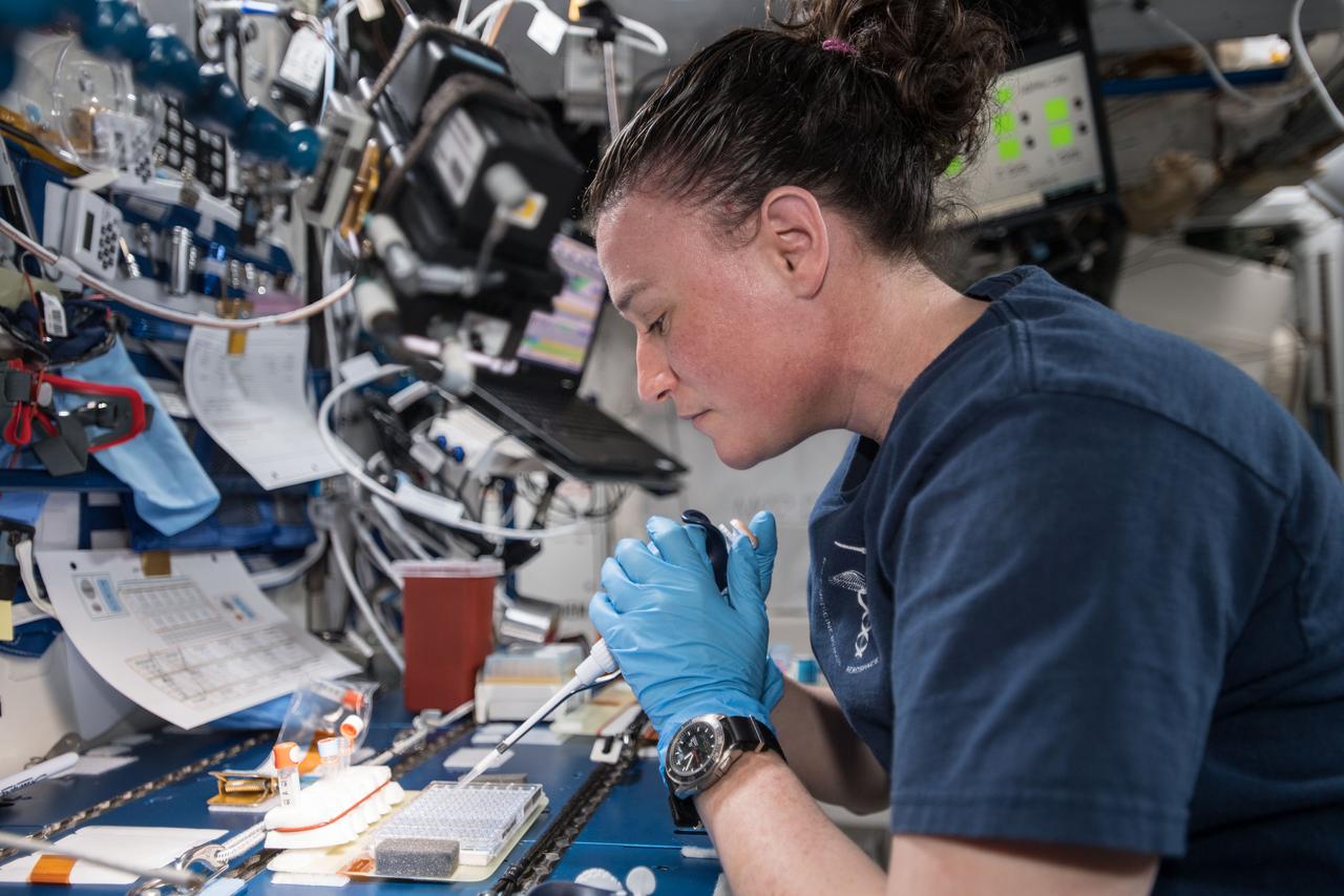
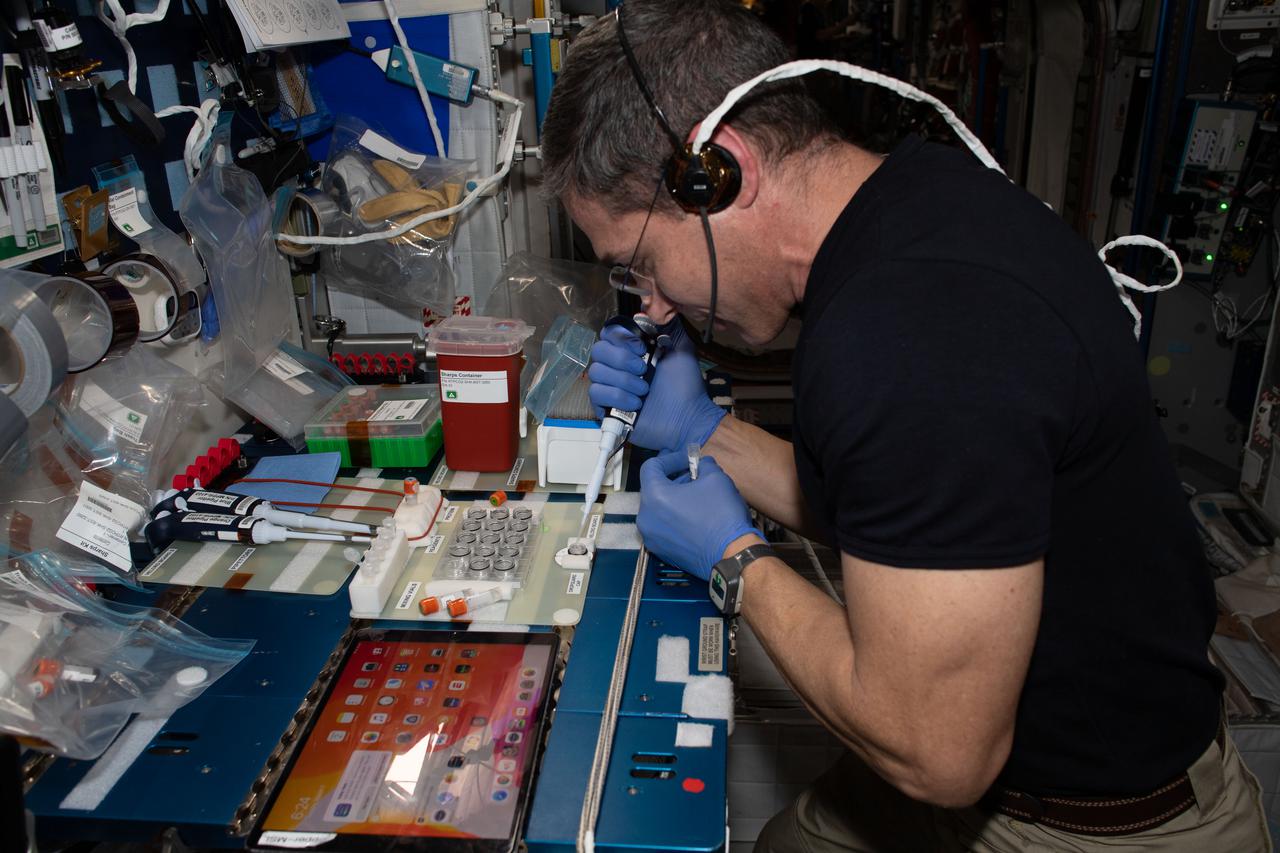
iss057e074528 (Nov. 9, 2018) --- NASA astronaut Serena Auñón-Chancellor is pictured in the Japanese Kibo lab module mixing protein crystal samples to help scientists understand how they work. BioServe Protein Crystalography-1 (BPC-1) seeks to demonstrate the feasibility of conducting protein crystal growth in real time aboard the International Space Station. Crew members add solutions to the hardware, observe the crystals that form and adjust for follow-on experiments.