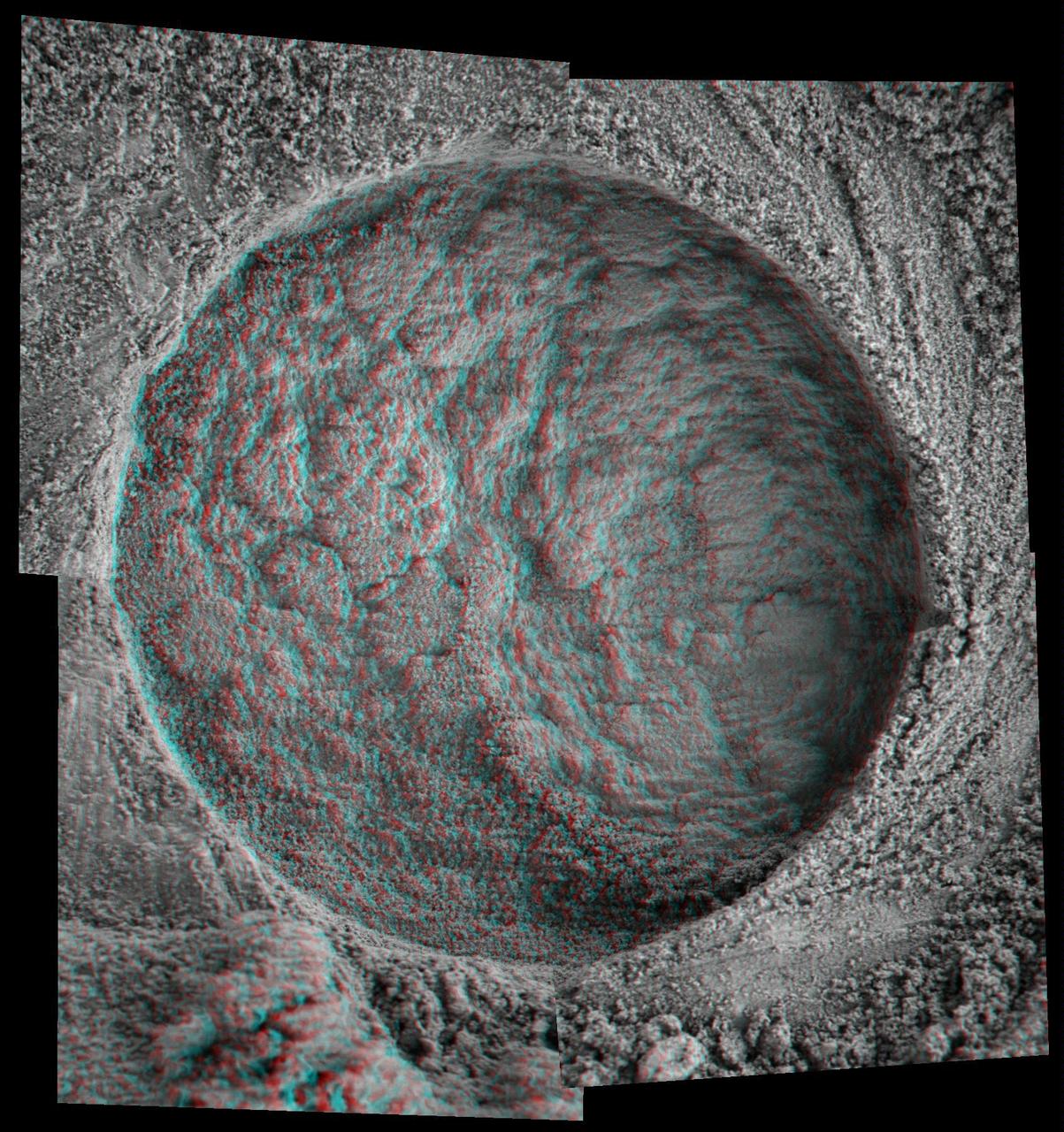
The Biggest Microscopic Image Ever
Microscopic Image Inside Endurance
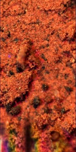
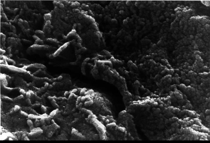
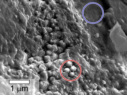
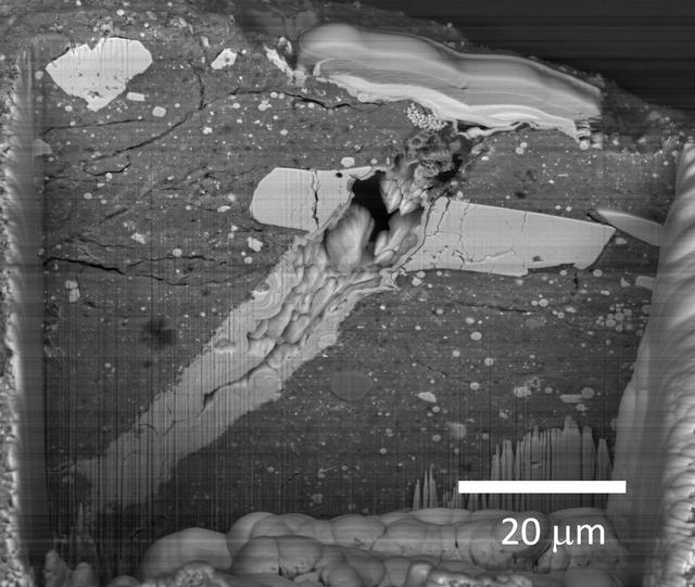
Microscope Image of Scavenged Particles
Microscopic Image of Martian Surface Material on a Silicone Substrate
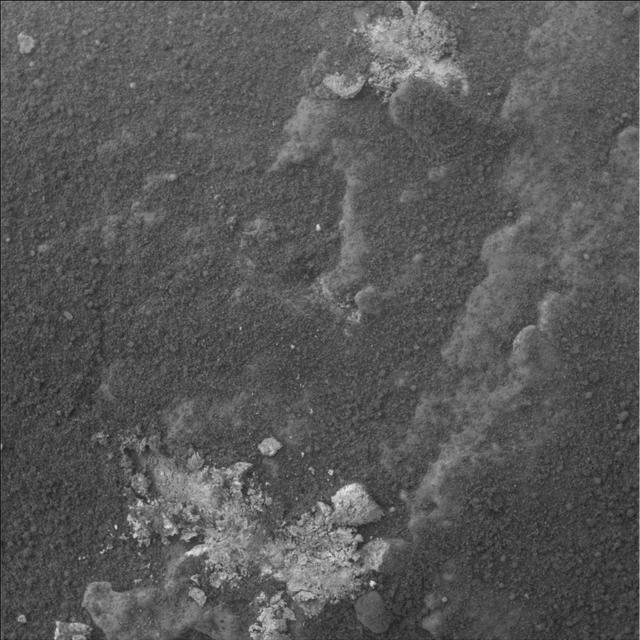
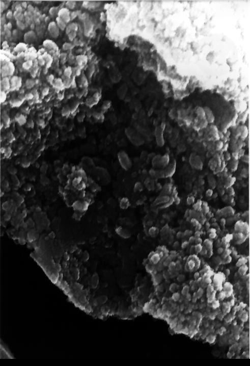
Microscope Image of a Martian Soil Surface Sample
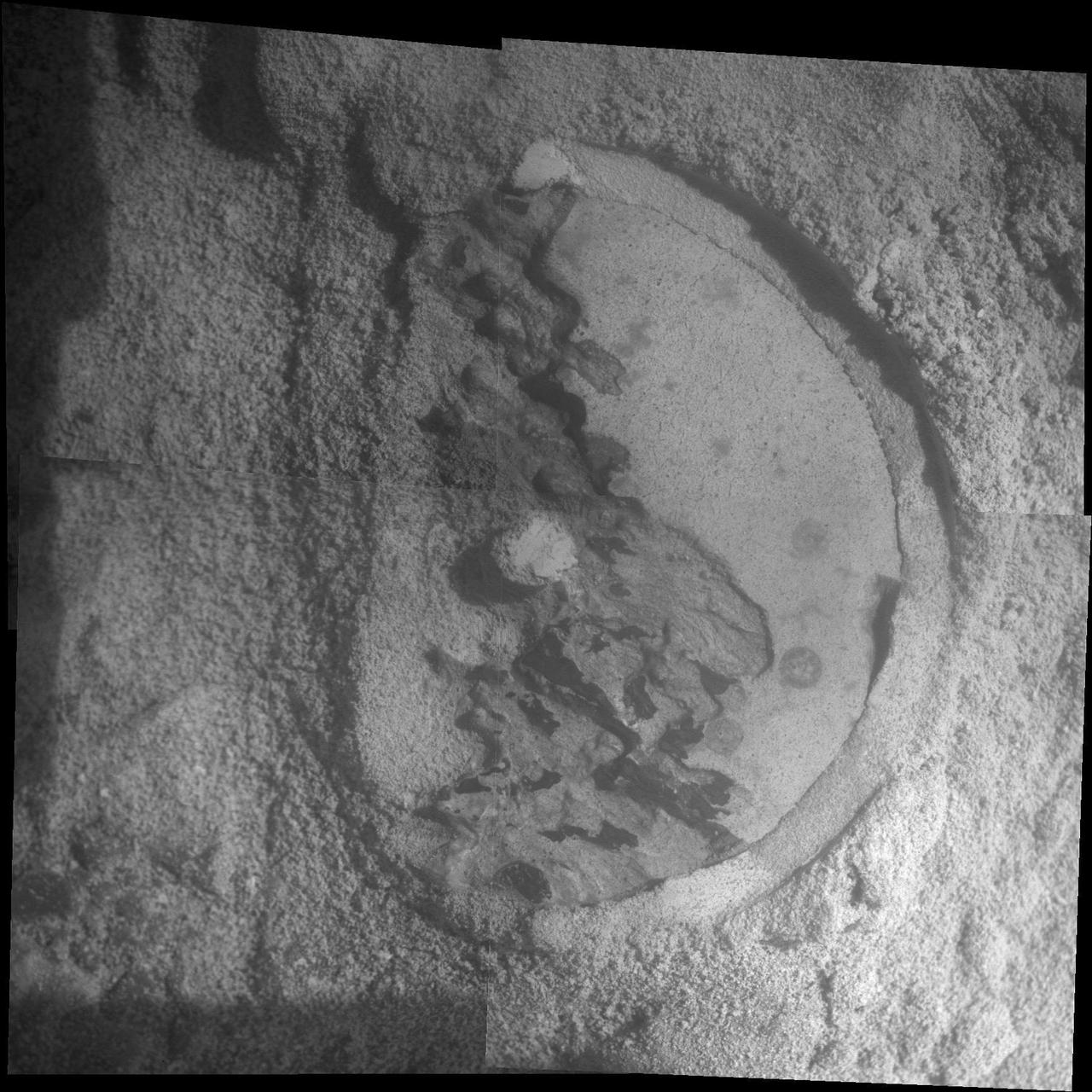
Spirit Examines Light-Toned Halley Microscopic Image
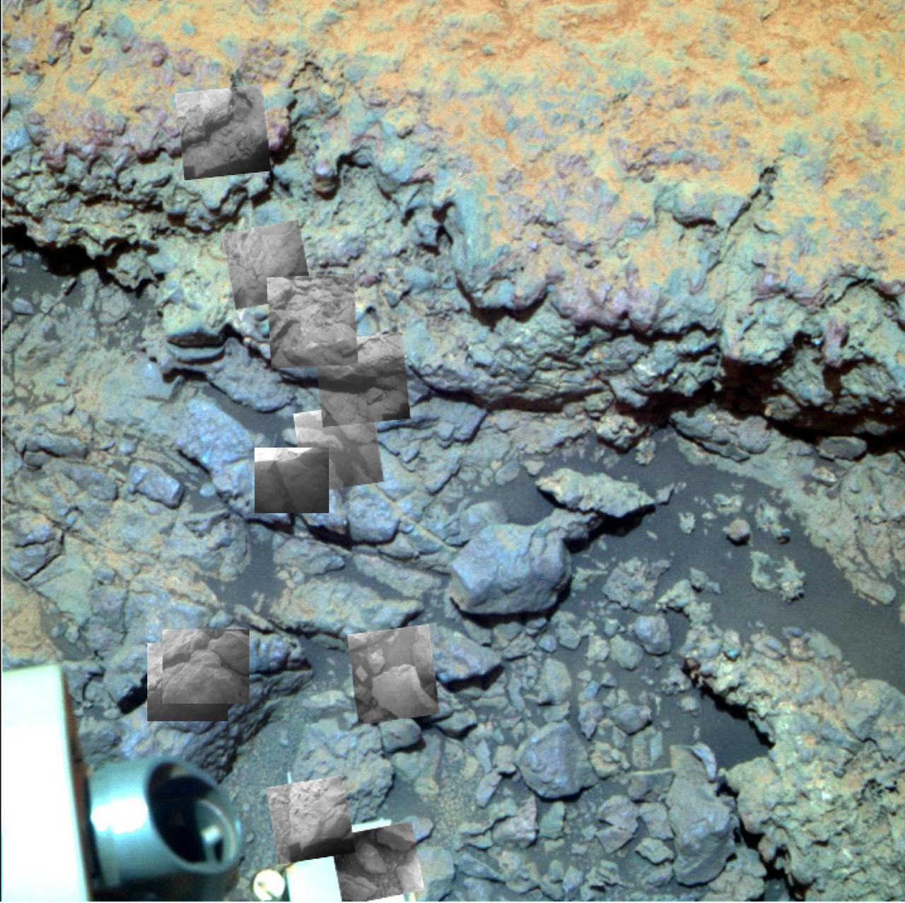
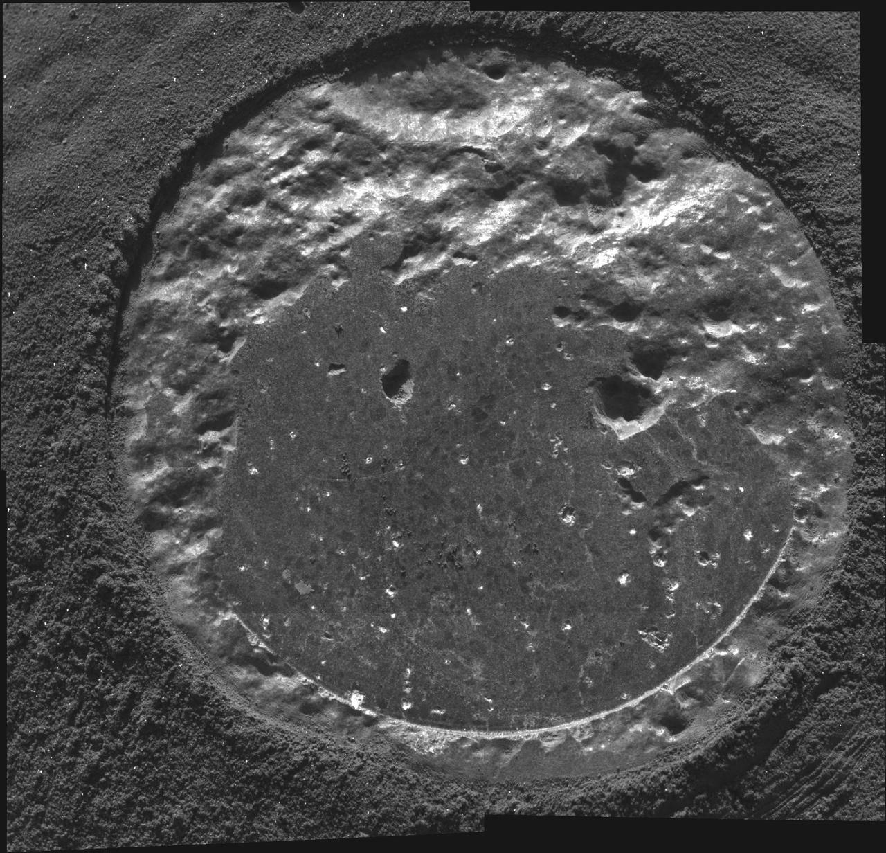
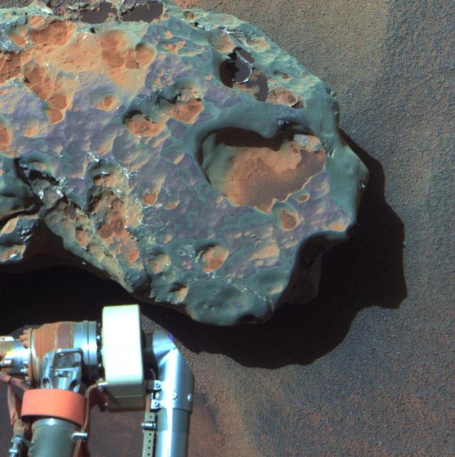
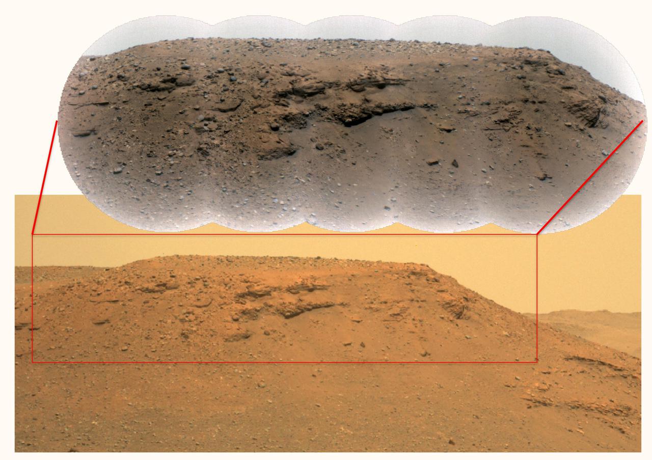
This image taken by the NASA Mars rover Opportunity shows locations of the microscopic imager observations on a rock informally named Tisdale 2.
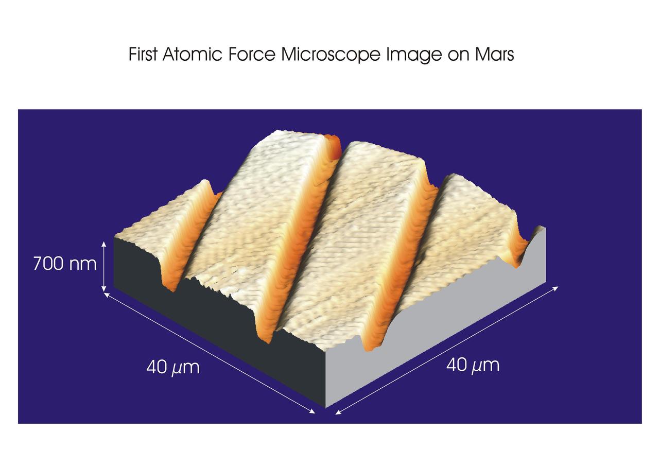
This calibration image presents three-dimensional data from the atomic force microscope on NASA Phoenix Mars Lander, showing surface details of a substrate on the microscope station sample wheel.
This image of a target called "Private Joseph Field" combines four images from the microscopic imager on the robotic arm of NASA's Mars Exploration Rover Opportunity, with enhanced color information added from the rover's panoramic camera. This target is within the "Marathon Valley" area of the western rim of Endeavour Crater. The component images were taken on May 29, 2016, during the 4,389th Martian day, or sol, of Opportunity's work on Mars. The mosaic shows an area spanning about 2 inches (5 centimeters). Geochemical data indicate the presence of magnesium and iron sulfates at this location, most likely corresponding to the white pebble visible near the center of the image. These sulfates may have formed by the interaction of acidic fluids with the rocks along the rim of Endeavour crater. http://photojournal.jpl.nasa.gov/catalog/PIA21142

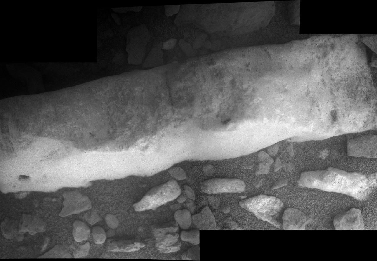
This relatively bright outcropping of rock, dubbed "Gasconade," was investigated by NASA's Mars Exploration Rover Opportunity while the rover was perched on "Spirit Mound" at the western edge of Mars' Endeavour Crater. This mosaic combines four frames taken by the microscopic imager on Opportunity's robotic arm on Oct. 2, 2016, during the 4,512st Martian day, or sol, of the rover's work on Mars. Enhanced color information from Opportunity's panoramic camera has been added to emphasize differences in the materials visible in the target. Figure A is a version with no color information added to the microscopic imager mosaic. The view covers an area about 2 inches (5 centimeters) wide. Opportunity's inspection found Gasconade to be a wind-etched outcrop with angular bits of darker rock within a lighter matrix, which may have been formed from fallout of the impact event that excavated the crater. This location of Spirit Mound, shown at PIA20854, is the deeper on the western rim of Endeavour Crater than any site visited previously by Opportunity. http://photojournal.jpl.nasa.gov/catalog/PIA21141
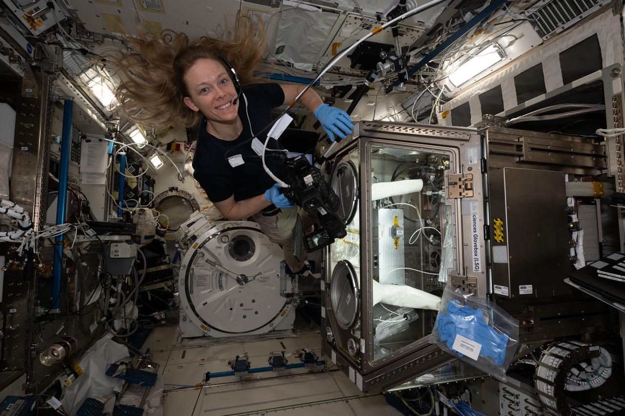
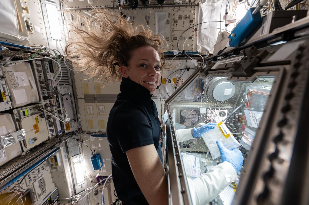
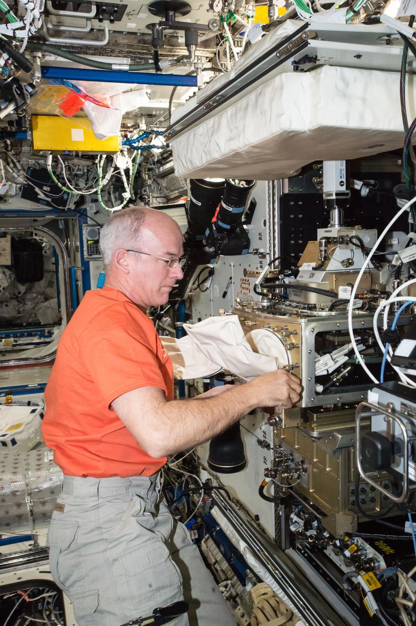
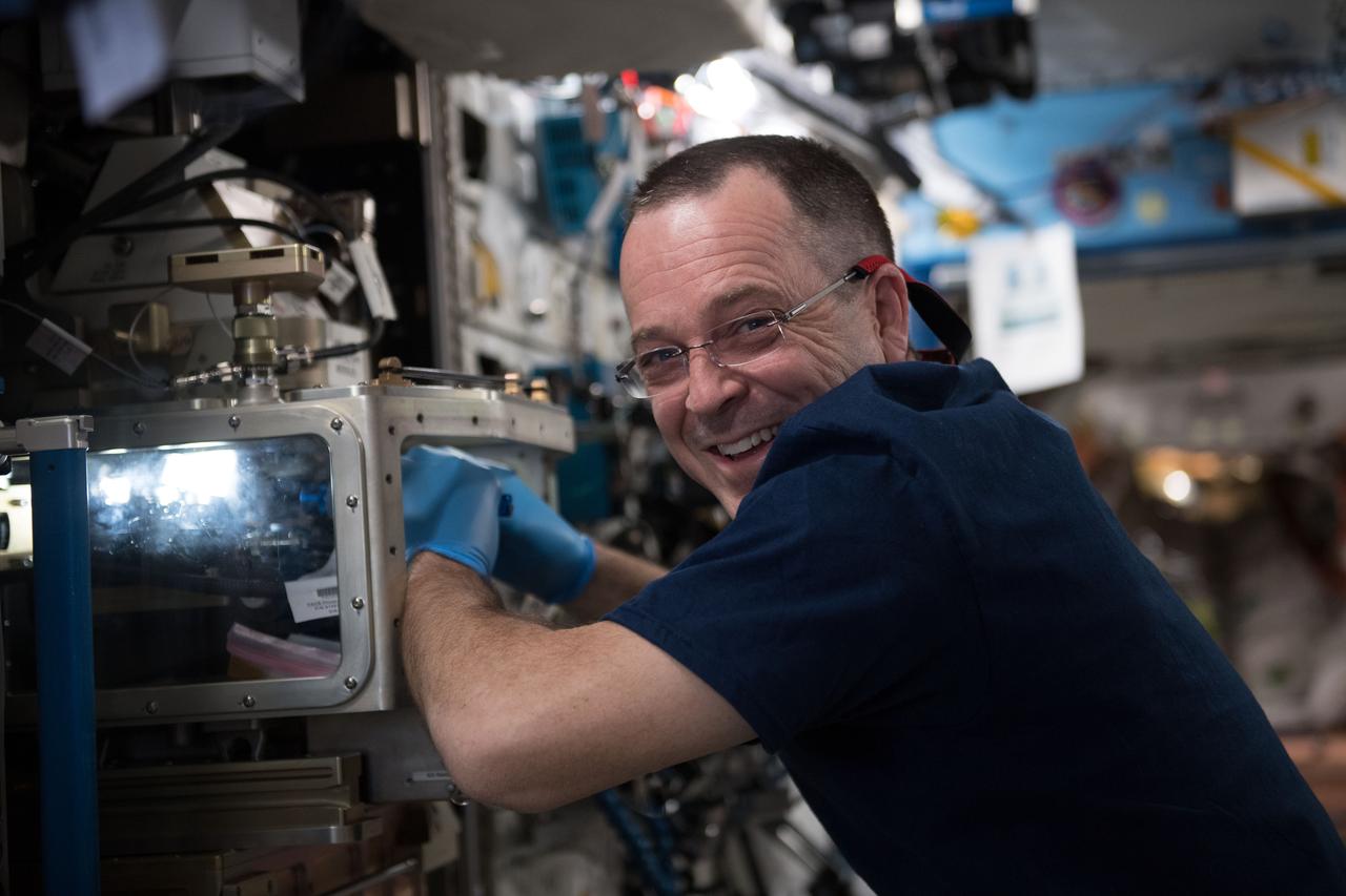
iss073e0134904 (June 5, 2025) --- NASA astronaut and Expedition 73 Flight Engineer Nichole Ayers works inside the Kibo laboratory module to test imaging operations of a 3D research microscope, also known as the Extant Life Volumetric Imaging System, or ELVIS. The specialized 3D imaging device, located in Kibo's Life Science Glovebox, could be used to monitor water quality, detect potentially infectious organisms, and study liquid mixtures and microorganisms in space and on Earth.
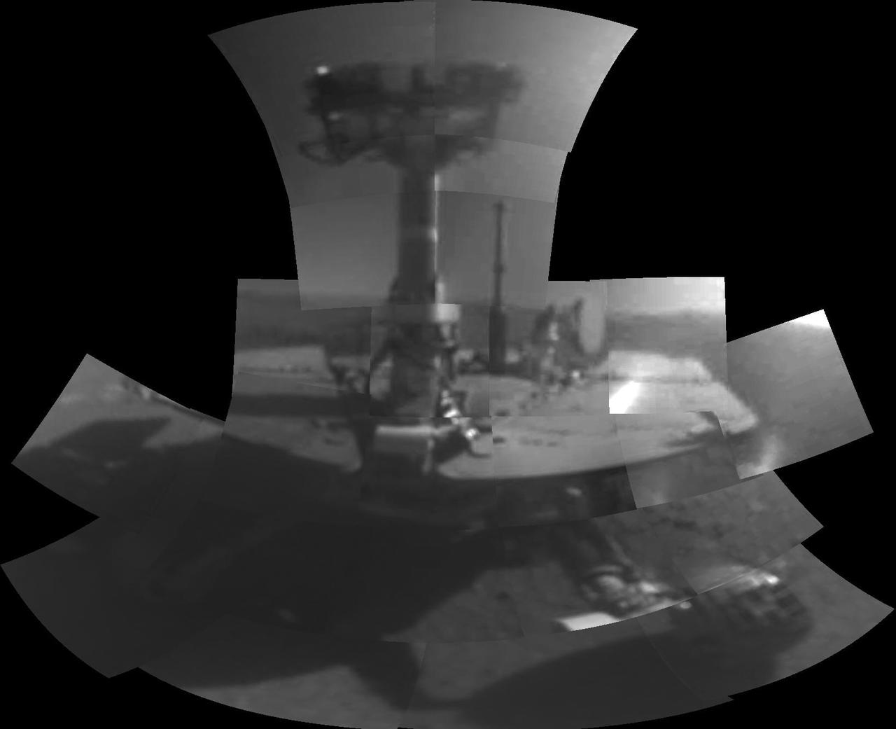
This mosaic image was taken with the microscopic imager on NASA Mars Exploration Rover Spirit to get a look underneath the rover.
This image taken by the microscopic imager on NASA Mars Exploration Rover Spirit shows the powdery soil of Mars in 3-D. 3D glasses are necessary to view this image.
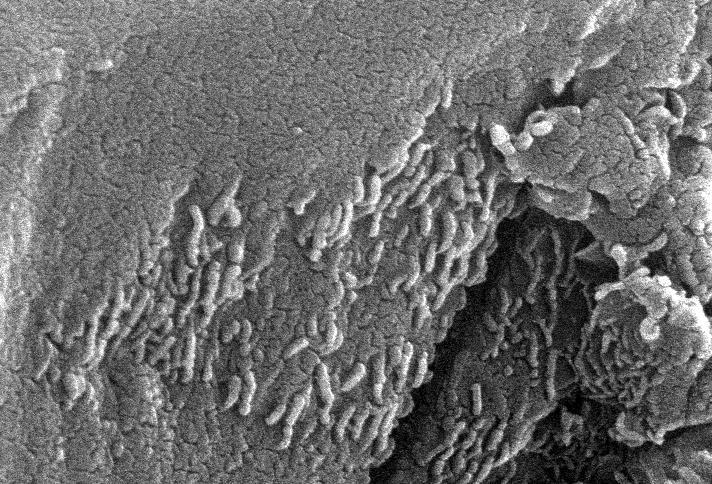
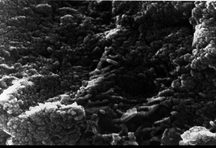
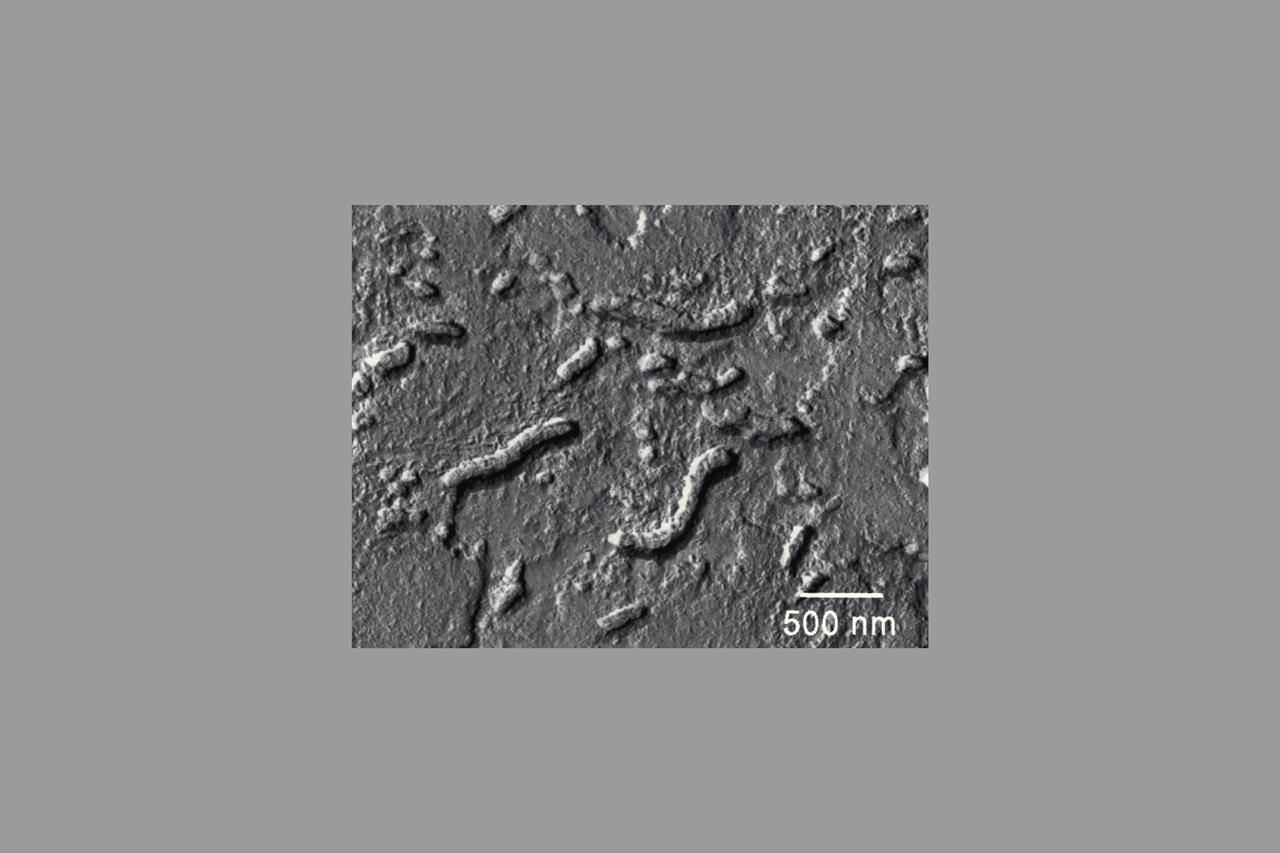
This electron microscope image shows extremely tiny tubular structures that are possible microscopic fossils of bacteria-like organisms that may have lived on Mars more than 3.6 billion years ago. http://photojournal.jpl.nasa.gov/catalog/PIA00285
This electron microscope image shows egg-shaped structures, some of which may be possible microscopic fossils of Martian origin as discussed by NASA research published in the Aug. 16, 1996. http://photojournal.jpl.nasa.gov/catalog/PIA00286
This 3D anaglyph, from NASA Mars Exploration Rover Spirit, shows a microscopic image taken of the rock called Adirondack. 3D glasses are necessary to view this image.
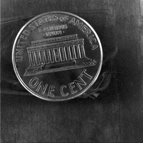
This close-up image of a penny shows the degree to which the microscopic imager on NASA Mars Exploration Rover Spirit can zoom in on a target.
This 3D anaglyph, from NASA Mars Exploration Rover Spirit, shows a microscopic image taken of the rock called Adirondack. 3D glasses are necessary to view this image.
This image shows the workings of the microscope station of the Microscopy, Electrochemistry and Conductivity Analyzer MECA instrument suite of NASA Phoenix Mars Lander.
This electron microscope image is a close-up of the center part of photo number S96-12301. http://photojournal.jpl.nasa.gov/catalog/PIA00284
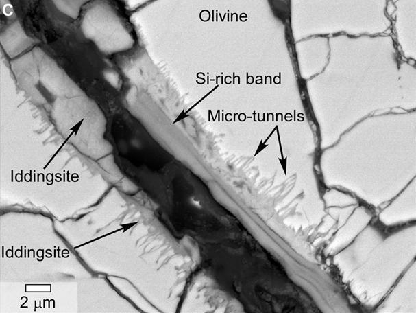
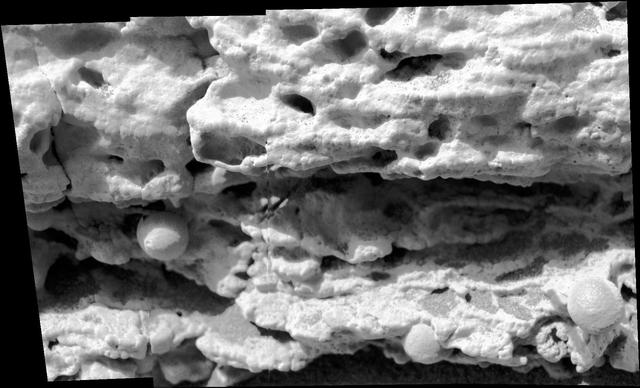
This scanning electron microscope image of a polished thin section of a meteorite from Mars shows tunnels and curved microtunnels.
In the center of this electron microscope image of a small chip from a meteorite are several tiny structures that are possible microscopic fossils of primitive, bacteria-like organisms that may have lived on Mars more than 3.6 billion years ago. http://photojournal.jpl.nasa.gov/catalog/PIA00283
iss073e0025978 (May 9, 2025) --- NASA astronaut and Expedition 73 Flight Engineer Nichole Ayers works in the Kibo laboratory module's Life Sciences Glovebox processing bacteria samples before viewing them inside a 3D imaging microscope called Extant Life Volumetric Imaging System, or ELVIS. The technology demonstration may enable applications for monitoring water quality, detecting infectious organisms on spacecraft, and researching colloids (suspensions of particles in a liquid) and microorganisms in microgravity.

iss073e0027806 (May 10, 2025) --- NASA astronaut and Expedition 73 Flight Engineer Anne McClain works in the Kibo laboratory module's Life Sciences Glovebox processing bacteria samples before viewing them inside a 3D imaging microscope called Extant Life Volumetric Imaging System, or ELVIS. The technology demonstration may enable applications for monitoring water quality, detecting infectious organisms on spacecraft, and researching colloids (suspensions of particles in a liquid) and microorganisms in microgravity.
This stereo view combines a pair of images taken two months apart by the microscopic imager on NASA Mars Exploration Rover Spirit. 3D glasses are necessary to view this image.
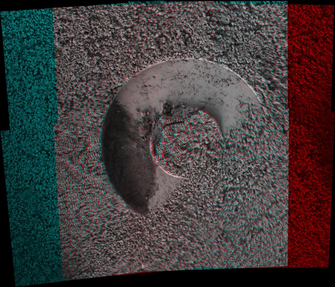
This 3-D image taken by the microscopic imager on NASA Mars Exploration Rover Opportunity shows a close-up of the center of the rock abrasion tool hole, ground into Bounce. 3D glasses are necessary to view this image.
This self-portrait of NASA's Opportunity Mars rover shows the vehicle at a site called "Perseverance Valley" on the slopes of Endeavour Crater. It was taken with the rover's Microscopic Imager to celebrate the 5000th Martian Day, or sol, of the rover's mission. The Microscopic Imager is a fixed-focus camera mounted at the end of the rover's robotic arm. Because it was designed for close inspection of rocks, soils and other targets at a distance of around 2.7 inches (7 cm), the rover is out of focus. The rover's self-portrait view is made by stitching together multiple images take on Sol 5,000 and 5,006 of the mission. Wrist motions and turret rotations on the arm allowed the Microscopic Imager to acquire the mosaic's component images. The resulting mosaic does not include the rover's arm. This simulation from planning software used to write commands for the rover shows the motion of the robotic arm, and an inset view of the Microscopic Imager. https://photojournal.jpl.nasa.gov/catalog/PIA22222
This scanning electron microscope image shows speroidal features embedded in a layer of iddingsite, a mineral formed by action of water, in a meteorite that came from Mars.
NASA Mars Exploration Rover Spirit used its microscopic imager to capture this spectacular, jagged mini-landscape on a rock called GongGong.
NASA Mars Exploration Rover microscopic imager onboard Spirit revealed a gap less than half an inch in the imprint left behind in the soil. 3D glasses are necessary to view this image.
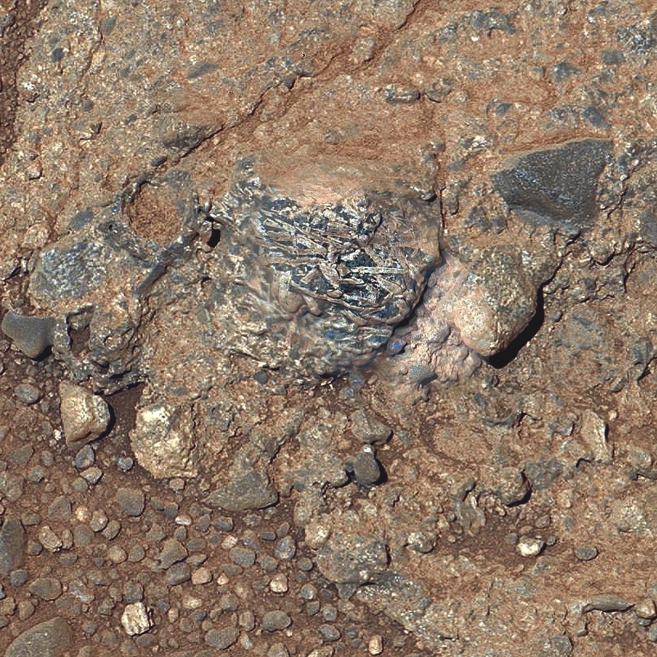
NASA Mars Exploration Rover Opportunity found this image of a meteorite. The science team used two tools on Opportunity arm, the microscopic imager and the alpha particle X-ray spectrometer, to inspect the rock texture and composition.
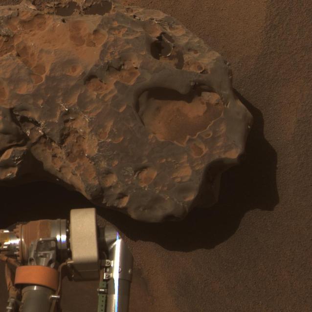
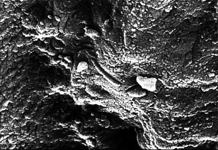
This image from the microscopic imager on NASA Mars Exploration Rover Opportunity shows details of the coating on a rock called Chocolate Hills, which the rover found and examined at the edge of a young crater called Concepción.
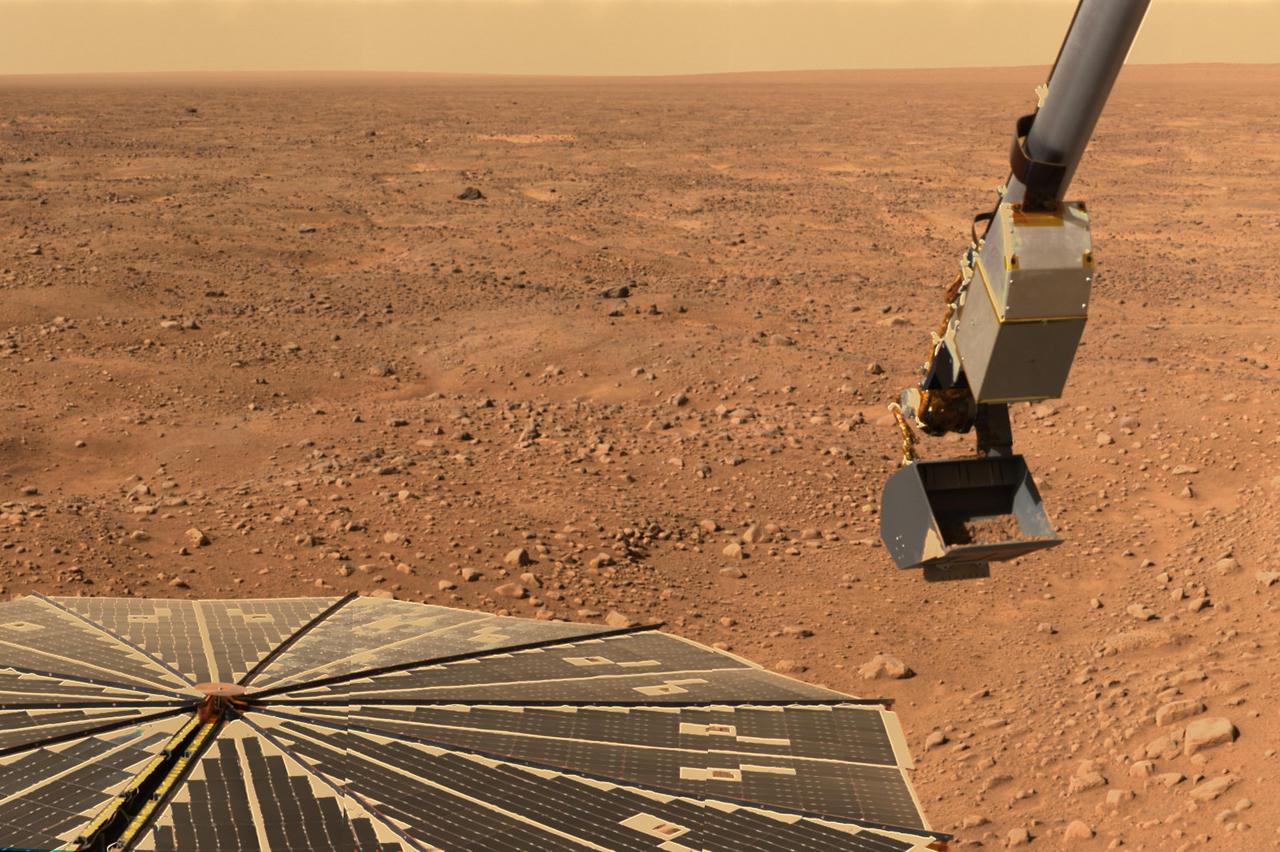
This panorama image of NASA’s Phoenix Mars Lander’s solar panel and the lander’s Robotic Arm with a sample in the scoop. The image was taken just before the sample was delivered to the Optical Microscope.
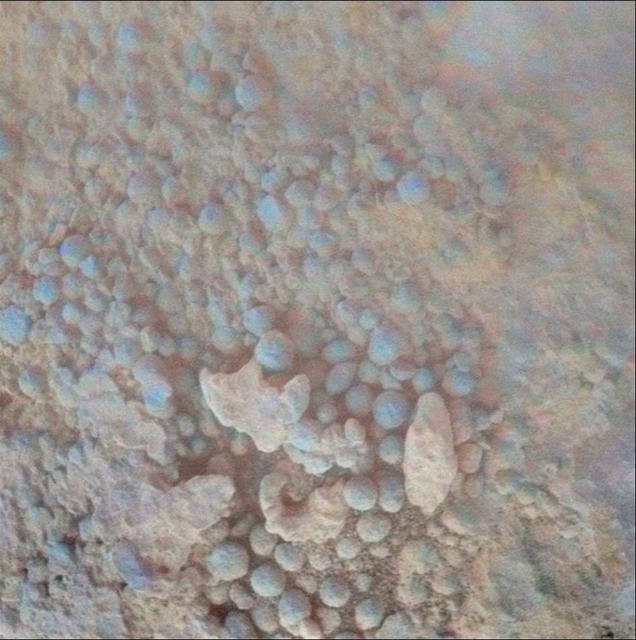
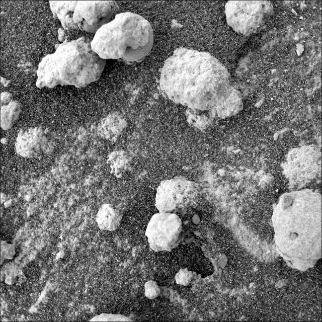
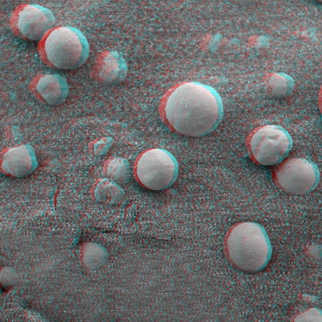
Small spherical objects fill the field in this mosaic combining four images from the Microscopic Imager on NASA Mars Exploration Rover Opportunity at an outcrop called Kirkwood in the Cape York segment of the western rim of Endeavour Crater.
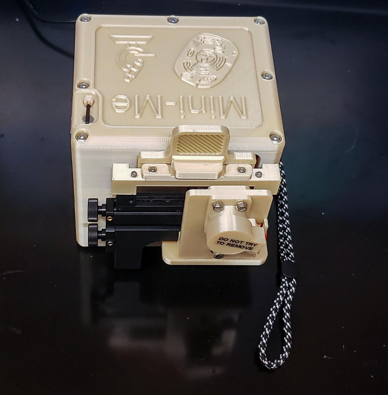
jsc2022e083574 (8/17/2022) --- A preflight image of the miniature microscope developed for the Moon Microscope investigation. Image courtesy of NASA’s Johnson Space Center Immunology/Virology Laboratory.
This stereo view combines a pair of images taken by the microscopic imager on NASA Mars Exploration Rover Spirit during the 1,925th Martian day sol of Spirit mission on Mars June 2, 2009. 3D glasses are necessary to view this image.
iss063e113776 (10/20/2020) --- A view of the CubeLab Microscope Imagery Tech Demo aboard the International Space Station (ISS). The CubeLab Microscope Imagery Technology Demonstration (CubeLab Microscope Imagery Tech Demo) tests enhanced microscope imagery capabilities for experiments aboard the space station. Images provide a primary way to document and analyze many microgravity investigations, and better quality images could lead to better results.
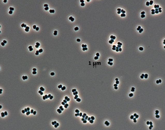
This microscopic image shows dozens of individual bacterial cells of the recently discovered species, Tersicoccus phoenicis, found in only two places: clean rooms in Florida and South America where spacecraft are assembled for launch.
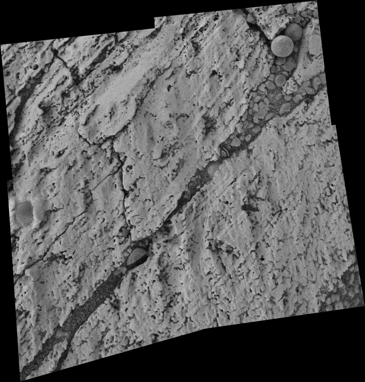
This close-up view of a mineral vein called Homestake comes from the microscopic imager on NASA Mars Exploration Rover Opportunity; the vein is found to be rich in calcium and sulfur, possibly the calcium-sulfate mineral gypsum.
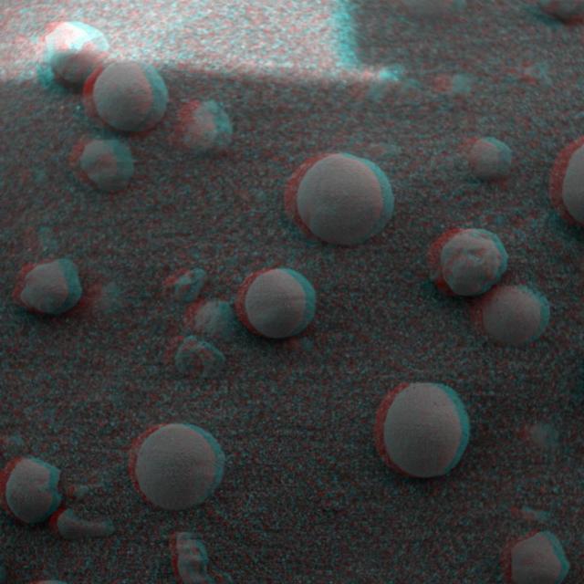
This view from the microscopic imager on NASA Mars Exploration Rover Opportunity shows a type of light-colored, rough-textured spherules scientists call popcorn in contrast to the darker, smoother spherules called blueberries.
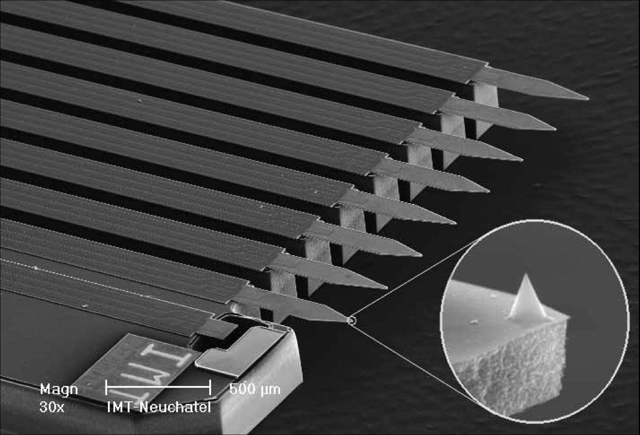
This image shows the eight sharp tips of the NASA Phoenix Mars Lander Atomic Force Microscope, or AFM. The AFM is part of Phoenix Microscopy, Electrochemistry, and Conductivity Analyzer, or MECA.
NASA Mars Exploration Rover Opportunity used its microscopic imager to get this view of the surface of a rock called Block Island during the 1,963rd Martian day, or sol, of the rover mission on Mars Aug. 1, 2009.
This mosaic shot by the microscopic imager on the robotic arm of NASA Mars Exploration Rover Opportunity shows a rock target called Esperance after some of the rock surface had been removed by Opportunity rock abrasion tool.
This view of a Martian rock target called /Harrison merges images from two cameras onboard NASA Curiosity Mars rover to provide both color and microscopic detail. The elongated crystals are likely feldspars, and the matrix is pyroxene-dominated.
This electron microscope image shows tubular structures of likely Martian origin. These structures are very similar in size and shape to extremely tiny microfossils found in some Earth rocks. http://photojournal.jpl.nasa.gov/catalog/PIA00287
NASA Mars Exploration Rover Opportunity found and examined this meteorite. The science team used two tools on Opportunity arm, the microscopic imager and the alpha particle X-ray spectrometer, to inspect the rock texture and composition.
jsc2022e031227 (5/4/2016) --- A preflight view of the BioServe Microscope. The BioServe Microscope facility allows astronauts to capture digital, full-color, high-definition microscopy images and videos of scientific investigations.
This 3-D anaglyph, from NASA Mars Exploration Rover Spirit, shows a microscopic image taken of soil featuring round, blueberry-shaped rock formations on the crater floor at Meridiani Planum, Mars. 3D glasses are necessary to view this image.
This 3-D anaglyph, from NASA Mars Exploration Rover Spirit, shows a microscopic image taken of soil featuring round, blueberry-shaped rock formations on the crater floor at Meridiani Planum, Mars. 3D glasses are necessary to view this image.
This high-resolution scanning electron microscope image shows an unusual tube-like structural form that is less than 1/100th the width of a human hair in size found in meteorite ALH84001, a meteorite believed to be of Martian origin. http://photojournal.jpl.nasa.gov/catalog/PIA00288
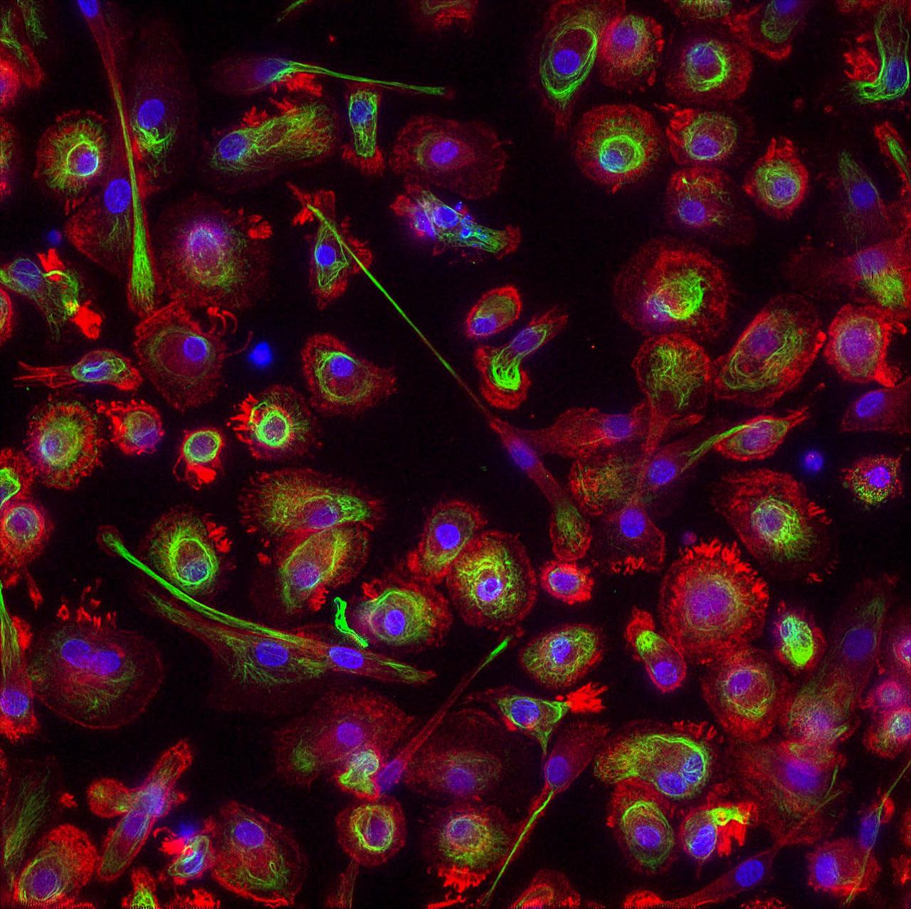
jsc2019e051831 --- Image of fixed macrophages using three chromophores created by the FLUMIAS-DEA miniaturized fluorescence microscope during Science Verification Test. Image courtesy of: Airbus

jsc2024e006100 (12/29/2022) --- A. platensis spiral trichomes under a microscope. Image courtesy of Katherine Fisher
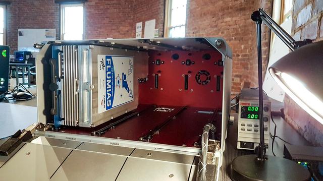
jsc2019e051830 --- FLUMIAS-DEA miniaturized fluorescence microscope loaded in TangoLab-2. Image courtesy of: Airbus
BioCell in BioServe’s ground unit of the on-orbit Microscope during in-situ imaging of the iPSC. (Credit: BioServe)
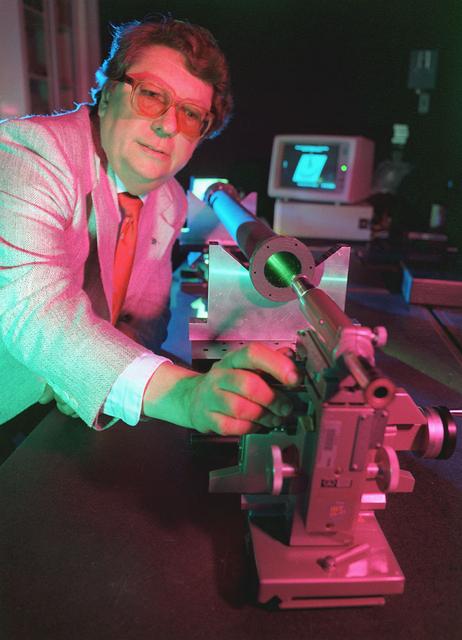
Marshall's 1992 Inventor of the Year demonstrates his multi-layer water window imaging x-ray microscope.
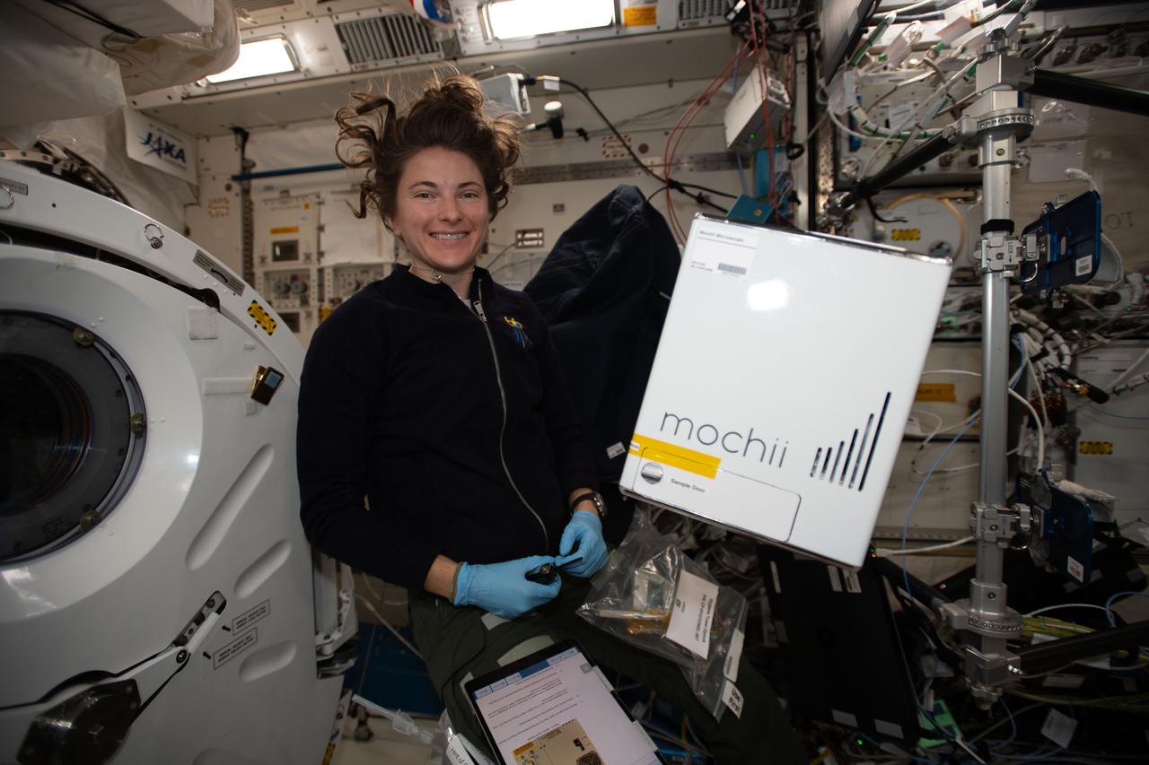
iss066e110531_alt (1/10/2022) --- NASA astronaut Kayla Barron sets up the Mochii microscope. Mochii is a miniature scanning electron microscope (SEM) with spectroscopy to conduct real-time, on-site imaging and compositional measurements of particles on the International Space Station (ISS).
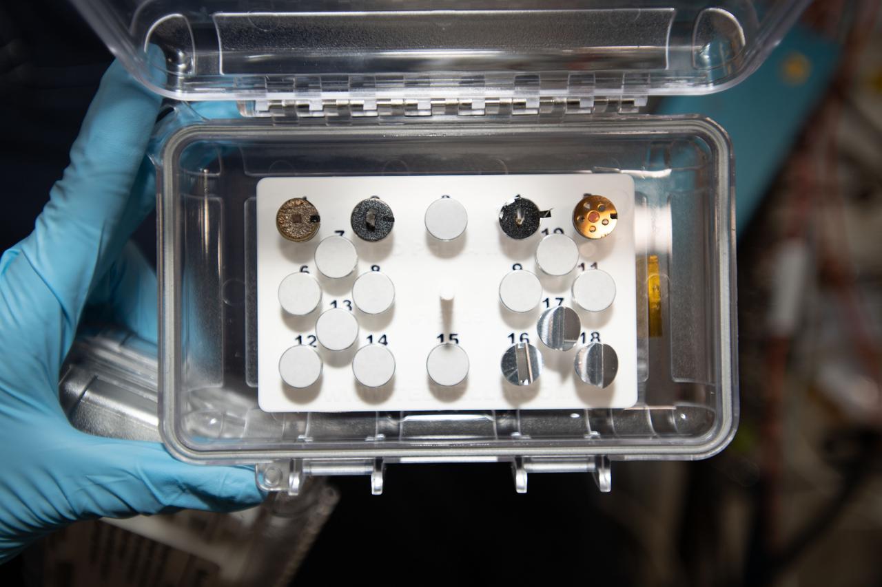
iss066e110547 (1/10/2022) --- A view of the Mochii microscope sample load aboard the International Space Station (ISS). Mochii is a miniature scanning electron microscope (SEM) with spectroscopy to conduct real-time, on-site imaging and compositional measurements of particles on the International Space Station (ISS)
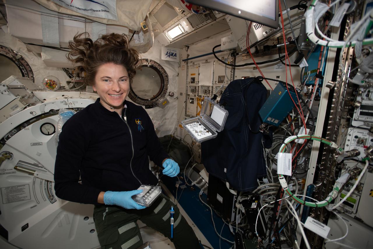
iss066e110556 (1/10/2022) --- NASA astronaut Kayla Barron sets up the Mochii microscope. Mochii is a miniature scanning electron microscope (SEM) with spectroscopy to conduct real-time, on-site imaging and compositional measurements of particles on the International Space Station (ISS).
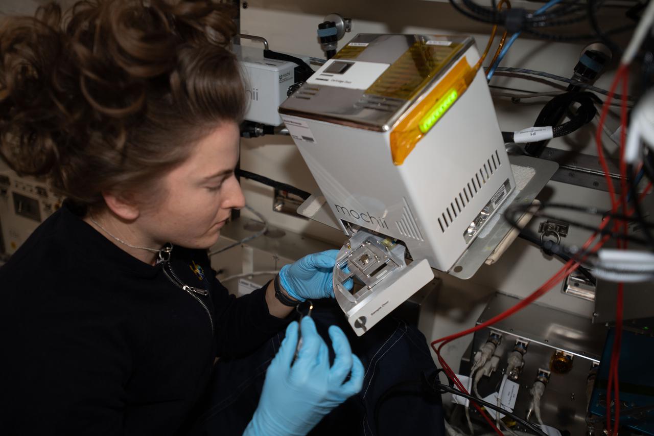
iss066e110566 (1/10/2022) --- NASA astronaut Kayla Barron sets up the Mochii microscope. Mochii is a miniature scanning electron microscope (SEM) with spectroscopy to conduct real-time, on-site imaging and compositional measurements of particles on the International Space Station (ISS).
A scanning electron microscope image of a micrometeorite impact crater in a particle of asteroid Bennu material. Scientists found microscopic craters and tiny splashes of once-molten rock – known as impact melts – on the surfaces of samples, signs that the asteroid was bombarded by micrometeorites.
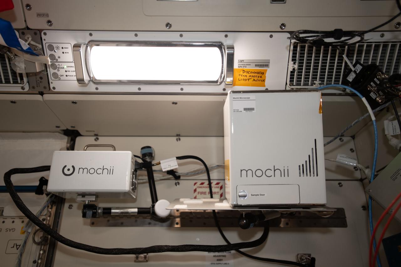
iss066e110545 (1/10/2022) --- A view of the Mochii microscope aboard the International Space Station (ISS. Mochii is a miniature scanning electron microscope (SEM) with spectroscopy to conduct real-time, on-site imaging and compositional measurements of particles on the International Space Station (ISS).
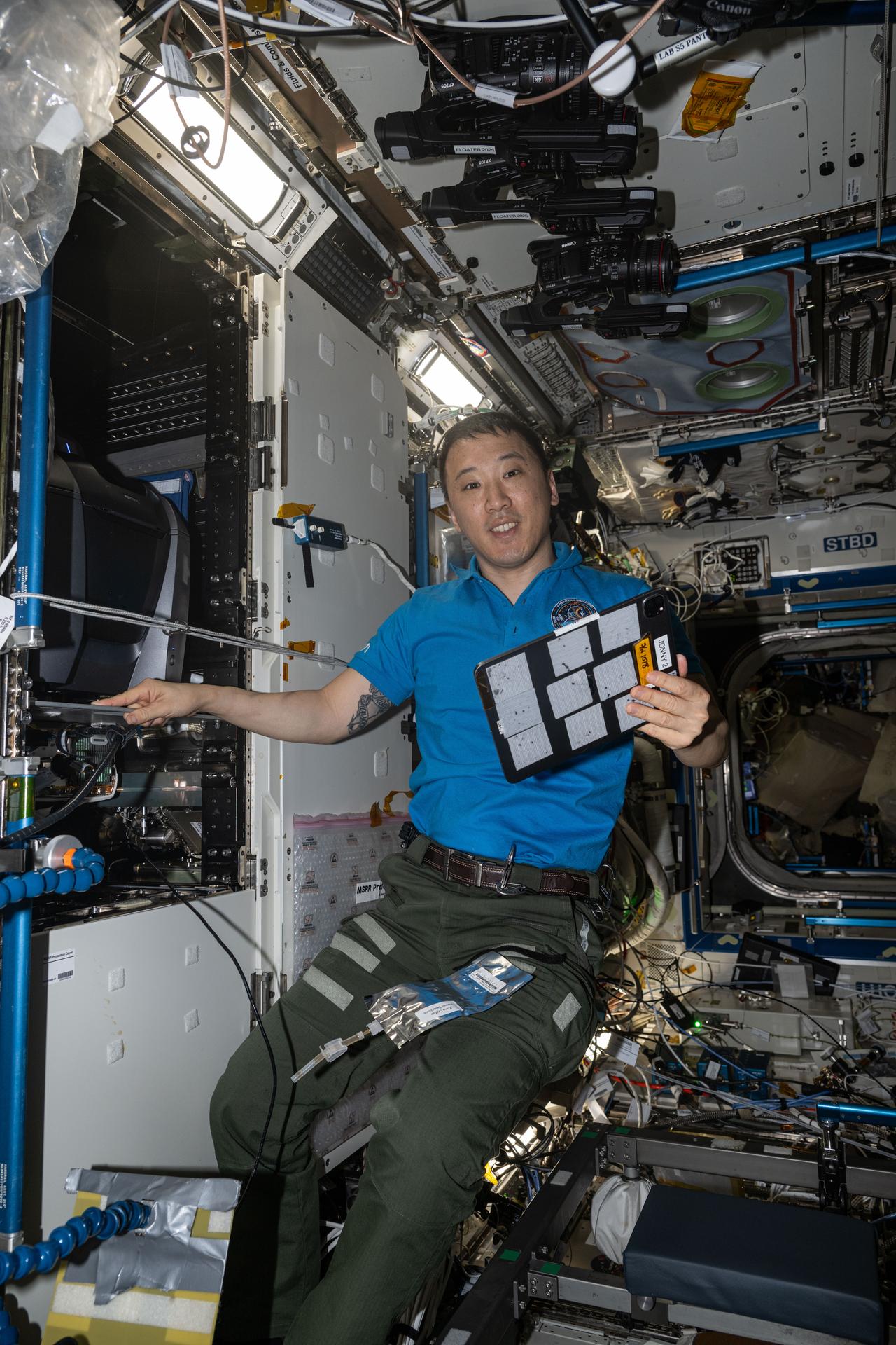
iss073e1049692 (Nov. 6, 2025) --- NASA astronaut and Expedition 73 Flight Engineer Jonny Kim poses for a portrait while servicing the KERMIT (Keyence Research Microscope Testbed) fluorescence microscope inside the Materials Science Research Rack aboard the International Space Station’s Destiny laboratory module. KERMIT is a commercial off-the-shelf microscope that provides researchers with essential imaging capabilities for biological, physical, and materials science research in microgravity.
This image shows a microscopic view of fine-grained material at the tip of the Robotic Arm scoop as seen by the Robotic Arm Camera RAC aboard NASA Phoenix Mars Lander on June 20, 2008, the 26th Martian day, or sol, of the mission.
This 3-D, microscopic imager mosaic of a target area on a rock called Diamond Jenness was taken after NASA Mars Exploration Rover Opportunity ground into the surface with its rock abrasion tool for a second time. 3D glasses are necessary.
jsc2021e044608 (9/23/2021) --- A microscopic image of a section of ZBLAN optical fiber produced by the second iteration of Fiber Optic Production (FOP1.5) aboard the International Space station (ISS). Image courtesy of Mercury Systems.
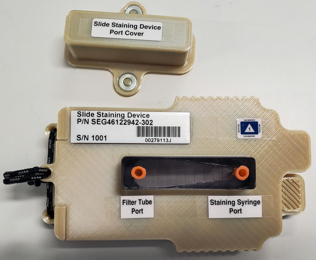
jsc2022e083575 (10/4/2022) --- A preflight image of the slide staining device developed for the Moon Microscope investigation. Image courtesy of NASA’s Johnson Space Center Immunology/Virology Laboratory.
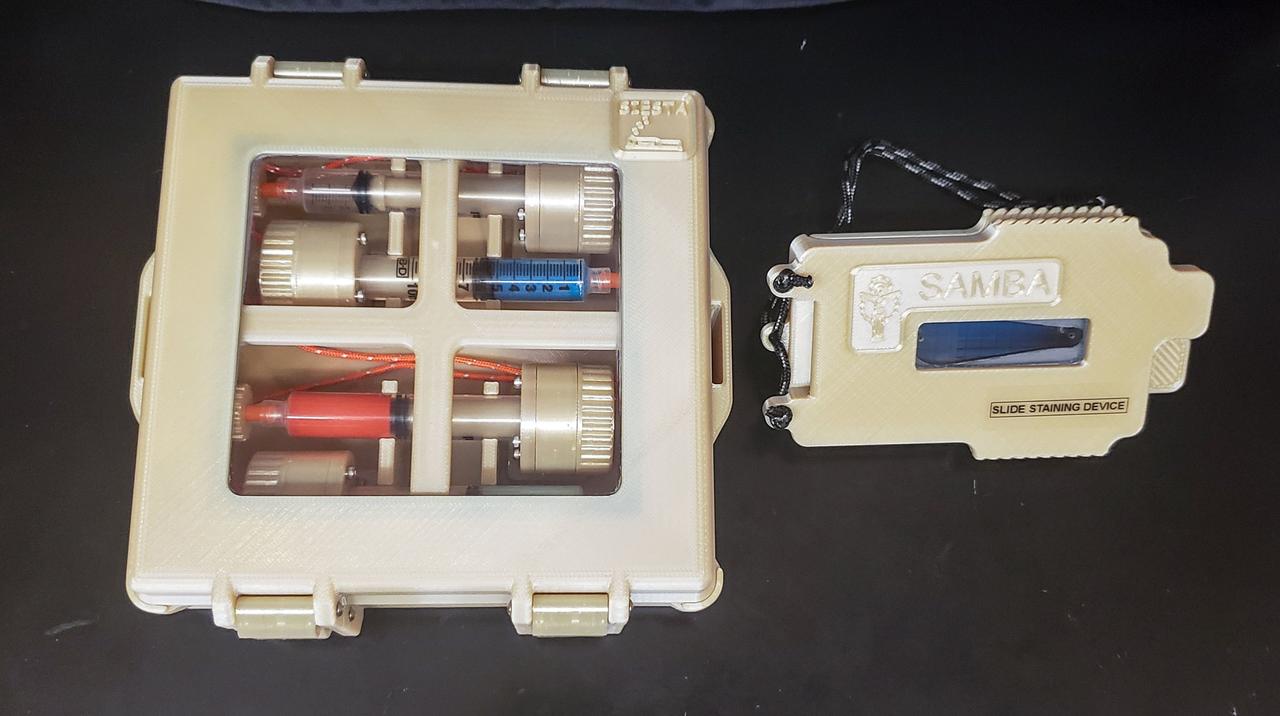
jsc2022e083573 (9/2/2022) --- A preflight image of staining reagents, casing, and staining device developed for the Moon Microscope investigation. Image courtesy of NASA’s Johnson Space Center Immunology/Virology Laboratory.
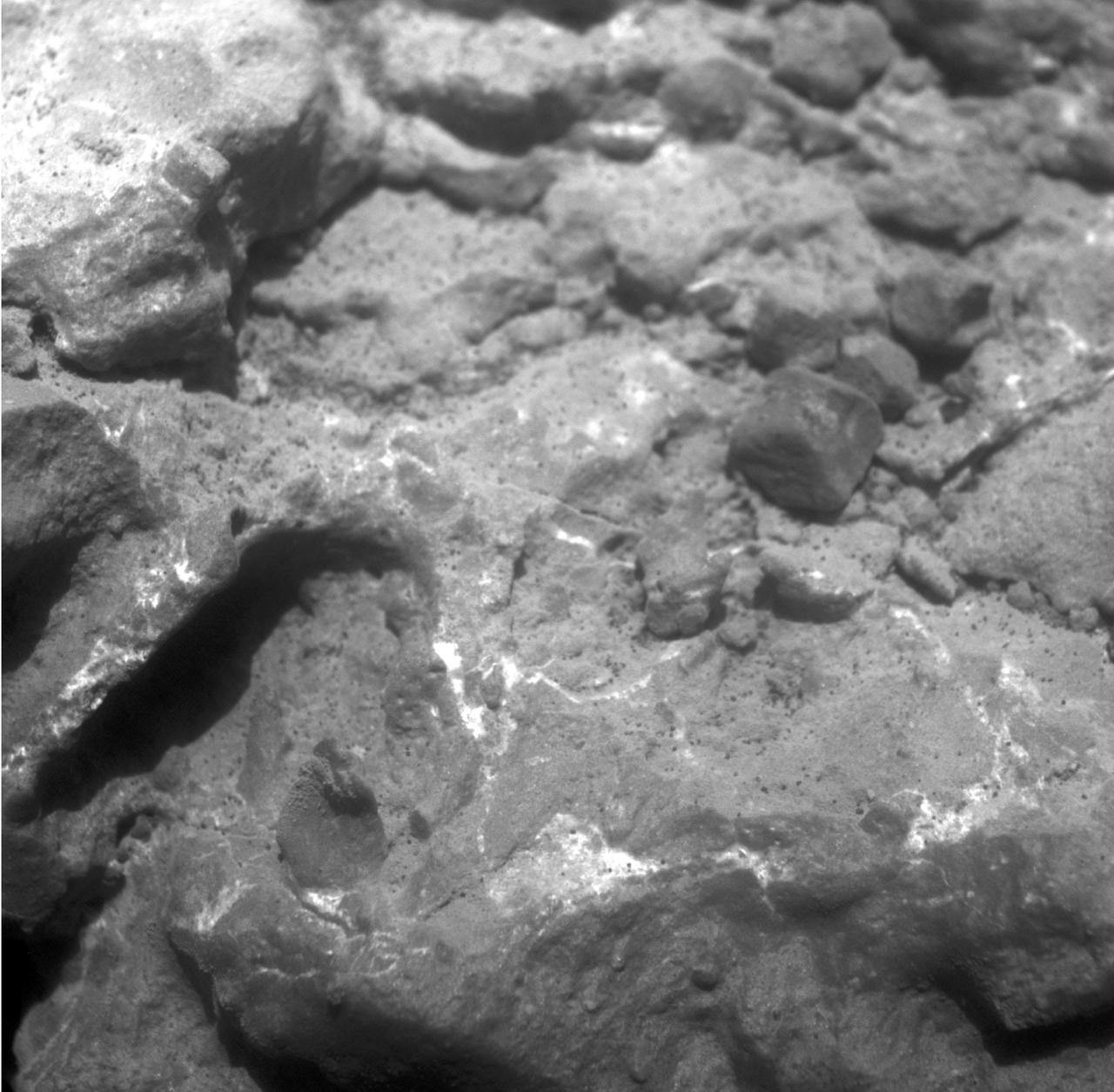
NASA Mars Exploration Rover Opportunity used its microscopic imager to record this close-up view of texture on part of a rock informally named Tisdale
Martian Meteorite (ALH84001): This high resolution transmission electron microscope image is of a cast, or replica, from a chip of a Martian meteorite, labeled ALH84001, that shows the outline of what are believed to be possible microscopic fossils of bacteria-like organisms that may have lived on Mars more than 3.6 billion years ago. The tubular features in this image are less than a micrometer in size, or about 1/500th the diameter of a human hair. (JSC ref: S96-12637)
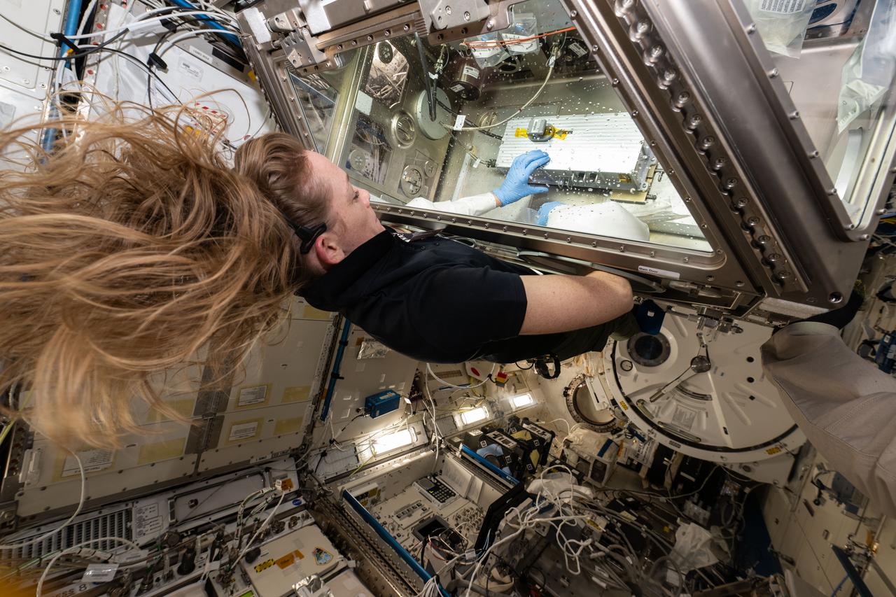
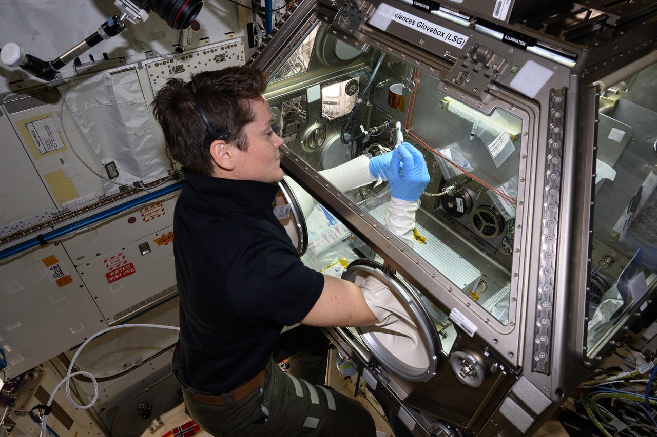
iss073e0025988 (5/9/2025) --- NASA astronaut Nichole Ayers works with the ELVIS investigation at the Life Sciences Glovebox (LSG), aboard the Kibo module of the International Space Station. Extant Life Volumetric Imaging System (ELVIS) is a microscope for 3D imaging of objects as small as bacteria, with the goal of making the technology available to anyone studying microscopic motion in space.
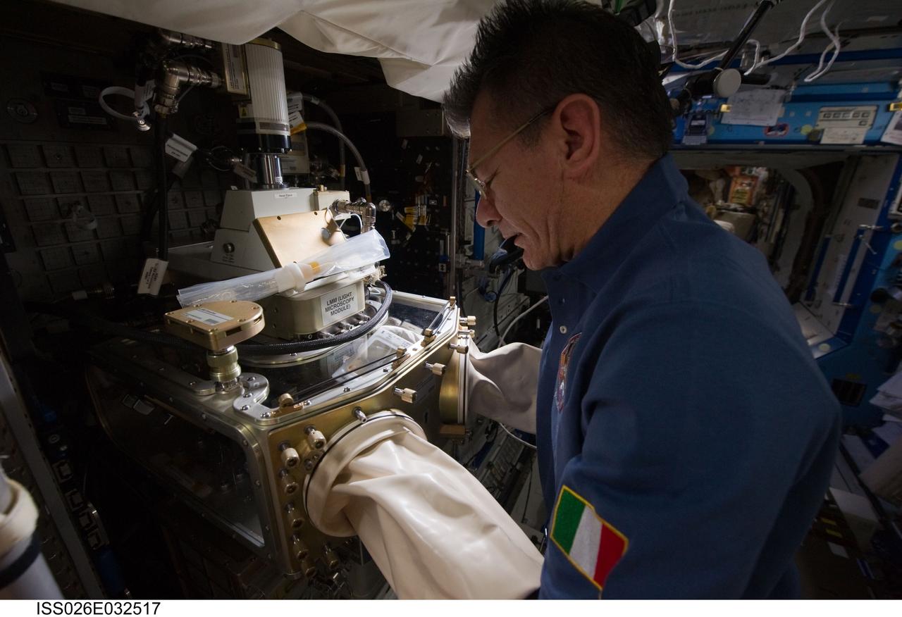
iss026e032517 (3/8/2011) --- European Space Agency (ESA) Paolo Nespoli works with the Light Microscopy Module (LMM) in the U.S. Laboratory. The Light Microscopy Module (LMM) is a modified commercial, highly flexible, state-of-the-art light imaging microscope facility that provides researchers with powerful diagnostic hardware and software onboard the International Space Station (ISS). The LMM enables novel research of microscopic phenomena in microgravity, with the capability of remotely acquiring and downloading digital images and videos across many levels of magnification.
ISS047e066551 (04/18/2016) --- NASA astronaut Jeff Williams configures the station’s Light Microscopy Module (LMM), a modified commercial, highly flexible, state-of-the-art light imaging microscope facility that provides researchers with powerful diagnostic hardware and software. The LMM enables novel research of microscopic phenomena in microgravity, with the capability of remotely acquiring and downloading digital images and videos across many levels of magnification.
This close-up view of a target rock called "Last Chance" was acquired by the microscopic imager on the arm of NASA's Mars Exploration Rover Opportunity on March 3, 2004, during the 39th Martian day, or sol, of Opportunity's work on Mars. The area covered in the view is about 2 inches (5 centimeters) across. The embedded spherules evident in this image reminded researchers of berries in a muffin, so they were nicknamed "blueberries." These mineral concretions and other textures in this rock provided evidence about wet environmental conditions in the ancient past at Opportunity's landing site in the Meridiani Planum region. http://photojournal.jpl.nasa.gov/catalog/PIA18885
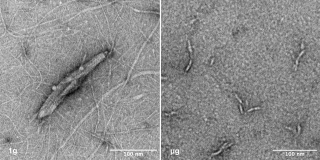
jsc2025e036194 (4/4/2025) --- Image of nanoparticles synthesized under 1g vs microgravity. Left: This transmission electron microscope image depicts the nano-scale structure of Janus Base Nanoparticles encapsulated with mRNA on ground. Right: This is a transmission electron microscope image of mRNA-encapsulated JBNp that was produced on ISS during the SpaceX CRS-31 mission. Here, you can see that the space-made JBNp is smaller and more uniform in size and shape with less background material, demonstrating the stark advantage that in-space manufacturing can provide JBNp: improved uniformity and drug loading. Image courtesy of University of Connecticut.
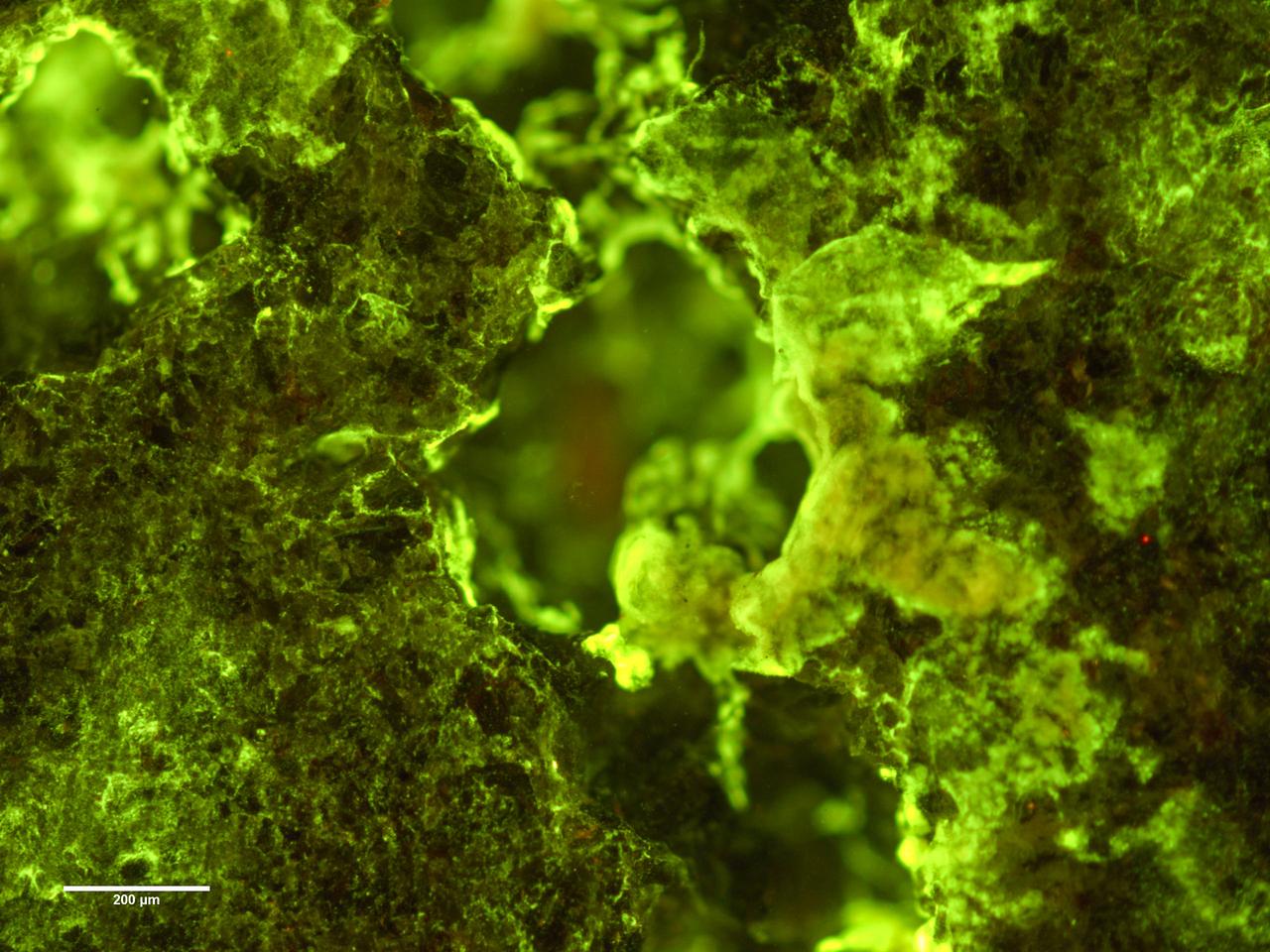
jsc2019e039825 (7/16/2019) --- Preflight Ffuorescence microscopy image of biofilm of Spingomonas desiccabilis growing over and into the surface of a basalt slide as part of BioRock experiment. Organisms are stained with DNA binding sye Sybr Gold. Growth can be seen into the rock cavities. The purpose of the Biorock investigation is to examine the effects of altered gravity on the rock/microbe/liquid system as a whole. (Image Courtesy of: ESA)
A scanning electron microscope captured this image of terresterial soil containing a phyllosilicate mineral from Koua Bocca, Ivory Coast, West Africa. This soil shares some similarities with Martian soil scooped by NASA Phoenix Lander.
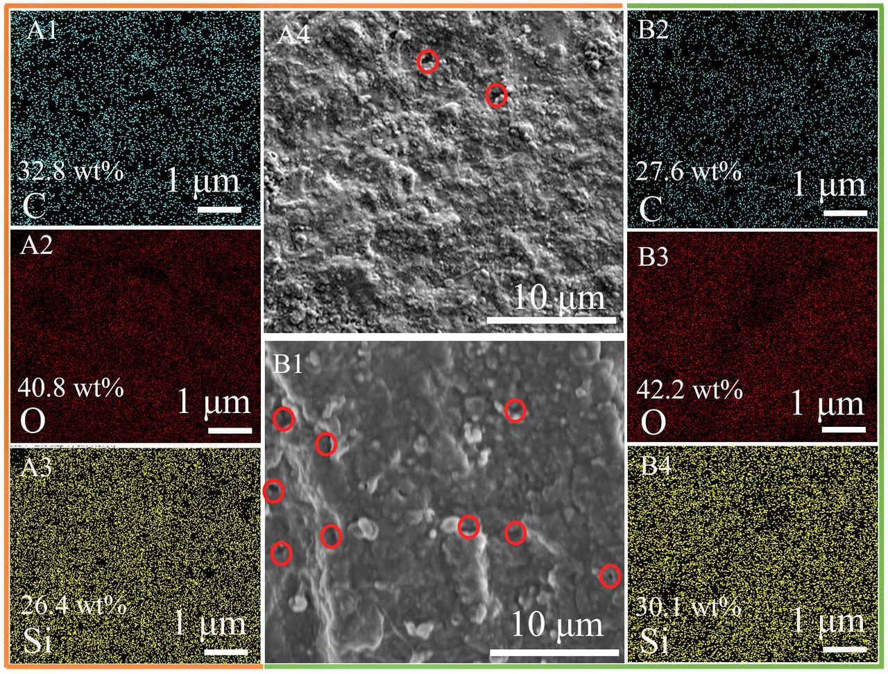
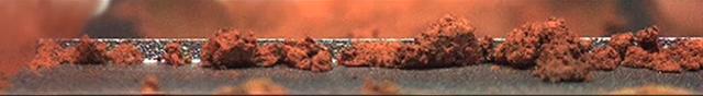
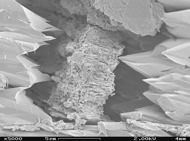
jsc2025e064333 (5/31/2024) --- Scanning electron microscope (SEM) image graphs and elemental mappings of the samples pyrolyzed at 1200 oC in the Ar (A1-A4) and microgravity (B1-B4). (The red circles in the SEM images represent obvious pores).
This mosaic image shows an extreme close-up of round, blueberry-shaped formations in the martian soil near a part of the rock outcrop at Meridiani Planum called Stone Mountain. Scientists are studying these curious formations for clues about the area's past environmental conditions. The image, one of the highest resolution images ever taken by the microscopic imager, an instrument located on the Mars Exploration Rover Opportunity's instrument deployment device or "arm." http://photojournal.jpl.nasa.gov/catalog/PIA05273
jsc2025e067418 (8/5/2025) --- Microscopic image of a semimetal-semiconductor composite (SSC) wafer extracted from one of four crystals grown in the International Space Station’s SUBSA facility during the first SUBSA-InSPA-SSCug mission. Credit: United Semiconductors LLC
iss055e035366 (April 16, 2018) --- NASA astronaut Ricky Arnold performs maintenance on the Advanced Colloids Experiment Module located inside the Light Microscopy Module which is a modified commercial, highly flexible, state-of-the-art light imaging microscope facility that provides researchers with powerful diagnostic hardware and software in microgravity.
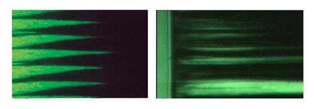
These are video microscope images of magnetorheological (MR) fluids, illuminated with a green light. Those on Earth, left, show the MR fluid forming columns or spikes structures. On the right, the fluids in microgravity aboard the International Space Station (ISS), formed broader columns.

jsc2025e067416 (8/5/2025) --- Microscopic image of a semimetal-semiconductor composite (SSC) wafer extracted from one of four crystals grown in the International Space Station’s SUBSA facility during the first SUBSA-InSPA-SSCug mission. Credit: United Semiconductors LLC

jsc2025e067417 (8/5/2025) --- Microscopic image of a semimetal-semiconductor composite (SSC) wafer extracted from one of four crystals grown in the International Space Station’s SUBSA facility during the first SUBSA-InSPA-SSCug mission. Credit: United Semiconductors LLC

jsc2026e014323 (March 23, 2026) --- Preflight microscopic image (4X) of hematopoietic stem cells in culture at high confluency as part of InSPA-StemCellEX-H2 investigation, which works upon prior research to produce stem cells in greater numbers in space with BioServe’s newly developed microgravity bioreactor. Credit. Mayo Clinic.
jsc2026e014322 (March 23, 2026) --- Preflight microscopic image (10X) of hematopoietic stem cells in culture as part of the InSPA-StemCellEX-H2 investigation, which works upon prior research to produce stem cells in greater numbers in space with BioServe’s newly developed microgravity bioreactor. Credit: Mayo Clinic.

jsc2025e067415 (8/5/2025) --- Microscopic image of a semimetal-semiconductor composite (SSC) wafer extracted from one of four crystals grown in the International Space Station’s SUBSA facility during the first SUBSA-InSPA-SSCug mission. Credit: United Semiconductors LLC
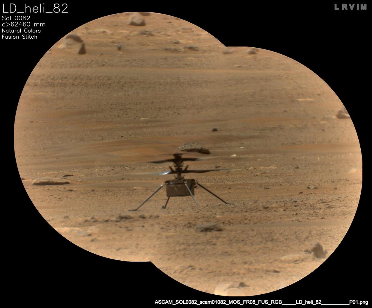
NASA's Ingenuity Mars Helicopter is viewed here through the Remote Microscopic Imager (RMI) camera, part of the SuperCam instrument aboard NASA's Perseverance rover. This image was taken on May 14, 2021, the 82nd Martian day, or sol, of the mission. The RMI is able to spot a softball from nearly a mile away, allowing scientists to take images of details from a long distance. It also provides fine details of nearby targets zapped by SuperCam's laser. A key objective for Perseverance's mission on Mars is astrobiology, including the search for signs of ancient microbial life. The rover will characterize the planet's geology and past climate, pave the way for human exploration of the Red Planet, and be the first mission to collect and cache Martian rock and regolith (broken rock and dust). Subsequent NASA missions, in cooperation with ESA (European Space Agency), would send spacecraft to Mars to collect these sealed samples from the surface and return them to Earth for in-depth analysis. The Mars 2020 Perseverance mission is part of NASA's Moon to Mars exploration approach, which includes Artemis missions to the Moon that will help prepare for human exploration of the Red Planet. https://photojournal.jpl.nasa.gov/catalog/PIA24665
This composite image of the "Delta Scarp" in Mars' Jezero Crater was generated using data from two imagers aboard NASA's Perseverance rover. Taken by the rover's Mastcam-Z, the bottom image shows both the base and plateau of the escarpment. The inset above, created from a mosaic of five Remote Microscopic Imager (RMI) pictures, zooms in on a 377-foot-wide (115-meter-wide) portion of the scarp, allowing closer inspection of some of its intriguing geologic features. Part of the rover's SuperCam instrument, the RMI is able to spot an object the size of a softball from nearly a mile away, allowing scientists to take images of details from a long distance. It also provides fine details of nearby targets zapped by SuperCam's laser. SuperCam is led by Los Alamos National Laboratory in New Mexico, where the instrument's Body Unit was developed. That part of the instrument includes several spectrometers, control electronics and software. The Mast Unit was developed and built by several laboratories of the CNRS (French National Centre for Scientific Research) and French universities under the contracting authority of CNES. Arizona State University in Tempe leads the operations of the Mastcam-Z instrument, working in collaboration with Malin Space Science Systems in San Diego. A key objective for Perseverance's mission on Mars is astrobiology, including the search for signs of ancient microbial life. The rover will characterize the planet's geology and past climate, pave the way for human exploration of the Red Planet, and be the first mission to collect and cache Martian rock and regolith (broken rock and dust). Subsequent NASA missions, in cooperation with ESA (European Space Agency), would send spacecraft to Mars to collect these sealed samples from the surface and return them to Earth for in-depth analysis. The Mars 2020 Perseverance mission is part of NASA's Moon to Mars exploration approach, which includes Artemis missions to the Moon that will help prepare for human exploration of the Red Planet. https://photojournal.jpl.nasa.gov/catalog/PIA24684
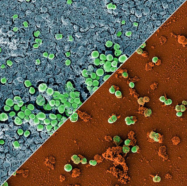
jsc2023e010179 (2/28/2023) --- This image is a composition of two scanning electron microscopic images of the bacterium Staphylococcus capitis on stainless steel versus antimicrobial copper. The image was colored to visualize the bacterial cells (green) either embedded in a biofilm matrix (blue), or covered with copper particles (red/orange). The ESA-Biofilms investigation studies bacterial biofilm formation and antimicrobial properties of different metal surfaces under spaceflight conditions in altered gravity. Both images were taken as part of the preflight experiments for ESA-Biofilms. Image courtesy of DLR, CC BY-NC-ND 3.0.
iss073e0027808 (May 10, 2025) --- NASA astronaut and Expedition 73 Flight Engineer Anne McClain works in the Kibo laboratory module's Life Sciences Glovebox processing bacteria samples before viewing them inside a 3D imaging microscope called the Extant Life Volumetric Imaging System, or ELVIS. The technology demonstration may enable applications for monitoring water quality, detecting infectious organisms on spacecraft, and researching colloids (suspensions of particles in a liquid) and microorganisms in microgravity.
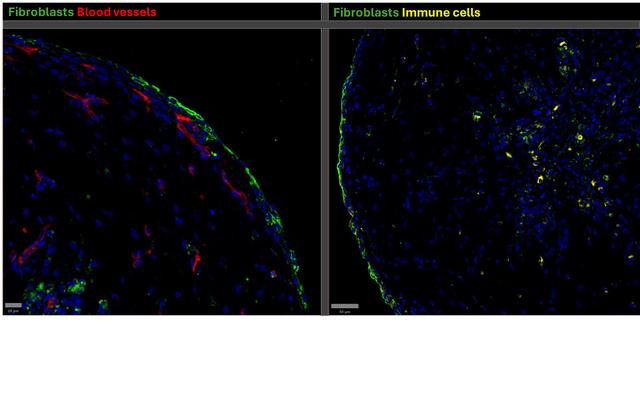
jsc2024e071691 (11/5/2024) --- Immunofluorescence staining of a synovial organoid shows CD68+ macrophages (immune cells) in yellow, CD31+ endothelial cells (blood vessels) in red, PDPN+ fibroblasts in green, and nuclei in blue is seen. This image was taken on a confocal microscope and is part of the ICE Cubes #9.4 – Aging in Microgravity investigation. Image courtesy of University of Oxford.
jsc2025e036195 (4/4/2025) --- A confocal microscope image shows a human cartilage cell, with its nucleus stained in blue, following delivery of Janus Base Nanoparticles (JBNp) and the subsequent release of bioactive mRNA (pink) that was translated into function protein (green). Biomimetic Fabrication of Multi-Functional DNA-Inspired Nanomaterials via Controlled Self-assembly in Space (DNA Nano Therapeutics-Mission 2) continues prior research on in-space manufacturing of nanomaterials that mimic DNA and have applications for vaccines and regenerative medicine. Image courtesy of University of Connecticut.
jsc2022e083017 (4/23/2022) --- A preflight image of a BioCell developed by BioServe Space Technologies that contains 162 beating cardiac spheroids derived from induced pluripotent stem cells (iPSCs). These cells are incubated and put under the microscope in space as part of the Effect of Microgravity on Drug Responses Using Heart Organoids (Cardinal Heart 2.0) investigation. Image courtesy of Drs. Joseph Wu, Dilip Thomas and Xu Cao, Stanford Cardiovascular Institute.
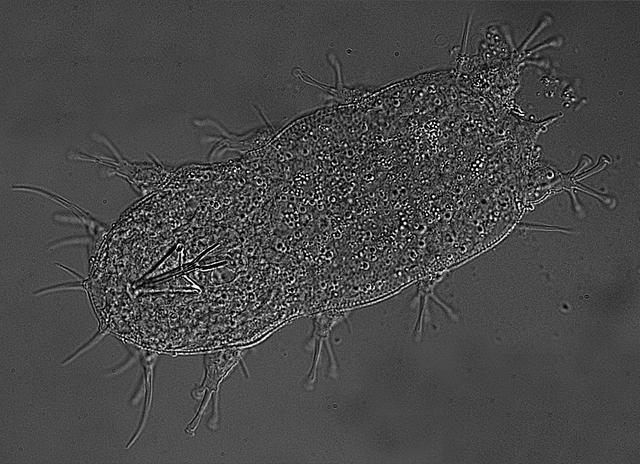
jsc2021e019950 (4/25/2014) --- A preflight photo of a marine tardigrade imaged at 40X magnification on a light microscope. The objective of the Using Water Bears to Identify Biological Countermeasures to Stress during Multigenerational (Cell Science-04) investigation is to characterize the molecular biology of short term and multigenerational survival in the space environment by identifying genes that are required for adaptation and survival in high stress environments. Image courtesy of Boothby Lab.
jsc2021e019397 (5/19/2021) --- The Nortis Organ Chip under a microscope in the laboratory of Edward Kelly in the University of Washington Department of Pharmaceutics. The image on the screen in the background shows a kidney cell tubule. Effects of Microgravity on the Structure and Function of Proximal and Distal Tubule MPS (Kidney Cells-02) uses a 3D kidney cell model or chip to study the effects of microgravity on formation of microcrystals in kidney tubules. Image courtesy of Alex Levine (UW School of Pharmacy).

jsc2022e083018 (10/26/2022) --- A preflight image of beating cardiac spheroid composed of iPSC-derived cardiomyocytes (CMs), endothelial cells (ECs), and cardiac fibroblasts (CFs). These cells are incubated and put under the microscope in space as part of the Effect of Microgravity on Drug Responses Using Heart Organoids (Cardinal Heart 2.0) investigation. Image courtesy of Drs. Joseph Wu, Dilip Thomas and Xu Cao, Stanford Cardiovascular Institute.

jsc2023e010177 (4/7/2022) --- This image is a scanning electron microscopic image of one of the ESA-Biofilms sample plates from the first launch to the ISS. The sample plate in this image is made of copper, which naturally has antimicrobial properties. This surface has a 3 µm laser structure engraved to the surface which improves antimicrobial efficacy. On the surface, only few cells of the bacterial species Staphylococcus capitis are attached. The cells appear small and are not actively dividing. The ESA-Biofilms investigation studies bacterial biofilm formation and antimicrobial properties of different metal surfaces under spaceflight conditions in altered gravity. Image courtesy of DLR, CC BY-NC-ND 3.0.

jsc2023e010175 (2/28/2023) --- This image shows a monospecies biofilm through the view of a scanning electron microscope. The image was colored to visualize the bacterial cells (orange) embedded in the biofilm matrix (blue). The biofilm was formed by a strain of the bacterial species Staphylococcus capitis that was isolated from the International Space Station. The ESA-Biofilms investigation studies bacterial biofilm formation and antimicrobial properties of different metal surfaces under spaceflight conditions in altered gravity. The image was taken as part of the preflight experiments for ESA-Biofilms together with the Robert Koch Institute in Berlin, Germany. Image courtesy of DLR, CC BY-NC-ND 3.0.
![jsc2025e047405 (5/28/2025) --- Image of the preparative zone of cultured tobacco cells with visualized microtubules (yellow: microtubules [preparative zone], magenta: nuclei). For the Effects of the Space Environment on Cell Division in Plants (Plant Cell Division) investigation, plant samples are collected that are fixed and frozen for analysis of microstructures, microtubules, proteomes, and transcriptome and imaged using the JAXA Confocal Microscope (COSMIC). The Plant Cell Division investigation provides researchers with a better understanding of how gravity affects the body plan of plants could support production of food crops on future spaceflight missions. Image courtesy of University of Toyama.](https://images-assets.nasa.gov/image/jsc2025e047405/jsc2025e047405~thumb.jpg)
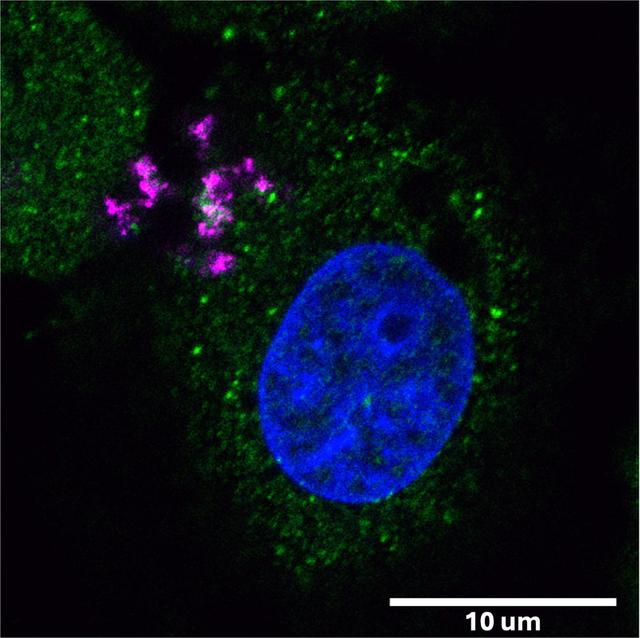
jsc2025e047405 (5/28/2025) --- Image of the preparative zone of cultured tobacco cells with visualized microtubules (yellow: microtubules [preparative zone], magenta: nuclei). For the Effects of the Space Environment on Cell Division in Plants (Plant Cell Division) investigation, plant samples are collected that are fixed and frozen for analysis of microstructures, microtubules, proteomes, and transcriptome and imaged using the JAXA Confocal Microscope (COSMIC). The Plant Cell Division investigation provides researchers with a better understanding of how gravity affects the body plan of plants could support production of food crops on future spaceflight missions. Image courtesy of University of Toyama.
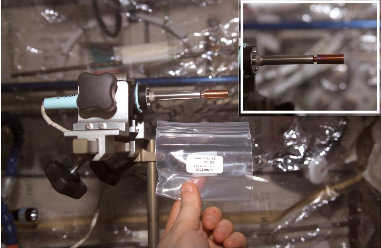
This soldering iron has an evacuated copper capsule at the tip that contains a pellet of Bulk Metallic Glass (BMG) aboard the International Space Station (ISS). Prior to flight, researchers sealed a pellet of bulk metallic glass mixed with microscopic gas-generating particles into the copper ampoule under vacuum. Once heated in space, such as in this photograph, the particles generated gas and the BMG becomes a viscous liquid. The released gas made the sample foam within the capsule where each microscopic particle formed a gas-filled pore within the foam. The inset image shows the oxidation of the sample after several minutes of applying heat. Although hidden within the brass sleeve, the sample retained the foam shape when cooled, because the viscosity increased during cooling until it was solid.